

Abstract
Although ovarian cancer usually responds well to platinum- and taxane-based first-line chemotherapy, most patients develop recurrence and chemoresistance. Regenerating gene 4 (REG4) is a secretory protein involved in cell differentiation and proliferation. We found higher REG4 expression in ovarian cancer than in normal tissues (p < .05). Regenerating gene 4 expression was negatively associated with overall, progression-free or post-progression survival rates of patients with ovarian cancer receiving platinum or paclitaxel treatment (p < .05) according to a Kaplan–Meier plotter. Regenerating gene 4 overexpression resulted in either cisplatin or paclitaxel resistance, and apoptosis resistance in CAOV3 ovarian cancer cells (p < .05). REG4-transfected ovarian cancer cells showed stronger migration and invasion treated with cisplatin or paclitaxel (p < .05). Additionally, cisplatin or paclitaxel exposure led to the overexpression of phosphorylated phosphoinositide 3-kinase (p-PI3K), p-Akt, phosphorylated mammalian target of rapamycin (p-mTOR), glutathione S-transferase-π, survivin, and B-cell lymphoma 2 in REG4 transfectants compared with control cells (p < .05). These findings suggested that REG4 expression was up-regulated in ovarian cancer, and associated with poor survival and chemotherapy resistance. REG4 promoted the occurrence, development, and chemotherapy resistance of ovarian cancer by regulating cell proliferation, apoptosis, migration, and invasion, and PI3K/Akt/m-TOR signalling pathways.
What is already known on this subject? REG4 mRNA expression is up-regulated in many digestive cancers. High REG4 expression was associated with an adverse prognosis, high tumour and nodal stages, poor differentiation, and hepatic and peritoneal metastases of digestive cancers. REG4 expression conferred cancer cells with increased resistance to chemoradiotherapy, especially 5-FU-based treatment, by activating the MAPK/Erk/Bim signalling pathway.
What do the results of this study add? REG4 was highly expressed in ovarian cancer. The expression of p-PI3K, p-AKT, p-mTOR, GST-π, survivin, and Bcl-2 was increased in REG4-overexpressing cells. High REG4 expression was significantly associated with inferior OS, PFS, and PPS rates in patients with ovarian cancer receiving platinum chemotherapy. REG4 mediated cisplatin and paclitaxel resistance in CAOV3 ovarian cancer cells. The percentage of apoptotic cells was markedly lower in REG4-transfected compared to mock-transfected cells after cisplatin or paclitaxel treatment.
What are the implications of these findings for clinical practice and/or further research? This study aimed to evaluate the prognostic significance of REG4 expression in ovarian cancer treated with platinum and paclitaxel, to explore REG4 chemoresistance mechanisms to platinum and paclitaxel, and to provide a scientific experimental basis for the clinical treatment and outcome evaluation of ovarian cancer. In order to provide comprehensive clinical treatment of ovarian cancer, it is helpful to improve our understanding of multi-drug resistance and identify new cancer diagnostic biomarkers.
IMPACT STATEMENT
Keywords:
Introduction
Ovarian cancer is one of the most common cancers in women, with an incidence that has been increasing (Siegel et al. Citation2018; Torre et al. Citation2018). More importantly, the mortality rate of ovarian cancer ranks first among gynaecologic malignancies (Bray et al. Citation2018). The early diagnosis of ovarian cancer is very difficult due to atypical early clinical manifestations. More than 70% of patients with ovarian cancer were not diagnosed until the disease had progressed to stage III (51%) or IV (29%), of which the 5-year survival rate was less than 30% (Torre et al. Citation2018; Lheureux et al. Citation2019). One of the important problems regarding ovarian cancers is relapse after surgery and first-line chemotherapy. Usually, recurrence of the disease responds poorly to first-, and sometimes even second-line chemotherapy. Inherent or acquired resistance to ovarian cancer may be due to weak immunosurveillance and drug-resistant cells (Laganà et al. Citation2015; Bizzarri et al. Citation2021). Accumulating evidence suggests that neoadjuvant chemotherapy and/or primary debulking surgery of ovarian cancer should be personalised according to the performance status of the patient, especially in elderly patients (Vitale et al. Citation2019; Fagotti et al. Citation2020). Therefore, in order to provide comprehensive clinical treatment of ovarian cancer, it is helpful to improve our understanding of multi-drug resistance and identify new cancer diagnostic biomarkers.
The regenerating gene (REG) family was originally found in regenerated islets (Chen et al. Citation2019), and includes REG1 (REG1α, REG1β), REG2, REG3 (REG3α, REG3β, REG3γ, and REG3δ) and REG4, which belong to the calcium-dependent lectin superfamily (C-type lectin), and have anti-apoptotic properties (Zhao et al. Citation2013). They stimulate regeneration in pancreatic β and epithelial cells of the digestive tract, and are related to the proliferation and differentiation of gastrointestinal tract, liver and pancreatic cells (Takasawa Citation2016). Regenerating gene 4 was initially identified from the cDNA library analysis of samples from patients with inflammatory bowel disease (IBS) (Takasawa Citation2016), suggesting its initiating role in the multi-step tumorigenesis of IBS-associated colorectal cancer. Regenerating gene 4 is located on human chromosome 1q12-q21, includes an open reading frame of 477 bp, and encodes an 18-kDa 158aa-containing protein (Hartupee et al. Citation2001). Regenerating gene 4 is a potent activator of the epidermal growth factor receptor (EGFR)/Akt/activator protein-1 pathway in colorectal cancer cells and enhances the expression of anti-apoptotic B-cell lymphoma (Bcl)-xL, Bcl-2, and survivin proteins (Bishnupuri et al. Citation2006). Either REG4 treatment or its overexpression protects intestinal crypt cells from radiation-mediated apoptotic resistance by causing the overexpression of anti-apoptotic Bcl-xL, Bcl-2, and survivin. Regenerating gene 4 overexpression has been associated with resistance to irradiation-induced apoptosis in colorectal cancer cells (Bishnupuri et al. Citation2010). Bishnupuri et al. (Citation2014) found that REG4 induced mitogenesis of colorectal cancer cells by an Akt–glycogen synthase kinase 3β-β-catenin-transcription factor-4 pathway. He et al. (Citation2012) found that REG4 contributed to the invasiveness and proliferation of pancreatic adenocarcinoma cells by up-regulating matrix metalloproteinase (MMP0-7 and −9 expression. Recently, REG4 expression was reported to phosphorylate EGFR and suppress 5-fluorouracil (FU)-induced apoptosis in gastric adenocarcinoma cells (Mitani et al. Citation2007).
Regenerating gene 4 expression was stimulated by transforming growth factor-α, basic fibroblast growth factor, epidermal growth factor, and hepatocyte growth factor in colorectal cancer cells (Oue et al. Citation2007). Regenerating gene 4 mRNA expression was up-regulated in gastric, colorectal, hepatocellular, pancreatic, and prostate cancers, and was higher in metastatic than primary prostate cancers (Zheng et al. Citation2010). Regenerating gene 4 protein was highly expressed in epithelial cells, which was characterised by neuroendocrine and mucin production (Oue et al. Citation2005). Zhang et al. (Citation2019) revealed that REG4 expression was associated with an adverse prognosis, high tumour and nodal stages, poor differentiation, and hepatic and peritoneal metastasis of cancers. Regenerating gene 4 expression conferred cancer cells with increased resistance to chemoradiotherapy, especially 5-FU-based treatment. Zhang et al. (Citation2020) found that exposure to a single-chain antibody against REG4 suppressed tumour growth and enhanced 5-FU-induced cell death in gastric cancer cells. Jin et al. (Citation2017) demonstrated that REG4 enhanced the resistance of gastric cancer to 5-FU by activating the mitogen activated protein kinase/extracellular-signal-regulated kinase/Bim signalling pathway. This study aimed to evaluate the prognostic significance of REG4 expression in ovarian cancer treated with platinum and paclitaxel, and to explore the chemoresistance mechanisms of REG4 to platinum and paclitaxel, in order to provide a scientific experimental basis for the clinical treatment and outcome evaluation of ovarian cancer.
Materials and methods
Oncomine database
The Oncomine database (https://www.oncomine.org/resource/login.html) is a data visualisation platform, established by physicians, scientists, and software engineers at the University of Michigan, that can link genes, drug development and clinical significance. It integrates databases such as Gene Expression Omnibus Database (GEO), The Cancer Genome Atlas (TCGA), and published cancer gene DNA and RNA microarray data. The screening conditions were as follows: ‘search: REG4’; ‘Visualize: Gene Summary View’; ‘Affyid: 223447_at’; ‘Gene: REG4’; ‘threshold (p value): 0.05’; ‘threshold (fold change): 2’; ‘threshold (gene ranking): top 10%’; ‘data type: all’.
TIMER database
The TIMER web server (https://cistrome.shinyapps.io/timer/) is a comprehensive resource to systematically analyse immune infiltration. The screening conditions were as follows: ‘Gene Symbol: REG4’; ‘Cancer Types: (OV) ovarian serous cystadenocarcinoma’.
Kaplan–Meier plotter database
The effect of REG4 expression on the survival of patients with ovarian cancer treated with platinum or paclitaxel was investigated using Kaplan–Meier plotter analysis. The y-axis represented the impact of REG4 expression on relapse-free survival (RFS), post-progression survival (PPS), and overall survival (OS), separately, and the x-axis represented the observation time. The two groups of patients were compared by Kaplan–Meier curves. The hazard ratios with 95% confidence intervals (CI) and p values were calculated as well. The screening conditions were as follows: ‘Cancer: Ovarian Cancer’; ‘Gene symbol: REG4’; ‘Affyid: 223447_at’.
Cell culture, transfection, and screening
Ovarian cancer cells (CAOV3) were routinely cultured in DMEM medium (Gibco, ThermoFisher Scientific, Waltham, MA, USA) containing 10% foetal bovine serum. CAOV3 cells in the logarithmic phase of growth were inoculated into a 6-cm2 culture plate. The supernatant was discarded, and cells were washed twice with phosphate buffered saline (PBS; Gibco) when the cells reached approximately 60% confluence. A pcDNA3.1–REG4 expression plasmid was provided by Prof. Akira Sugawara from the Department of Advanced Biological Sciences for Regeneration, Tohoku University, Japan. Serum-free and double-antibody-free medium (4 mL) was added to the 6-cm2 culture plate. Plasmid (2.4 μg) and transfection reagent (9 μL; QIAGEN, Hilden, Germany) were added to 200 μL of serum-free medium, respectively, and the mixture was added to the culture dish after incubation at 37 °C for 15 min. The medium was changed after 16 h. Monoclonal cell lines were screened by 500 μg/mL of geneticin (G418, Solarbio, Beijing, China). The expression level of REG4 was detected by real-time reverse transcriptase (RT)–PCR and western blotting. Monoclonal cell lines showing high expression of REG4 were used for subsequent experiments. CAOV3 cells stably transfected with pcDNA3.1–REG4 were used as the REG4 group, and cells stably transfected with pcDNA3.1 empty vector were used as the mock group.
Proliferation assay
Logarithmic-phase cells were seeded into 96-well plates (2 × 10³/well) for routine culture. After cell adherence, either cisplatin or paclitaxel was added at different concentrations at 0 h, respectively. After 0 and 48 h, 20 μL MTT (tetrazolium; 5 mg/mL, Sigma, St Louis, MI, USA) solution was added to each well. After incubation for 4 h, 150 μL dimethylsulfoxide (Solarbio) was added to each well to dissolve the MTT product and cells were shaken for 10 min. Absorbance values were measured at 490 nm by a spectrometer and the proliferation rate was calculated. A proliferation curve was drawn, with time as the horizontal axis and the proliferation rate as the vertical axis.
Apoptosis by flow cytometry
Cells in both mock and REG4 groups at a logarithmic phase of growth were collected, trypsinised, and centrifuged routinely; cells were resuspended in complete medium. The cells were counted and inoculated in three 6-cm2 plates (2 × 10⁶ cells per plate). After cell adhesion, different concentrations of cisplatin and paclitaxel were added to two plates per group; one untreated plate per group was used a control. After 48 h of culture, cells and supernatants were collected, washed twice with PBS, and cells resuspended as single-cell suspensions, with annexin V-fluorescein isothiocyanate (FITC) combined buffer, to a concentration of 1 × 10⁶/mL. Annexin V-FITC dye (5 μL; Keygen Biotech, Nanjing, China) was added to each cell suspension, which was incubated in the dark at room temperature for 15 min, and then incubated with 5 μL propidium iodide dye for 5 min. Flow cytometry was used for detection and analysis.
Transwell assay
For the migration assay, cells (3 × 10⁴) were suspended in 100 μL of serum-free DMEM and inoculated in the upper chamber of each Transwell® (BD, Franklin Lakes, NJ, USA). DMEM (600 μL) with 10% foetal bovine serum was added to each bottom chamber. After cells attached, cisplatin and paclitaxel were added to each upper chamber. For the invasion assay, the membrane in the upper chamber was coated with Matrigel for 4 h in advance, and the rest of the assay was identical to migration experiments. After 48 h, cells that had not migrated through the membrane in the upper chamber were wiped off with cotton swabs. The cells adhering to the lower side of the membrane were washed with PBS, fixed with 4% paraformaldehyde, and stained with 0.1% crystal violet. After cleaning and drying, the membranes were cut with a knife and sealed on a slide with neutral resin. Five random fields of view were selected for photography and cells counted under a microscope (100×).
Real-time RT–PCR
Total RNA was extracted from cells in a logarithmic phase of growth from both mock- and REG4-transfected groups according to the instructions of a QIAGEN RNase mini kit. Total RNA was synthesised using SMART M-MLV (Moloney Murine Leukaemia Virus; Takara, Shiga, Japan) reverse transcriptase and random primers. The volume was 50 μL, and the final template concentration was 100 ng/μL. A 30 μL pre-denatured sample containing template, primer, and diethylpolycarbonate (DEPC) water was pre-denatured at 70 °C for 10 min. Subsequently, 5 × M-MLV buffer, M-MLV, dNTP, RNA enzyme inhibitor, and DEPC water were added to final 50 μL volume according to instructions for reverse transcription. The procedure was: 30 °C for 10 min, 42 °C for 60 min, and 72 °C for 10 min. Real-time RT–PCR was performed according to a SYBR Premix Ex Taq TM II kit (Takara, Japan) instructions. The REG4 gene amplification primers used were: upstream 5′-TAACTTGGAGCAG CAACGAATG-3′, downstream 5′-GGGCTAGCAGAAAGGAAGGA-3′. GAPDH primers: upstream 5′-CAATGACCTTCATTGACC-3′, downstream 5′-TGGAAGATGGTGATGGGATT-3′. Reaction conditions were: 94 °C for 1 min, 60 °C for 1 min, 72 °C for 1 min, and for 34 cycles. The relative expression of mRNA was calculated by 2−ΔΔCt method.
Western blot
Logarithmic-phase mock and REG4 cells were trypsinised and centrifuged. The cells were resuspended in complete medium and counted. Six 10-cm2 plates were inoculated in each of the two groups at 3 × 10⁶ cells per plate. After cell adhesion, cells in two plates per group were treated with cisplatin and paclitaxel, and the remaining two untreated plates were used as controls. After 48 h of culture, total protein was extracted using radioimmunoprecipitation lysis buffer, and the absorbance was measured at 595 nm on a spectrometer using a Coomassie brilliant blue method. The protein concentration was calculated according to a standard curve. For electrophoresis, proteins were denatured at 95 °C for 5 min after dilution. The proteins in samples were separated by polyacrylamide gel electrophoresis at 110 V/90 min, transferred to a PVDF membrane at 60 V/150 min, and blocked in 5% skim milk for 1 h at room temperature. Phosphorylated proteins were blocked with 5% bovine serum albumin (Sigma). Membranes were incubated with different primary antibodies () overnight at 4 °C. Membranes were washed three times with tris-buffered saline/Tween 20 for 10 min, then incubated with secondary antibodies for 1 h at room temperature, and washed again. Target proteins were detected by ECL chemiluminescence kit.
Table 1. Primary antibodies used in the present study.
Statistical analysis
GraphPad Prism 8.0 software was employed for statistical analysis. Continuous variables with normal distribution were presented as mean ± SD and compared by Student’s t-test. Non-normal variables were expressed as median plus interquartile range and compared by either Mann–Whitney U or Kruskal–Wallis tests. A log-rank test was used to compare Kaplan–Meier curves. p < .05 was considered statistically significant.
Results
Prognostic value of REG4 in ovarian cancer treated with platinum or paclitaxel
The Oncomine database revealed 285 studies on REG4 gene expression in various tumours and normal tissues. Twenty-one studies showed significant differences in REG4 expression between normal and tumour tissues (p < .05), including 14 showing high expression and seven showing low expression. One study showed that REG4 was highly expressed in ovarian cancer (, p < .05). A positive relationship existed between REG4 mRNA expression and cell purity in ovarian cancer (, p < .05). To estimate the prognostic significance of REG4 expression in ovarian cancer treated with platinum or paclitaxel, we plotted a Kaplan–Meier survival curve of patients showing low or high expression of REG4 using an online Kaplan–Meier plotter database. We found that increased REG4 mRNA expression markedly correlated with worse overall survival (OS) and post-progression survival (PPS) in ovarian cancer treated with taxol (, p < .05). As shown in , high REG4 mRNA expression was significantly associated with inferior OS, progression-free survival (PFS), and PPS in patients with ovarian cancer who received platinum chemotherapy (p < .05). and show that REG4 had significant prognostic values in patients with ovarian cancer at different clinical stages and grades, and with serous subtype and TP53 mutations (p < .05).
Figure 1. Expression of REG4 mRNA in ovarian cancer. (A) Regenerating gene 4 (REG4) mRNA expression was observed in various cancers according to an Oncomine database. (B) A positive correlation existed between REG4 mRNA expression and cell purity in ovarian cancer according to a Timer database. (C) The prognostic significance of REG4 mRNA expression was analysed in ovarian cancer treated with taxol chemotherapy using a Kaplan–Meier plotter. (D) The prognostic significance of REG4 mRNA expression was analysed in ovarian cancer treated with platinum (platin) chemotherapy using a Kaplan–Meier plotter.
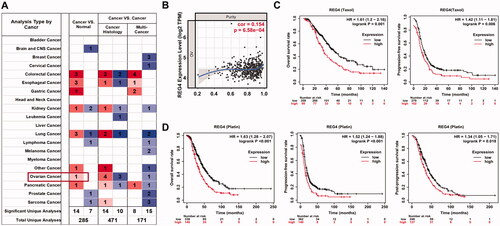
Table 2. Prognostic value of REG4 in subtype ovarian cancer treated with platin.
Table 3. Prognostic value of REG4 in ovarian cancer treated with paclitaxel.
The effects of REG4 on phenotypes of ovarian cancer cells
To investigate the role of REG4 in the chemoresistance of ovarian cancer, we established REG4-overexpressing cell models in the human serous ovarian cancer cell line, CAOV3. As shown in , REG4 mRNA expression was much higher in the REG4-transfected compared to mock-transfected group by real-time RT–PCR (p < .001). Western blotting showed that REG4 protein expression was significantly higher in the REG4-transfected compared to mock-transfected group (, p < .05). Thus REG4-transfected cells showed stable and high expression of REG4 compared with the mock-transfected cells.
Figure 2. Effects of REG4 on cellular phenotypes of ovarian cancer cells. (A) After transfection of a regenerating gene 4 (REG4)-overexpressing plasmid, REG4 mRNA was detected in CAOV3 cells by real-time reverse transcriptase (RT)–PCR. (B) After transfection of REG4-overexpressing plasmid, REG4 protein was detected in CAOV3 cells by western blot. (C) The effect of REG4 expression on the proliferation of CAOV3 cells treated with cisplatin or taxol. (D) The effect of REG4 expression on apoptosis in CAOV3 cells treated with cisplatin or taxol. (E) The effect of REG4 expression on migration and invasion by CAOV3 cells treated with cisplatin or taxol. Note: compared with control, *p < .05; **p < .01; ***p < .001.
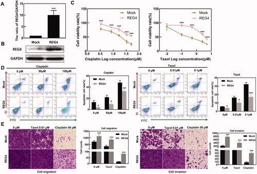
To delineate the role of REG4 in chemoresistance, we overexpressed REG4 in CAOV3 cells by transfecting REG4 expression plasmids and then treated cells with different concentrations of cisplatin or taxol, followed by an MTT assay to measure cell proliferation. After chemical exposure, CAOV3 cancer cells overexpressing REG4 showed a higher proliferative capacity than cells of the mock-transfected group (, p < .05).
To better understand the role of REG4 overexpression in cisplatin- or paclitaxel-sensitivities in ovarian cancer cells, we examined the apoptosis rate induced by cisplatin or taxol. Apoptosis was measured by flow cytometry, which showed that the percentage of apoptotic cells was markedly lower in REG4-transfected cells than mock-transfected cells after cisplatin or taxol treatment (, p < .05).
Transwell assays were used to assess the effect of REG4 on migration and invasion in CAOV3 cells treated with cisplatin or taxol. It was found that the number of invading cells were higher in the REG4 transfection group than in the mock-transfected group after cisplatin or taxol treatment (, p < .05).
The effects of REG4 on the expression of drug resistance-related molecules in ovarian cancer cells after chemo-treatment
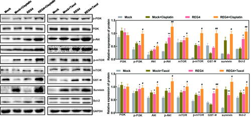
Western blots revealed that after either cisplatin or taxol treatment, the expression of phosphorylated phosphoinositide 3-kinase (p-PI3K), Akt, p-Akt, mammalian target of rapamycin (mTOR), p-mTOR, glutathione S-transferase (GST)-π, and Bcl-2 was increased in REG4-overexpressing cells compared with those in the mock-transfected group (, p < .05).
Figure 3. Effect of REG4 expression on expression of drug-resistant proteins in ovarian cancer cells. Western blotting was used to screen chemoresistance-related proteins in ovarian cancer cells or their regenerating gene 4 (REG4) transfectants treated with cisplatin or taxol. Note: REG4-transfected cells were compared with mock-transfected cells, *p < .05; **p < .01; compared with cisplatin or taxol, #p < .05; ##p < .01.
Discussion
Regenerating gene 4 expression was found to positively correlate with the depth of invasion, clinical stage, dedifferentiation, distant metastasis, and inherent resistance to 5-FU of gastric cancer (Ying et al. Citation2013). In patients with rectal cancer treated with neoadjuvant concurrent chemoradiotherapy (CCRT), REG4 overexpression was closely related to a poor response, adverse outcomes, and aggressive phenotypes; this showed that REG4 was a surrogate marker that predicted CCRT resistance (He et al. Citation2014). Zhou et al. (Citation2013) also found that REG4 knockdown reduced gastric cancer cells’ stemness and increased the effectiveness of cell death after radiochemotherapy. Eguchi et al. (Citation2009) demonstrated that either serum REG4 concentration or REG4 overexpression was a useful biomarker to predict the clinical outcome of patients with pancreatic cancer who received preoperative chemoradiotherapy. Cells that overexpressed REG4 appeared highly resistant to radiotherapy but showed a modest resistance to gemcitabine. Taken together, we concluded that REG4 expression might play an important role in the radiochemoresistance of cancers.
The serum REG4 level or REG4 mRNA and protein expression was shown to be potential biomarkers of ovarian mucinous adenocarcinoma (Chen et al. Citation2015; Lehtinen et al. Citation2016). The expression of REG4 was closely linked as an independent factor to short OS or relapse-free survival (RFS) in patients with ovarian cancer (Chen et al. Citation2015). Here, REG4 overexpression was found to be an adverse prognostic factor for OS, PFS, and PPS in ovarian cancer treated with cisplatin, while REG4 overexpression was a poor prognostic factor for OS and PFS in ovarian cancer treated with paclitaxel. Patients with serous, advanced and well-differentiated ovarian cancer, and high levels of REG4, all responded poorly to platinum and paclitaxel chemotherapy indicating that REG4 was related to resistance to these therapies and was a poor predictor. Hwang et al. (Citation2020) reported that REG4 promoted cancer stemness via Wnt/β-catenin signalling as evidenced by tumour organoid systems, intestinal tumours from Apcmin/+/KrasG12D LA2 mice, and an NSG mouse xenograft system. Sasaki et al. (Citation2016) found that deep REG4-positive crypt secretory cells functioned as an epithelial niche for intestinal leucine-rich repeat-containing G-protein coupled receptor 5-positive stem cells. These findings indicated that REG4 mRNA expression was closely linked to a poor prognosis in ovarian cancer treated with cisplatin or paclitaxel due to the effects of REG4 on cancer stemness.
Sun et al. (Citation2019) revealed that silenced REG4 expression significantly reduced cancer cell proliferation, inhibited tumorigenesis, and arrested the cell cycle by targeting the G2/M checkpoint and E2F. Wang et al. (Citation2016) found that REG4 promoted peritoneal metastasis of gastric cancer cells via G-protein coupled receptor 37, and triggered a positive feedback loop. Chen et al. (Citation2015) demonstrated that REG4 overexpression and recombinant REG4 weakened cellular apoptosis, and strengthened G2/S progression, proliferation, migration, and invasion of ovarian cancer cells. Rafa et al. (Citation2010) found that 24-h treatment with REG4 stimulated cell growth in a paracrine manner, and REG4 overexpression promoted migration and invasion of cancer cells in either an autocrine or paracrine manner, which was significantly blocked by an anti-REG4 antibody. In the present study, we found that the cellular activities of proliferation, anti-apoptosis, migration, and invasion of REG4-overexpressing CAOV3 ovarian cancer cells were stronger than those of the mock-transfected group after cisplatin and taxol treatment. This indicates that REG4 may promote resistance to cisplatin and taxol by promoting the proliferation, migration and invasion of ovarian cancer cells and by inhibiting apoptosis.
Anticancer drugs such as platin and paclitaxel play a crucial role in the treatment of cancer. However, inherent or acquired chemotherapy resistance often severely limits their efficacy. Recognition of mechanisms of cell resistance has progressed, and includes reducing drug intake, increasing drug efflux, changing drug targets, inactivating drugs, increasing DNA repair, changing the expression of oncogenes, and the activation and inactivation of several signalling pathways (Zheng Citation2017). The PI3K/Akt/mTOR signalling cascade is the most common pathway in human cancer and is crucial to cancer development and chemoresistance (Nakayama et al. Citation2006; Zheng Citation2017). The carcinogenesis of the PI3K/Akt/mTOR signalling pathway in ovarian cancer is very complex, but changes in this produce the same endpoints: tumour formation, cancer cell migration, invasion, and increased chemoradiotherapy resistance (Philp et al. Citation2001; Huang et al. Citation2011; Cheaib et al.Citation2015). The expression of p-PI3K, p-Akt, and P-mTOR in REG4-overexpressing CAOV3 cells was increased compared with mock-transfected cells after cisplatin and paclitaxel treatment, indicating that REG4 might activate the PI3K/Akt/mTOR signalling pathway allowing CAOV3 cells to become resistant to cisplatin and taxol.
In addition, we found that the expression of GST-π, survivin, and Bcl-2 in REG4-overexpressing CAOV3 cells treated with cisplatin and paclitaxel were up-regulated compared with mock-transfected cells. Glutathione S-transferases are a family of isoenzymes that detoxify electrophiles by conjugation to thiol-reduced glutathione. Therefore, they are essential to protect cells from toxins (drugs, pesticides, carcinogens) and oxidative stress (Chatterjee and Gupta Citation2018). Glutathione S-transferases in human cells can be divided into three subclasses: alkaline (α), neutral (μ), and acidic (π); GST-π is the most closely related to drug resistance in tumours. The main function of GST is to catalyse the combination of glutathione with chemical drugs, thereby reducing their cytotoxic effect (Matsui et al. Citation2020). Therefore, REG4 may also increase the expression of GST-π and Bcl-2 allowing ovarian cancer cells to become resistant to cisplatin and paclitaxel.
This study highlighted the prognostic significance of REG4 mRNA in patients with ovarian cancer receiving chemotherapy, and the effects and related molecular mechanisms of REG4 overexpression on chemoresistance, proliferation, apoptosis, migration, and invasion by ovarian cancer cells. However, knowledge of the relationship between REG4 expression and chemotherapy efficacy and the prognostic significance of REG4 expression in patients with ovarian cancer remains unelucidated and is a limitation of this study.
In summary, REG4 was associated with platin and paclitaxel chemotherapy resistance in ovarian cancer. The overexpression of REG4 is an important predictor of a poor prognosis in patients with ovarian cancer treated with platin and paclitaxel chemotherapy. Our data showed that REG4 overexpression was closely associated with an invasive phenotype, and led to cisplatin and paclitaxel resistance by activating the PI3K/Akt/mTOR signalling pathway and up-regulating the expression of GST-π, survivin, and Bcl-2.
Ethical approval
This study was approved by the Ethics committee of Chengde Medical University Affiliated Hospital (No. CYFYLL2022166). All methods were performed in accordance with the relevant guidelines and regulations.
Consent to participate statement
Informed consent was obtained from all subjects and/or their legal guardian(s).
Disclosure statement
No potential conflict of interest was reported by the authors.
Data availability statement
There is no data needed to be deposited. The datasets generated and/or analysed during the current study are available from the corresponding author on reasonable request. This study was performed according to the Enhancing the QUAlity and Transparentcy of health Research (EQUATOR) network guideline.
Additional information
Funding
References
- Bishnupuri KS, Luo Q, Murmu N, Houchen CW, Anant S, Dieckgraefe BK. 2006. Reg IV activates the epidermal growth factor receptor/Akt/AP-1 signaling pathway in colon adenocarcinomas. Gastroenterology 130:137–149.
- Bishnupuri KS, Luo Q, Sainathan SK, Kikuchi K, Sureban SM, Sabarinathan M, et al. 2010. Reg IV regulates normal intestinal and colorectal cancer cell susceptibility to radiation-induced apoptosis. Gastroenterology 138:616–626.e1-2.
- Bishnupuri KS, Sainathan SK, Bishnupuri K, Leahy DR, Luo Q, Anant S, et al. 2014. Reg4-induced mitogenesis involves Akt-GSK3β-β-catenin-TCF-4 signaling in human colorectal cancer. Molecular Carcinogenesis 53(Suppl 1):E169–E180.
- Bizzarri N, du Bois A, Fruscio R, De Felice F, De Iaco P, Casarin J, et al. 2021. Is there any therapeutic role of pelvic and para-aortic lymphadenectomy in apparent early stage epithelial ovarian cancer? Gynecologic Oncology 160:56–63.
- Bray F, Ferlay J, Soerjomataram I, Siegel RL, Torre LA, Jemal A; Global Cancer Statistics. 2018. GLOBOCAN estimates of incidence and mortality worldwide for 36 cancers in 185 countries. CA: A Cancer Journal for Clinicians 68:394–424.
- Chatterjee A, Gupta S. 2018. The multifaceted role of glutathione S-transferases in cancer. Cancer Letters 433:33–42.
- Cheaib B, Auguste A, Leary A. 2015. The PI3K/Akt/mTOR pathway in ovarian cancer: therapeutic opportunities and challenges. Chinese Journal of Cancer 34:4–16.
- Chen S, Gou WF, Zhao S, Niu ZF, Zhao Y, Takano Y, Zheng HC. 2015. The role of the REG4 gene and its encoding product in ovarian epithelial carcinoma. BMC Cancer 15:471.
- Chen Z, Downing S, Tzanakakis ES. 2019. Four decades after the discovery of regenerating islet-derived (Reg) proteins: current understanding and challenges. Frontiers in Cell and Developmental Biology 7:235.
- Eguchi H, Ishikawa O, Ohigashi H, Takahashi H, Yano M, Nishiyama K, et al. 2009. Serum REG4 level is a predictive biomarker for the response to preoperative chemoradiotherapy in patients with pancreatic cancer. Pancreas 38:791–798.
- Fagotti A, Ferrandina MG, Vizzielli G, Pasciuto T, Fanfani F, Gallotta V, et al. 2020. Randomized trial of primary debulking surgery versus neoadjuvant chemotherapy for advanced epithelial ovarian cancer (SCORPION-NCT01461850). International Journal of Gynecological Cancer 30:1657–1664.
- Hartupee JC, Zhang H, Bonaldo MF, Soares MB, Dieckgraefe BK. 2001. Isolation and characterization of a cDNA encoding a novel member of the human regenerating protein family: Reg IV. Biochimica et Biophysica Acta 1518:287–293.
- He HL, Lee YE, Shiue YL, Lee SW, Lin LC, Chen TJ, et al. 2014. Overexpression of REG4 confers an independent negative prognosticator in rectal cancers receiving concurrent chemoradiotherapy. Journal of Surgical Oncology 110:1002–1100.
- He XJ, Jiang XT, Ma YY, Xia YJ, Wang HJ, Guan TP, et al. 2012. REG4 contributes to the invasiveness of pancreatic cancer by upregulating MMP-7 and MMP-9. Cancer Science 103:2082–2091.
- Huang J, Zhang L, Greshock J, Colligon TA, Wang Y, Ward R, et al. 2011. Frequent genetic abnormalities of the PI3K/AKT pathway in primary ovarian cancer predict patient outcome. Genes, Chromosomes & Cancer 50:606–618.
- Hwang JH, Yoon J, Cho YH, Cha PH, Park JC, Choi KY. 2020. A mutant KRAS-induced factor REG4 promotes cancer stem cell properties via Wnt/β-catenin signaling. International Journal of Cancer 146:2877–2890.
- Jin J, Lv H, Wu J, Li D, Chen K, Zhang F, et al. 2017. Regenerating family member 4 (Reg4) enhances 5-fluorouracil resistance of gastric cancer through activating MAPK/Erk/Bim signaling pathway. Medical Science Monitor 23:3715–3721.
- Laganà AS, Colonese F, Colonese E, Sofo V, Salmeri FM, Granese R, et al. 2015. Cytogenetic analysis of epithelial ovarian cancer’s stem cells: an overview on new diagnostic and therapeutic perspectives. European Journal of Gynaecological Oncology 36:495–505.
- Lehtinen L, Vesterkvist P, Roering P, Korpela T, Hattara L, Kaipio K, et al. 2016. REG4 is highly expressed in mucinous ovarian cancer: a potential novel serum biomarker. PLoS One 11:e0151590.
- Lheureux S, Braunstein M, Oza AM. 2019. Epithelial ovarian cancer: evolution of management in the era of precision medicine. CA: A Cancer Journal for Clinicians 69:280–304.
- Matsui R, Ferran B, Oh A, Croteau D, Shao D, Han J, et al. 2020. Redox regulation via glutaredoxin-1 and protein S-glutathionylation. Antioxidants & Redox Signaling 32:677–700.
- Mitani Y, Oue N, Matsumura S, Yoshida K, Noguchi T, Ito M, et al. 2007. Reg IV is a serum biomarker for gastric cancer patients and predicts response to 5-fluorouracil-based chemotherapy. Oncogene 26:4383–4393.
- Nakayama K, Nakayama N, Kurman RJ, Cope L, Pohl G, Samuels Y, et al. 2006. Sequence mutations and amplification of PIK3CA and AKT2 genes in purified ovarian serous neoplasms. Cancer Biology & Therapy 5:779–785.
- Oue N, Kuniyasu H, Noguchi T, Sentani K, Ito M, Tanaka S, et al. 2007. Serum concentration of Reg IV in patients with colorectal cancer: overexpression and high serum levels of Reg IV are associated with liver metastasis. Oncology 72:371–380.
- Oue N, Mitani Y, Aung PP, Sakakura C, Takeshima Y, Kaneko M, et al. 2005. Expression and localization of Reg IV in human neoplastic and non-neoplastic tissues: Reg IV expression is associated with intestinal and neuroendocrine differentiation in gastric adenocarcinoma. The Journal of Pathology 207:185–198.
- Philp AJ, Campbell IG, Leet C, Vincan E, Rockman SP, Whitehead RH, et al. 2001. The phosphatidylinositol 3′-kinase p85alpha gene is an oncogene in human ovarian and colon tumors. Cancer Research 61:7426–7429.
- Rafa L, Dessein AF, Devisme L, Buob D, Truant S, Porchet N, et al. 2010. REG4 acts as a mitogenic, motility and pro-invasive factor for colon cancer cells. International Journal of Oncology 36:689–698.
- Sasaki N, Sachs N, Wiebrands K, Ellenbroek SI, Fumagalli A, Lyubimova A, et al. 2016. Reg4+ deep crypt secretory cells function as epithelial niche for Lgr5+ stem cells in colon. Proceedings of the National Academy of Sciences of the United States of America 113:E5399–E5407.
- Siegel RL, Miller KD, Jemal A. 2018. 2019. Cancer statistics. CA: A Cancer Journal for Clinicians 68:7–30.
- Sun S, Hu Z, Huang S, Ye X, Wang J, Chang J, et al. 2019. REG4 is an indicator for KRAS mutant lung adenocarcinoma with TTF-1 low expression. Journal of Cancer Research and Clinical Oncology 145:2273–2283.
- Takasawa S. 2016. Regenerating gene (REG) product and its potential clinical usage. Expert Opinion on Therapeutic Targets 20:541–550.
- Torre LA, Trabert B, DeSantis CE, Miller KD, Samimi G, Runowicz CD, et al. 2018. Ovarian cancer statistics. CA: A Cancer Journal for Clinicians 68:284–296.
- Vitale SG, Capriglione S, Zito G, Lopez S, Gulino FA, Di Guardo F, et al. 2019. Management of endometrial, ovarian and cervical cancer in the elderly: current approach to a challenging condition. Archives of Gynecology and Obstetrics 299:299–315.
- Wang H, Hu L, Zang M, Zhang B, Duan Y, Fan Z, et al. 2016. REG4 promotes peritoneal metastasis of gastric cancer through GPR37. Oncotarget 7:27874–27888.
- Ying LS, Yu JL, Lu XX, Ling ZQ. 2013. Enhanced RegIV expression predicts the intrinsic 5-fluorouracil (5-FU) resistance in advanced gastric cancer. Digestive Diseases and Sciences 58:414–422.
- Zhang J, Zhu Z, Miao Z, Huang X, Sun Z, Xu H, et al. 2020. The clinical significance and mechanisms of REG4 in human cancers. Frontiers in Oncology 10:559230.
- Zhang XQ, Yu LT, Du P, Yin TQ, Zhang ZY, Xu Y, et al. 2019. Single-chain antibody against Reg4 suppresses gastric cancer cell growth and enhances 5-FU-induced cell death in vitro. Anti-Cancer Agents in Medicinal Chemistry 19:610–619.
- Zhao J, Wang J, Wang H, Lai M. 2013. Reg proteins and their roles in inflammation and cancer of the human digestive system. Advances in Clinical Chemistry 61:153–173.
- Zheng HC. 2017. The molecular mechanisms of chemoresistance in cancers. Oncotarget 8:59950–59964.
- Zheng HC, Xu XY, Yu M, Takahashi H, Masuda S, Takano Y. 2010. The role of Reg IV gene and its encoding product in gastric carcinogenesis. Human Pathology 41:59–69.
- Zhou W, Sun M, Wang DL, Wang Y, Jin F, Zhang YY, et al. 2013. Silencing of RegIV by shRNA causes the loss of stemness properties of cancer stem cells in MKN45 gastric cancer cells. Oncology Reports 30:2685–2690.