

Abstract
To clarify the clinicopathological importance of REG4 mRNA expression, we used GEO, TCGA, xiantao, UALCAN, and Kaplan-Meier plotter for a bioinformatics analysis in breast, cervical, endometrial and ovarian cancers. Compared to normal tissues, REG4 expression was found to be upregulated in breast, cervical, endometrial, and ovarian cancers (p < 0.05). Breast cancer had a higher level of REG4 methylation than normal tissues (p < 0.05), which was negatively correlated with its mRNA expression. REG4 expression was positively correlated with oestrogen and progesterone receptor expression, and aggressiveness of PAM50 classification of breast cancer patients (p < 0.05). Breast infiltrating lobular carcinomas expressed more REG4 than ductal carcinomas (p < 0.05). The REG4-related signal pathways mainly included peptidase, keratinisation, brush border and digestion and so forth in gynecological cancers. Our results indicated that REG4 overexpression was associated with gynecological carcinogenesis and their histogenesis, and may be used as a marker for aggressive behaviour and prognosis of breast or cervical cancer.
What is already known on this subject? REG4 encodes a secretory c-type lectin and plays an essential role in inflammation, carcinogenesis, apoptotic and radiochemotherapeutic resistance.
What do the results of this study add? As a standalone predictor, REG4 expression was positively correlated with progression-free survival. Expression of REG4 mRNA was positively associated with the T stage and adenosquamous cell carcinoma of cervical cancer. The top signal pathways related to REG4 included smell and chemical stimulus, peptidase, intermediate filament, and keratinisation in breast cancer; ligand-receptor interaction, metabolism of hormone, xenobiotic and retinol, peptidase, brush border and digestion in cervical and ovarian cancers; bile secretion, intermediate filament, chemical carcinogenesis, brush border and keratinisation in endometrial cancer. REG4 mRNA expression was positively correlated with DC cell infiltration in breast cancer, positively with Th17 cells, TFH, cytotoxic cells and T cells in cervical and endometrial cancers, and negatively with DC cell infiltration, cytotoxic cells and T cells in ovarian cancer. The top hub genes mainly included small proline rich protein 2B in breast cancer; fibrinogens and apoproteins in cervical, endometrial and ovarian cancers.
What are the implications of these finding for clinical practice and/or further research? Our study has showed that REG4 mRNA expression is a potential biomarker or therapeutic target for gynaecologic cancers.
IMPACT STATEMENT
1. Introduction
REG4 was discovered in the cDNA library of ulcerative colitis. It encodes an 18-kDa secretory glycoprotein with a 22aa signal peptide and has a calcium-dependent lectin domain (Zheng et al. Citation2022). Transcriptionally, CDX, GLI1 and ATF2 are discovered to activate REG4 expression by interacting with the REG4 gene’s promoter (Wang et al. Citation2011, Naito et al. Citation2012, Xiao et al. Citation2019), while miR-363 inhibits the translation of GATA6, which transcriptionally induce REG4 expression (Kawasaki et al. Citation2015). Translationally, miR-24 inhibited gastric cancer growth by downregulating REG4 (Duan et al. Citation2014). Reportedly, recombinant human REG4 (rhREG4) inhibited apoptosis of colorectal cancer cells and protected normal intestinal crypt cells from irradiation-induced apoptosis by increasing the expression of Bcl-2 and Bcl-xL via activation of the EGFR/Akt pathway (Bishnupuri et al. Citation2006, Citation2010), while Li et al. (Citation2011) discovered that proteoglycan from Phellinus linteus inhibited the growth of colorectal cancer cells by inhibiting the REG4/EGFR/Akt pathway. Additionally, REG4 was found to interact with mannan and heparin (Ho et al. Citation2010), CD44 to activate the intramembrane proteolysis by γ-secretase (Bishnupuri et al. Citation2022), and GPR37 to mediate EGFR signal (Wang et al. Citation2016).
REG4 immunostaining appeared remarkably lower in the uterus than in the ovary, where REG4 protein was identified strongly in oocytes, granulosa, and interstitial cells. In contrast, its expression was weak in the luminal epithelium and endometrial glands (Du et al. Citation2013). REG4 is overexpressed in inflammatory bowel conditions and gastrointestinal carcinomas, and is considered a biomarker for signet ring cells and neuroendocrine tumours (Oue et al. Citation2005, Sentani et al. Citation2008, Zheng et al. Citation2022).
In terms of cancer risk, women are particularly susceptible to breast, vulvar, vaginal, cervical, endometrial, and ovarian cancers (Paepke et al. Citation2020, Aquil et al. Citation2021, Segev et al. Citation2021, Seland et al. Citation2022). According to the statistical data of comprehensive global cancer in 2020 by the International Agency for Research on Cancer, gynaecologic cancers accounted overall for 16.5% of 8.2 million estimated new cancer cases in women, and have an ongoing source of concern due to their high morbidity and mortality (Golia et al. Citation2023). Accumulating evidence suggests that obesity and metabolic diseases may play a key role in increasing the risk of cancers, modulating pivotal cross-talk pathways for cell proliferation and differentiation (Giannini et al. Citation2022; Vargiu et al. Citation2022). Although significant therapeutic advances in surgical, radiochemotherapeutic, and target intervention took place in recent years, patients’ outcomes remained poor because of lack in novel strategies to detect all gynaecologic tumours as early as possible, especially new biomarkers (Giannini et al. Citation2022; Golia et al. Citation2023; Vargiu et al. Citation2022). Therefore, we sought to define the clinical, pathological, and prognostic significances of REG4 mRNA expression and the relevant signalling pathways in breast, cervical, endometrial, and ovarian cancers using TCGA, GEO, xiantao, UALCAN, and Kaplan-Meier plotter datasets as described previously.
2. Material and methods
2.1. TCGA database analysis
Via R software (4.2.1) (R package: ggplot2 3.3.6, stats 4.2.1, car), the expression data (RNA-seqV2) and clinicopathological data of breast cancer (n = 1171) were acquired from the Cancer Genome Atlas (TCGA, https://cancergenome.nih.gov/) database by TCGA-assembler (Chen et al. Citation2015). We evaluated REG4 mRNA expression in breast cancer and compared it to the clinicopathological characteristics of the cancer patients. In the sense of contrast, we used Student’s t-test. Using log-rank statistics, Kaplan-Meier survival plots were created with survival curves compared. Multivariate analysis was carried out using the Cox proportional hazards model. Two-sided p < 0.05 was statistically significant. The SPSS 17.0 program was used to examine all of the data.
2.2. GEO analysis
R software (4.2.1) (R package: ggplot2 3.3.6, stats 4.2.1, car) was used to investigate differences in REG4 mRNA expression between cancer and normal tissues. The mRNA expression profiles of GSE63514 (platform: Affymetrix-GPL570, cervical cancer) and GSE18520 (platform: Affymetrix-GPL570, ovarian cancer) were downloaded from the NCBI GEO database (https://www.ncbi.nlm.nih.gov/geo/). All data statistical methods adopt Wilcoxon rank sum test and visualised with ggplot2 3.3.6 package.
2.3. KM plotter analysis
After inputting the target gene REG4 (ID: 223447_at) at Kaplan-Meier plotter (http://kmplot.com), we analysed the prognostic significance of REG4 mRNA in cases of breast (n = 4929). In this process, we splitted patients by median, used all probe sets per gene, excluded biased arrays and analysed all the subtypes. All data were analysed by univariate COX regression analysis, subjected to logrank test and visualised using R (4.2.1) (R package: survival 3.3.1, survminer, ggplot2 3.3.6).
2.4. UALCAN analysis
Using the UALCAN database (http://ualcan.path.uab.edu), we examined the expression and methylation of the REG4 gene by entering REG4 gene in the box of breast, cervical, endometrial, and ovarian malignancies cancer. In addition, they were compared to the clinicopathological and prognostic parameters of breast, cervical, endometrial, and ovarian malignancies. We used Welch T test to evaluate the significant difference in expression level between normal and primary tumours or tumour subgroups according to clinicopathological characteristics and used a similar method to display the promoter DNA methylation status and the significance of the hypomethylation/hypermethylation status measured. All data statistical methods visualised with ggplot2 3.3.6 package.
2.5. Xiantao analysis
The expression and methylation of the REG4 gene (ID: ENSG00000134193.15) were examined by R (4.2.1) (R package: ggplot2 3.3.6, stats 4.2.1, car) in xiantao platform (https://www.xiantao.love/). Wilcoxon rank sum test method is adopted for statistics and the data was visualised with ggplot2 3.3.6 package. Using xiantao platform, we discovered the differential and associated genes of REG4 by extracting target molecules and total data, and carrying out pairwise correlation analysis on the target molecules and all other molecules. All data were analysed using Pearson correlation, and p-value was corrected by Benjamini and Hochberg FDR methods. Important hub genes were chosen after constructing a PPI network with differentially-expressed genes. We analysed protein interactions using a STRING database (https://string-db.org). KEGG analysis was performed on these genes by R (4.2.1) (R package: clusterProfiler 4.4.4) to develop signalling networks. After ID conversion of the entered molecular list, enrichment analysis was performed using the clusterProfiler package.
2.6. Ethical approval statement
The ethical committee of the Affiliated Hospital of Chengde Medical University authorised the research plan. Project Ethics number: CYFYLL2022064.
2.7. Reporting statement declaration
We used the STREGA checklist when writing our report (Little et al. Citation2009).
3. Results
3.1. Clinicopathological and prognostic significance of REG4 mRNA expression in breast cancer
According to xiantao (), UALCAN () and TCGA () databases, breast cancer had increased REG4 expression in comparison to normal tissue (p < 0.05). As shown in , REG4 expression was positively linked with ER (oestrogen receptor), PR (progesterone receptor) expression, the aggressiveness of PAM50 classification, and long progression-free survival of breast cancer patients (p < 0.05). Infiltrating lobular carcinomas expressed more REG4 mRNA than infiltrating ductal ones (p < 0.05). Moreover, REG4 mRNA and methylation showed a negative connection (TSS-46) according to xiantao (, p < 0.05). On the other hand, REG4 methylation was lower in breast cancer tissues than in normal tissues (, p < 0.05).
Figure 1. The clinicopathological and prognostic significances of REG4 mRNA expression in breast cancer. According to xiantao (A), UALCAN(B) and TCGA(C) data, REG4 hyper-expression was detectable in breast cancer, compared with normal breast tissue (p < 0.05). The negative relationship between REG4 expression and methylation was analysed in breast cancer using xiantao database (D). Its methylation was lower in breast cancer than normal tissues (E). The correlation between REG4 expression and overall, or recurrence-free survival rate of the patients with breast cancer, even stratified by different clinicopathological parameters (F, p < 0.05). Note: N, normal tissue; T, tumour; S, staging; HR, hazard ratio; *, p < 0.05; **, p < 0.01; ***, p < 0.001.
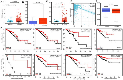
Table 1. The relationship between REG4 mRNA expression and clinicipathological characteristics of breast cancer by xiantao.
Our findings of the Kaplan-Meier plotter revealed that long overall survival was seen in female, black/African, stage 3 and 4 cancer patients with high REG4 expression compared to those with low expression (, p < 0.05). It was the same for the overall survival rate in cancer patients with high mutation burden or low neoantigen load (, p < 0.05). REG4 expression and recurrence-free survival in breast cancer patients were positively correlated in breast cancer patients (, p < 0.05). In terms of the xiantao database, univariate analysis indicated that T stage, N stage, and M stage were negatively associated with progression-free survival, but ER, PR and REG4 expression was positively linked to progression-free survival (, p < 0.05). Multivariate analysis showed that only T stage, N stage, M stage, and REG4 expression were independent predictors for progression-free survival (, p < 0.05).
Table 2. The progression-free survival analysis of breast cancer patients by xiantao.
3.2. Clinicopathological and prognostic significances of REG4 mRNA expression in cervical, endometrial and ovarian cancers
We conducted bioinformatic analysis using the GEO, xiantao, and UALCAN databases and discovered that REG4 expression was decreased in cervical (Supplementary Figure 1(A), p < 0.05), but increased in endometrial (Supplementary Figure 1(B, C), p < 0.05) and ovarian cancer than in normal tissues (Supplementary Figure 1(D,E) p < 0.05). According to , expression of REG4 mRNA was positively linked to T stage and adenosquamous cell carcinoma of cervical cancer (p < 0.05).
Table 3. The relationship between REG4 mRNA expression and clinicipathological characteristics of cervical cancer by xiantao.
3.3. Relationship between REG4 mRNA expression and infiltrating immune cells in gynecological cancers
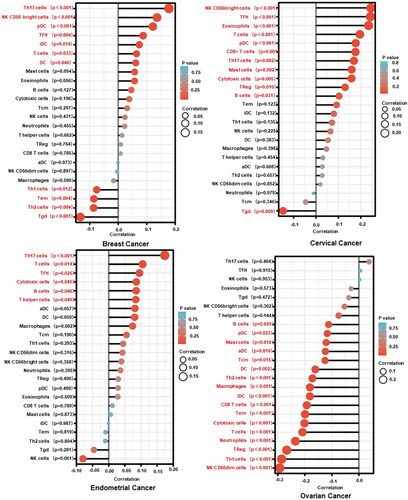
In line with xiantao, Th17, NK CD56bright cells, pDC, TFH, iDC, T cell and DC in breast cancer were strongly correlated with REG4 mRNA expression (, p < 0.05). Meanwhile, it was associated favourably with the infiltration of NK CD56bright cells, TFH, eosinophils, T cells, pDC, CD8+ T cells, Th17 cells, mast cells, cytotoxic cells and Treg, B cells and negatively with Tgd in cervical cancer (, p < 0.05). It was also favourably associated with the infiltration of Th17 cells, T cells, TFH, cytotoxic cells, B cells and T helper cells in endometrial cancer (, p < 0.05). Moreover, it was negatively related to the infiltration of B cells, pDC, mast cells, aDC, Tcm, DC, Th2 cells, macrophages, iDC, CD8 T cells, Tem, cytotoxic cells, T cells, neutrophils, Treg, Th1 cells, and NK CD56dim cells in ovarian cancer (, p < 0.05).
Figure 2. The relationship between REG4 mRNA expression and immune infiltration in gynecological cancers. The enrichment of immune cells was explored between low and high expression of REG4 in breast, cervical, endometrial, and ovarian cancers using xiantao. Note: DC, dendritic cell; Treg, regulatory T cell; Tcm, central memory T cells; Tem, effector memory T cell; TFH, follicular helper T cell; NK, natural killer. Tdg, γδ T cells.
3.4. REG4-related genes and pathways in gynecological cancers
We found the differently-expressed genes about REG4 gene that were in gynecological malignancies using the xiantao platform. KEGG analysis revealed the leading signal pathways of these differential genes included peptidase, intermediate filament and keratinisation in breast cancer (Supplementary Fig. 2A p < 0.05). The leading signalling pathways did ligand-receptor interaction, metabolism of hormone, xenobiotic and retinol, peptidase, brush border and digestion in cervical cancer (Supplementary Figure 2(A), p < 0.05). In endometrial cancer, the differential genes of REG4 were involved in bile secretion, intermediate filament, keratinisation and so on (Supplementary Figure 2(A), p < 0.05). In ovarian cancer, they did ligand-receptor interaction, metabolism of hormones, hormone, xenobiotic and retinol, digestion and extracellular matrix (Supplementary Figure 2(A), p < 0.05). Furthermore, STRING was employed to find the PPI pairings, and cytoscape was used to rank the top 10 nodes according to their connection (Supplementary Figure 2(B). The top hub genes mainly included small proline rich protein 2B in breast cancer, apolipoproteins in cervical cancer, fibrinogens and apoproteins in endometrial cancer, and fibrinogens and apoproteins in ovarian cancer (Supplementary Figure 2(B)).
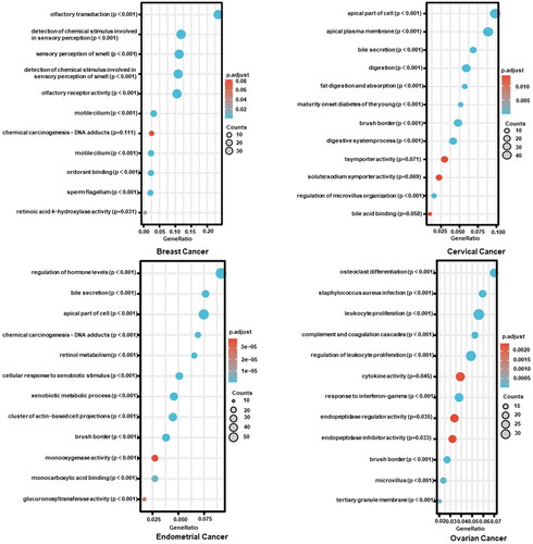
The REG4-related genes were analysed in cancers by KEGG using xiantao database (). The REG4-correlated genes were found to be important in smell sense, detection of chemical stimulus, chemical carcinogenesis, motile cilium, and sperm flagellum in breast carcinoma (p < 0.05). They included apical part of cell or plasma membrane, brush border, bile secretion and digestion, and symporter in cervical cancer (p < 0.05). In endometrial cancer, the top signal pathway was composed of hormonal regulation, bile secretion, chemical carcinogenesis, brush border, and so forth (p < 0.05). In ovarian cancer, REG4-related genes consisted of cellular differentiation, leukocyte proliferation, cytokine activity, endopeptidase, brush border and microvillus (p < 0.05).
Figure 3. The REG4-related signal pathways in gynecological cancers. The top REG4-related genes were screened and classified into the signal pathways of KEGG using xiantao database.
4. Discussion
REG4 mRNA and protein levels were elevated in ovarian tumours compared to normal ovaries, and in mucinous carcinomas compared to serous carcinomas (Chen et al. Citation2015, Xiang et al. Citation2022). REG4 was used as a possible biomarker with specificity for the subtype of mucinous ovarian cancer (2016). For patients with ovarian cancer, the expression of REG4 was either an overall or relapse-free poor prognostic predictor (Chen et al. Citation2015). In our study group, the survival of ovarian cancer patients undergoing platinum or paclitaxel therapy was found to inversely correlate with REG4 expression. REG4 overexpression resulted in chemical and apoptotic resistance of ovarian cancer cells via Akt/mTOR pathway (Xiang et al. Citation2022). As compared to normal tissues, REG4 mRNA was considerably up-regulated in gastric cancer (Zheng et al. Citation2010, Tao et al. Citation2011) and glioma (Wang et al. Citation2012). However, there have been no reports about REG4 expression and breast, cervical and endometrial cancers until now. In line with the immunohistochemical observation in gastric cancer (Zheng et al. Citation2010), colorectal cancer (Zheng et al. Citation2011), and glioma (Wang et al. Citation2012), serum REG4 level was higher in gastric (Mitani et al. Citation2007, Kobayashi et al. Citation2010, Zheng et al. Citation2010), pancreatic (Takayama et al. Citation2010, Saukkonen et al. Citation2018), and gallbladder (Tamura et al. Citation2009) cancers than healthy volunteers. In the present study, we discovered that the breast, endometrial and ovarian cancers had up-regulated levels of REG4 mRNA, which was inversely linked to its promoter methylation in breast cancer with REG4 hypomethylation. These results demonstrated that up-regulated expression of REG4 mRNA is closely linked to the carcinogenesis of gynecological cancers, possibly due to its promoter methylation, which will be analysed in a future study.
Reportedly, REG4 mRNA was positively linked to diffuse-type, poor differentiation, lymph node metastasis, distant metastasis, and TNM stage of gastric cancer (2011), and to TNM stage, distant metastasis, and histologic grade of colorectal cancer (2015). He et al. (Citation2012) discovered that via upregulating MMP-7 and MMP-9, REG4 promoted the in vitro proliferation and invasiveness of pancreatic cancer cells. In line with the findings in pancreatic cancer (Takehara et al. Citation2006), either REG4 overexpression or rhREG4 therapy increased proliferation, G2/S progression, anti-apoptosis, migration, and invasion of ovarian cancer cells (Chen et al. Citation2015). Here, we demonstrated that REG4 expression was positively related to ER and PR expression, aggressiveness of PAM50 classification, and infiltrating lobular carcinomas in breast cancer, and to T stage and adenosquamous cell carcinoma of cervical cancer. REG4 expression was positively associated with progression-free survival of breast cancer patients as an independent predictor as well. These data indicated that REG4 expression may be used to reflect the aggressiveness, prognosis and histogenesis of breast or cervical cancer.
Wang et al. (Citation2016) found that REG4 promoted peritoneal metastasis of gastric cancer through the REG4-GRP37 complex by enhancing adhesion ability. Because TGF-α stimulated SP1 to transcriptionally up-regulate REG4 expression, a REG4-triggered positive feedback loop was also discovered to amplify REG4 through EGFR transactivation, consisting of GPR37, ADAM17, TGF-α EGFR, SP1 and REG4. Our bioinformatic analysis showed that the top signal pathways related to REG4 included smell and chemical stimulus, peptidase, intermediate filament, and keratinisation in breast cancer; ligand-receptor interaction, metabolism of hormone, xenobiotic and retinol, peptidase, brush border and digestion in cervical and ovarian cancers; bile secretion, intermediate filament, chemical carcinogenesis, brush border, keratinisation in endometrial cancer. These findings provide novel clues about the roles and molecular mechanisms by which REG4 plays an important role in the tumorigenesis and progression of gynecological cancers, which should be investigated in future work.
Lu et al. (Citation2013) found that c-type lectins influence inflammatory/immune responses and contribute to immunological escape of infections and malignancies and REG4 belongs to c-type lectins. Wang et al. (Citation2022) discovered that REG4 expression was elevated in pancreatic ductal adenocarcinoma and that its knockdown triggered innate immune activation and decreased tumour development in the Zebrafish xenograft model. Jin et al. (Citation2017) demonstrated that REG4 increased gastric cancer’s resistance to 5-fluorouracil by stimulating the MAPK/Erk/Bim signalling pathway. Gao et al. (Citation2021) found that REG4 immunopositivity might be used as a biomarker for radiochemotherapeutic sensitivity for colorectal cancer. Additionally, the inhibition of either immune surveillance or activation is closely linked to chemoresistance. In the current research, we discovered that REG4 mRNA expression was positively associated with the infiltration of DC cells in breast cancer, positively with Th17 cells, TFH, cytotoxic cells, and T cells in cervical and endometrial cancers, but negatively with the infiltration of DC, cytotoxic cells, and T cells in ovarian cancer. These results demonstrated that REG4’s potential role in immune surveillance and therapy, and tumour-associated immune responses to gynecological cancers. In ovarian cancer, the specific association between REG4 expression and the infiltration of immune cells should be further investigated in future work.
Oue et al. (Citation2005) found that REG4 expression was linked to both the mucin phenotype and neuroendocrine differentiation of the intestine. Bishnupuri et al. (Citation2014) discovered that REG4-induced mitogenesis in human colorectal cancer was initiated by Akt-GSK3-β-catenin-TCF-4 signalling. Kuniyasu et al. (Citation2009) found that REG4 expression decreased nitric oxide-induced apoptosis, and increased peritoneal metastasis of gastric cancer cells by activating EGFR/Akt pathway. To improve our understanding of the biological effect of REG4 and identify the diagnostic biomarkers of gynecological cancers, we first clarified analysed the clinicopathological and prognostic significance of REG4 mRNA expression in breast, cervical and endometrial cancers, and their related signal pathways. However, this study could not validate these bioinformatic data utilising real-time RT-PCR, even followed by laser capture dissection. Recently, we have started to explore the relationships between promoter methylation, chemoresistance, immune surveillance and REG4 mRNA expression.
5. Conclusion
Up-regulated REG4 mRNA expression is closely linked to gynecological carcinogenesis and histogenesis, and can be used as a potential marker of the aggressiveness or prognosis of either breast or cervical cancer. In gynecological cancer, REG4-related signal pathways included bile secretion, brush border and digestion, fibrinogens and apoproteins, ligand-receptor interaction, and metabolism of hormone, xenobiotic and retinol, and peptidase.
Author contributions
All authors approved the final manuscript as submitted and agree to be accountable for all aspects of the work. Hua-chuan Zheng: Project development, Study design, and Manuscript writing. Cong-yu Zhang: Revised the manuscript, Data analysis and interpreted the data. Li Zhang: Project development, Revised the manuscript. Zi-mo Wang, Dong-hui Ren: Critically reviewed the manuscript.
Supplemental Material
Download Zip (2.6 MB)Disclosure statement
The authors declare that there are no competing interests associated with this study. Registration: not applicable.
Data availability statement
The datasets generated and analysed in the present study are available from the corresponding author on reasonable request.
Additional information
Funding
References
- Aquil, A., et al., 2021. Predictors of mental health disorders in women with breast and gynecological cancer after radical surgery: a cross-sectional study. Annals of Medicine and Surgery (2012), 65, 102278.
- Bishnupuri, K.S., et al., 2006. Reg IV activates the epidermal growth factor receptor/Akt/AP-1 signaling pathway in colon adenocarcinomas. Gastroenterology, 130 (1), 137–149.
- Bishnupuri, K.S., et al., 2010. Reg IV regulates normal intestinal and colorectal cancer cell susceptibility to radiation-induced apoptosis. Gastroenterology, 138 (2), 616–626.
- Bishnupuri, K.S., et al., 2014. Reg4-induced mitogenesis involves Akt- GSK3β-β-catenin-TCF-4 signaling in human colorectal cancer. Molecular Carcinogenesis, 53 Suppl 1 (0 1), E169–80.
- Bishnupuri, K.S., et al., 2022. Reg4 interacts with CD44 to regulate proliferation and stemness of colorectal and pancreatic cancer cells. Molecular Cancer Research, 20 (3), 387–399.
- Chen, S., et al., 2015. The role of the REG4 gene and its encoding product in ovarian epithelial carcinoma. BMC Cancer, 15, 471.
- Du, F., et al., 2013. The expression patterns of Reg IV gene in normal rat reproduction system. Journal of Experimental Zoology. Part A, Ecological Genetics and Physiology, 319 (1), 32–38.
- Duan, Y., et al., 2014. Tumor suppressor miR-24 restrains gastric cancer progression by downregulating RegIV. Molecular Cancer, 13, 127.
- Gao, L., et al., 2021. REG4 is a potential biomarker for radiochemotherapy sensitivity in colorectal cancer. OncoTargets and Therapy, 14, 1605–1611.
- Giannini, A., et al., 2022. Advances on prevention and screening of gynecologic tumors: are we stepping forward? Healthcare, 10 (9), 1605.
- Golia, D. T., et al., 2023. Novel insights into molecular mechanisms of endometrial diseases. Biomolecules, 13 (3), 499.
- He, X.J., et al., 2012. REG4 contributes to the invasiveness of pancreatic cancer by upregulating MMP-7 and MMP-9. Cancer Science, 103 (12), 2082–2091.
- Ho, M.R., et al., 2010. Human RegIV protein adopts a typical C-type lectin fold but binds mannan with two calcium-independent sites. Journal of Molecular Biology, 402 (4), 682–695.
- Jin, J., et al., 2017. Regenerating family member 4 (Reg4) enhances 5-fluorouracil resistance of gastric cancer through activating MAPK/Erk/Bim signaling pathway. Medical Science Monitor : international Medical Journal of Experimental and Clinical Research, 23, 3715–3721.
- Kawasaki, Y., et al., 2015. REG4 is a transcriptional target of GATA6 and is essential for colorectal tumorigenesis. Scientific Reports, 5, 14291.
- Kobayashi, Y., et al., 2010. Serum tumor antigen REG4 as a useful diagnostic biomarker in gastric cancer. Hepato-gastroenterology, 57 (104), 1631–1634.
- Kuniyasu, H., et al., 2009. Reg IV enhances peritoneal metastasis in gastric carcinomas. Cell Proliferation, 42 (1), 110–121.
- Li, Y.G., et al., 2011. Anti-tumor effects of proteoglycan from Phellinus linteus by immunomodulating and inhibiting Reg IV/EGFR/Akt signaling pathway in colorectal carcinoma. International Journal of Biological Macromolecules, 48 (3), 511–517.
- Little, J., et al., 2009. STrengthening the REporting of genetic association studies (STREGA)–an extension of the STROBE statement. Genetic Epidemiology, 33 (7), 581–598.
- Lu, S., et al., 2013. Genetic variants in c-type lectin genes are associated with colorectal cancer susceptibility and clinical outcome. International Journal of Cancer, 133 (10), 2325–2333.
- Mitani, Y., et al., 2007. Reg IV is a serum biomarker for gastric cancer patients and predicts response to 5-fluorouracil-based chemotherapy. Oncogene, 26 (30), 4383–4393.
- Naito, Y., et al., 2012. Reg IV is a direct target of intestinal transcriptional factor CDX2 in gastric cancer. PloS One, 7 (11), e47545.
- Oue, N., et al., 2005. Expression and localization of Reg IV in human neoplastic and non-neoplastic tissues: Reg IV expression is associated with intestinal and neuroendocrine differentiation in gastric adenocarcinoma. The Journal of Pathology, 207 (2), 185–198.
- Paepke, D., et al., 2020. Prevalence and predictors for nonuse of complementary medicine among breast and gynecological cancer patients. Breast Care, 15 (4), 380–385.
- Saukkonen, K., et al., 2018. Prognostic and diagnostic value of REG4 serum and tissue expression in pancreatic ductal adenocarcinoma. Tumour Biology : the Journal of the International Society for Oncodevelopmental Biology and Medicine, 40 (3), 1010428318761494.
- Segev, Y., et al., 2021. Correlation between an integrative oncology treatment program and survival in patients with advanced gynecological cancer. Supportive Care in Cancer : official Journal of the Multinational Association of Supportive Care in Cancer, 29 (7), 4055–4064.
- Seland, M., et al., 2022. Distress, problems and unmet rehabilitation needs after treatment for gynecological cancer. Acta Obstetricia et Gynecologica Scandinavica, 101 (3), 313–322.
- Sentani, K., et al., 2008. Immunohistochemical staining of Reg IV and claudin-18 is useful in the diagnosis of gastrointestinal signet ring cell carcinoma. The American Journal of Surgical Pathology, 32 (8), 1182–1189.
- Takayama, R., et al., 2010. Serum tumor antigen REG4 as a diagnostic biomarker in pancreatic ductal adenocarcinoma. Journal of Gastroenterology, 45 (1), 52–59.
- Takehara, A., et al., 2006. Tumor marker REG4 detected in serum of patients with resectable pancreatic cancer and feasibility for antibody therapy targeting REG4. Cancer Science, 97 (11), 1191–1197.
- Tamura, H., et al., 2009. Reg IV expression and clinicopathologic features of gallbladder carcinoma. Human Pathology, 40 (12), 1686–1692.
- Tao, H.Q., et al., 2011. Evaluation of REG4 for early diagnosis and prognosis of gastric cancer. Human Pathology, 42 (10), 1401–1409.
- Vargiu, V., et al., 2022. Impact of obesity on sentinel lymph node mapping in patients with apparent early-stage endometrial cancer: the ObeLyX study. Gynecologic Oncology, 165 (2), 215–222.
- Wang, F., et al., 2011. Identification of RegIV as ael GLI1 target gene in human pancreatic cancer. PLOS One, 6 (4), e18434.
- Wang, H., et al., 2016. REG4 promotes peritoneal metastasis of gastric cancer through GPR37. Oncotarget, 7 (19), 27874–27888.
- Wang, Q., et al., 2012. Oncogenic reg IV is a novel prognostic marker for glioma patient survival. Diagnostic Pathology, 7, 69.
- Wang, X., et al., 2022. Zebrafish xenograft model for studying pancreatic cancer-instructed innate immune microenvironment. International Journal of Molecular Sciences, 23 (12), 6442.
- Xiang, L.W., et al., 2022. The effects of REG4 expression on chemoresistance of ovarian cancer. Journal of Obstetrics and Gynaecology, 42 (7), 3149–3157.
- Xiao, Y., et al., 2019. Deficiency in intestinal epithelial Reg4 ameliorates intestinal inflammation and alters the colonic bacterial composition. Mucosal Immunology, 12 (4), 919–929.
- Zheng, H.C., et al., 2010. The role of Reg IV gene and its encoding product in gastric carcinogenesis. Human Pathology, 41 (1), 59–69.
- Zheng, H.C., et al., 2011. Expression profile of the REG gene family in colorectal carcinoma. The Journal of Histochemistry and Cytochemistry : official Journal of the Histochemistry Society, 59 (1), 106–115.
- Zheng, H.C., et al., 2022. REG4 promotes the proliferation and anti-apoptosis of cancer. Frontiers in Cell and Developmental Biology, 10, 1012193.