

Abstract
Background
Asiaticoside (AS) has been reported to improve the changes induced by high glucose stimulation, and it may have potential therapeutic effects on gestational diabetes mellitus (GDM). This study aims to explore the effect of AS on the cell model of GDM and the action mechanism of the PI3K/AKT pathway.
Methods
The GDM model was established in HTR-8/Svneo cells with a high glucose (HG) medium. After the cytotoxicity assay of AS, cells were divided into the control group, HG group and HG + AS group to conduct control experiment in cells. The cell proliferation and migration were detected by CCK-8 assay and scratch test, respectively. The mRNA levels of PI3K, AKT2, mTORC1, and GLUT4 in PI3K/AKT signalling pathway were measured by RT-PCR, and the protein expressions of these signalling molecules were monitored by western blot.
Results
AS showed a promotion effect on the cell proliferation rate of HTR-8/Svneo cells, and 80 μmol/L AS with a treatment time of 48 h had no cytotoxicity. The cell proliferation rate, migration rate, mRNA levels and protein expressions of PI3K, AKT2, mTORC1, and GLUT4 in the HG group were significantly lower than those in the control group, which were significantly increased in the HG + AS group (p < 0.05).
Conclusions
AS can facilitate the cell proliferation and migration in the cell model of GDM, and might play a role in GDM treatment via PI3K/AKT pathway.
PLAIN LANGUAGE SUMMARY
Asiaticoside possesses various pharmacological effects and has been reported to show a beneficial effect on the treatment of diabetes mellitus. This research firstly investigated the effect and mechanism of asiaticoside on gestational diabetes mellitus, and found that asiaticoside could facilitate the cell proliferation and migration of HTR-8/Svneo cells treated with high glucose, and affect the signalling molecules of PI3K/AKT pathway. Therefore, asiaticoside may be a novel useful therapeutic drug in the treatment of gestational diabetes mellitus.
Introduction
Gestational diabetes mellitus (GDM) is the occurrence of abnormal glucose metabolism with onset or firstly detected during pregnancy, which is closely associated with adverse pregnancy outcomes, including birth trauma, stillbirth, and macrosomia (Farahvar et al. Citation2019, He et al. Citation2021). Women with GDM are more likely to develop type 2 diabetes after childbirth, and their offspring are at increased risk of developing metabolic syndrome such as diabetes and obesity in childhood and adulthood (Bernstein et al. Citation2018, Herrera-Martínez et al. Citation2018). Therefore, timely treatment for GDM is crucial to reduce the risk of these adverse pregnancy outcomes.
The pathogenesis of GDM is very complex. The placenta is the bridge between mother and foetus, playing an important role in the maintenance of pregnancy (Liuet al, Citation2021). On the other hand, the placenta has been found to participate in the occurrence and development of GDM by inhibiting proliferation and cell cycle progression, promoting apoptosis of trophoblast cells (Lin et al. Citation2022). Studies also have found that the incidence of GDM in patients with low maturity of the placenta is 52.6 times higher than that in patients with high maturity (Liemet al., Citation2017). It is suggested that the low survival rate and high apoptosis rate of trophoblasts may be one of the pathogeneses of GDM, and trophoblast cells are widely used to construct cell models of GDM (Bai et al. Citation2021, Liu et al, Citation2021).
Notably, insulin resistance (IR), referring to a reduction of the sensitivity of target organs to insulin, is an important factor in the pathophysiology of GDM (Wang et al. Citation2022). The placental lactogen, sex hormone and adrenocortical hormone induced by pregnancy have significant effects on IR, thus reducing the sensitivity of insulin-targeting tissues to insulin, and ultimately leading to the occurrence of GDM (Molinaro et al. Citation2019). The phosphoinositide 3-kinase (PI3K)/protein kinase B (AKT) signalling pathway, as a main insulin-signalling pathway, is widely investigated in the analysis of diabetes pathogenesis, and has been proven to be closely related to the occurrence and development of GDM (Ramenzoni et al. Citation2019). Studies have shown that abnormal expression or incorrect phosphorylation of signalling molecules in PI3K/AKT signalling pathway, may lead to the decrease of IR and glucose transport functions, and then induce GDM (Du et al. Citation2022).
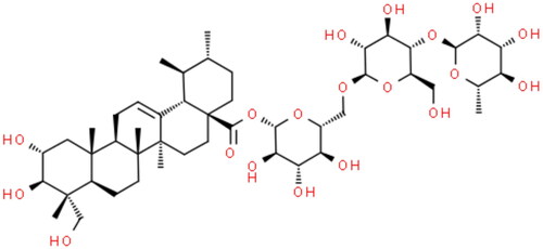
Centella asiatica, first recorded in the book of Shennong Classic of Materia Medica, is sweet in tasty and cool in nature, benefitting for clearing heat and removing toxicity, inducing diuresis and reducing edoema, stopping bleeding and promoting blood circulation. Asiaticoside (AS, C48H78O19, CAS: 16830-15-2) is a triterpenoid compound extracted from Centella asiatica, and its molecular structure is shown in retrieved from www.ChemSpider.com. AS has been proven to possess the effects of immunoregulation, antioxidant, anti-inflammatory, promotion of wound healing, and inhibition of scar formation (Yin et al. Citation2015). At present, AS is mainly used for skin scar repair in diabetes mellitus, for example, it has been reported to accelerate diabetic cutaneous ulcers healing by activating Wnt/β-catenin signalling pathway in combination with nitric oxide, and its polymeric nanoparticles are effective for diabetic wound healing by increased collagen biosynthesis (Narisepalli et al. Citation2023, Nie et al. Citation2020). Moreover, AS has been indicated to play a role in diabetes-associated cognitive dysfunction, high glucose-induced hearing loss and diabetic kidney disease (Yin et al. Citation2015, Xing et al. Citation2017, Zhu et al. Citation2020). Recent studies have shown that AS can significantly reduce fasting blood glucose concentration in obese diabetic rats, and greatly promote insulin secretion under high glucose (HG) stimulation (Maulidiani et al. Citation2016, Zhu et al. Citation2020). Therefore, it is greatly needed to understand the effect and mechanism of AS on GDM. In our study, the cell model of GDM in HTR-8/Svneo cells was established to study the effect of AS on trophoblasts, and to explore the underlying mechanism of AS on PI3K/AKT signalling pathway during GDM.
Figure 1. Molecular structure of AS.
Methods
Cell culture and establishment of GDM model
The study was conducted following the Strengthening the Reporting of Genetic Association study (STREGA) statement checklist (Little et al. Citation2009). Human trophoblast HTR-8/Svneo were purchased from ATCC (No. CRL3271), and cultured in RPMI 1640 medium (Invitrogen, Shanghai, China) with 10% foetal bovine serum (Invitrogen, MA, USA) and 100 U/mL penicillin/streptomycin (Thermo Fisher, MA, USA) in an environment of 37 °C and 5% CO2. The GDM model in HTR-8/Svneo cells was established as described previously (He and Liu, 2021). The cells were cultured in a HG medium with 25 mmol/L glucose for 24 h to establish the GDM model in vitro.
Cytotoxicity assay and grouping
The cytotoxicity assay of AS was firstly determined by Cell counting kit-8 (CCK-8) against HTR-8/Svneo cells. The cells were cultured in 96-well plates at a density of 5 × 10³ cells/well for 24 h, and then treated with the different concentrations (0, 10, 20, 40, 60, 80, and 100 μmol/L) of AS. After 24 h or 48 h, the CCK-8 assay was performed to obtain the optical density (OD) value at 450 nm. The cell proliferation rate was calculated by the following formula: cell proliferation rate (%) = (OD of the experimental group- OD of blank)/(OD of the control group- OD of blank) × 100% (Luo et al. Citation2021). The proper concentration of AS and intervention time were determined based on the cell proliferation rate.
Whereafter, the cells were divided into three groups to conduct control experiment. The control group was cultured in a normal medium and treated with phosphate buffer solution (PBS). The HG group was cultured in the HG medium to establish GDM model and treated with PBS. The HG + AS group was cultured in the HG medium and treated with AS.
CCK-8 assay
CCK-8 assay was performed according to the instrument of CCK-8 assay kits (Dojindo Laboratories, Kyushu, Japan). Briefly, the HTR-8/Svneo cells were incubated in 96-well plates and received the intervention of the three groups. After cultured for 12, 24, 36, 48, and 60 h, the cells of each well were treated with 10 μL CCK-8, and further incubated for 2 h. After washing with PBS three times, the OD values at 450 nm were measured in a microtiter plate reader (Thermo Fisher Scientific, Waltham, USA).
Scratch test
Scratch test was performed to analyse cell migration in vitro (Gao et al. Citation2020, Jiang et al. Citation2016). The HTR-8/Svneo cells were seeded in 6-well plates and cultured with the HG medium to establish GDM cell model, then the cell layers were scratched with sterile tips. Subsequently, the cells in the HG + AS group were treated with AS in the HG medium, while the HG group was only cultured with the HG medium. The cells in the control group were cultured in a normal medium all the time. After the incubation for 0 (before AS treatment), 24, and 48 h, images of the cells were captured by microscopy, and analysed using the Olympus CellSens Dimension software to obtain the widths of the scratch. The ratio of migration area was expressed as a percentage relative to the area of the initial wound area.
Real-time reverse transcription polymerase chain reaction (RT-PCR)
The total RNAs in cells of the three groups were extracted by Trizol reagent (Invitrogen, USA), and used for cDNA synthesisation by PrimeScript II 1st strand cDNA Synthesis Kit (Takara, Shiga, Japan). Real-time RT-PCR was carried out in StepOnePlus Real-Time PCR (AB International, CA, USA) according to the instrument of SYBR Premix Ex Taq II (Takara, Shiga, Japan). To analyse the relative expression levels of various mRNAs, the 2 − ΔΔCt method was performed with GADPH as reference gene (Yao et al. Citation2016). The primer sequences were as follows: PI3K forward 5′-GAAGCACCTGAATAGGCAAGTCG-3′ and PI3K reverse 5′-CGCTGGTCTAAAGTACCTACGA-3′, AKT2 forward 5′-CATCCTCATGGAAGAGATCCGC-3′ and AKT2 reverse 5′-GTACCTCGTGTCCAAGAAGGAG-3′, mTORC1 forward 5′-AGCATCGGATGCTTAGGAGTGG-3′ and mTORC1 reverse 5′-GCAGAGGTTTCTAC TGACCGAC-3′, GLUT4 forward 5′-CCATCCTGATGACTGTGGCTCT-3′ and GLUT4 reverse 5′-GGTGAGGAACCAAGTAGCACCG-3′, GADPH forward 5′-CAATGACCCCTTCATTGACC-3′ and GADPH reverse 5′-GACTCTTGCCCTTCGAACAG-3′.
Western blot
Total proteins were extracted from the cells of the three groups using the Tissue or Cell Total Protein Extraction kit (Chundu Bio, Wuhan, China). After the determination of protein concentration by the BCA assay, 50 μg proteins were loaded on SDS-PAGE gels (Cosmo Bio, Tokyo, Japan) and then transferred to PVDF membranes (Millipore, MA, USA). The membranes were blocked by 5% fat-free milk for 2 h, and incubated with primary antibody, anti-PI3K, AKT2, GLUT4 (1: 500, Bioss, Beijing, China), and mTORC1 (1: 500, Abmart, Shanghai, China) at 4 °C overnight. After washing with TBST, the membranes were incubated with a secondary antibody (ZSGB-BIO, Beijing, China) at room temperature for 1 h. The target bands were visualised and quantified by the Tanon-5200 Image Analyser using GAPDH as control for normalisation.
Statistical analysis
SPSS software version 26.0 (SPSS Inc, Chicago, USA) was applied for statistical analysis and the data were presented as mean ± standard deviation. The difference between two groups was determined by Student’s t test and the difference among multiple groups was conducted by one-way analysis of variance. A P-value <0.05 was considered statistically significant.
Results
The cytotoxicity assay of AS with different concentrations and treatment times in HTR-8/Svneo cells is shown in . The cell proliferation rate was generally increased with the concentration of AS from 0 to 80 μmol/L, however, it was significantly decreased under the incubation with 100 μmol/L AS, suggesting that AS of 80 μmol/L not only showed no cytotoxicity in HTR-8/Svneo cells, but also had a good promotion effect on cell proliferation (p < 0.05). In addition, the cell proliferation rates with treatment time of 48 h were significantly higher than those of 24 h at all concentrations of AS, indicating AS with a treatment time of 48 h could promote cell proliferation (p < 0.05). These results indicated that 80 μmol/L AS with a treatment time of 48 h had no cytotoxicity on HTR-8/Svneo cells, which could even show a relatively good promotion effect on cell proliferation. Therefore, 80 μmol/L AS with a treatment time of 48 h was subsequently adopted in our later experiments.
Figure 2. AS promoted cell proliferation in cell model of GDM. (A) Cytotoxicity assay of AS with different concentrations and treatment times, (B) Cell proliferation in GDM cell model with AS treatment.
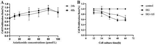
The cell proliferation rates were compared among the control group, HG group and HG + AS group to explore the effect of AS on cell proliferation in the cell model of GDM, as shown in . The cell proliferation rates in the HG group were significantly decreased at all the time points compared to the control group, however, the AS treatment could increase the cell proliferation rates in the HG group (p < 0.05).
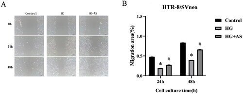
The cell migration in the GDM model of HTR-8/Svneo cells after AS treatment was evaluated by scratch test, which is shown in . The scratch areas in the three groups were generally decreased over time by microscopical observation. At both 24 and 48 h, the cell migration rates in the HG group were significantly lower than those in the control group, indicating HG stimulation could inhibit cell migration, however, the cell migration rates in the HG + AS group were significantly higher than those in the HG group, suggesting AS could promote cell migration (p < 0.05).
Figure 3. AS promoted cell migration in cell model of GDM. *p < 0.05 compared with control group; #p < 0.05 compared with HG group.
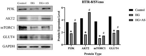
The mRNA levels and protein expressions of signalling molecules in PI3K/AKT signalling pathway, including PI3K, AKT2, mTORC1, and GLUT4, are shown in and , respectively. The mRNA levels and protein expressions of PI3K, AKT2, mTORC1, and GLUT4 in the HG group were significantly lower than those in the control group, indicating an inhibition effect of HG stimulation on PI3K/AKT pathway, however, the mRNA levels and protein expressions of PI3K, AKT2, mTORC1, and GLUT4 in the HG + AS group were significantly higher than those in the HG group, suggesting a promotion effect of AS on PI3K/AKT pathway (p < 0.05).
Figure 4. Effect of AS on mRNA levels of signalling molecules in PI3K/AKT pathway. *p < 0.05 compared with control group; #p < 0.05 compared with HG group.
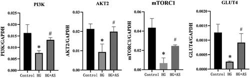
Figure 5. Effect of AS on protein expressions of signalling molecules in PI3K/AKT pathway. *p < 0.05 compared with control group; #p < 0.05 compared with HG group.
Discussion
Globally, the prevalence of GDM is on the rise, and has become one of the most common complications of pregnancy (Calzada et al. Citation2019). AS has a wide range of pharmacological effects, and it has been reported recently to show a beneficial effect on the treatment of diabetes mellitus (Maulidiani et al. Citation2016, Zhu et al. Citation2020). However, there is no research to elucidate the role of AS in GDM to our knowledge. In this study, we established a cell model of GDM to study the effect of AS in vitro.
The placenta can promote metabolic adaptations in pregnancy, and its aberrant structural and functional alterations contribute to the pathogenesis of GDM (Du et al. Citation2021). As the outer layer of the placenta, trophoblasts are the main cell type of the placenta, which also is crucial for a successful pregnancy as well as multiple gestational diseases. HTR-8/Svneo, derived from first-trimester extravillous cytotrophoblast cells, is often used to study trophoblast biology and functions in vitro (Msheik et al. Citation2020). In our study, the cytotoxicity assay was firstly performed in HTR-8/Svneo cells, and found AS with a concentration of 80 μmol/L and treatment time of 48 h had no cytotoxicity on HTR-8/Svneo cells, even promoted cell proliferation, laying a foundation for the follow-up experiments. To further explore the effect of AS in GDM, HTR-8/Svneo cells were cultured with the HG medium to induce GDM in vitro. Proliferation and migration are the foundation of the growth of trophoblast cells, thus affecting the development of GDM (Ke et al. Citation2022). It has been indicated that hyperglycaemia can reduce cell viability, proliferation, and migration in vitro, meanwhile, decrease trophoblast proliferation contributing to reduced placental growth in vivo (Ke et al. Citation2022, Tao et al. Citation2020). Our results showed that the cell proliferation and migration rates in the HG group were significantly decreased compared to the control group, which is consistent with previous studies. Study has shown that AS can increase cell proliferation and migration of human keratinocytes HaCaT (Anand et al. Citation2022, Liu et al. Citation2024). Moreover, AS has been reported to significantly promote the proliferation and migration of human foreskin fibroblasts HFF-1 induced by HG (Nie et al. Citation2020). Similarly, our study found that AS could facilitate the cell proliferation and migration of HTR-8/Svneo treated with HG, which might further contribute to the treatment of GDM.
Next, we investigated the possible mechanisms underlying the beneficial effect of AS on the cell model of GDM in HTR-8/Svneo cells. The insulin-signalling pathway is activated by the binding of insulin to insulin receptor, autophosphorylation of receptor tyrosine residues, and tyrosine phosphorylation of insulin receptor substrates (Li et al. Citation2022). PI3K, a heterodimeric lipase, can be activated by binding to the tyrosine phosphorylation site of insulin receptor substrate-1, and then leads to the recruitment of signalling proteins AKT (Wang et al. Citation2023). AKT further reduces the inhibitory effects of the tuberous sclerosis complex on the mammalian target of rapamycin complex 1 (mTORC1) to phosphorylate the downstream mTOR, which is involved in glucose, lipid, and protein metabolism (Lin et al. Citation2022, Wang et al. Citation2023). On the other hand, phosphorylation of AKT induces the translocation of glucose transporter 4 (GLUT4) to the cell membrane, thereby promoting glucose utilisation (Li et al. Citation2022). IR during GDM has a close link to the dysregulation of PI3K/AKT pathway, which can promote cellular glucose uptake and regulate cell growth (Du et al. Citation2022). Study has shown that the protein levels of PI3K/p-PI3K, AKT/p-AKT, mTOR/p-mTOR, and GLUT4 by western blot were significantly reduced in HTR-8/SVneo cells after the treatment with a glucose concentration of 25 mmol/L (Lin et al. Citation2022). In addition, compared to the healthy controls, the expressions of PI3K, AKT, and GLUT4 were significantly downregulated in the placenta tissues of women with GDM (Li et al. Citation2022). Similarly, our study indicated that the protein expression levels of PI3K, AKT2, mTORC1, and GLUT4 in the HG group were significantly lower than those in the control group. Moreover, the mRNA levels of signalling molecules in PI3K/AKT signalling pathway corresponded to their protein expression, suggesting the accuracy of our results. Further, AS has been indicated to promote synaptic proteins expression by PI3K/AKT pathway in human neuroblastoma SH-SY5Y cells exposed to HG medium (Yin et al. Citation2015). In our study, the mRNA and protein levels of PI3K, AKT2, mTORC1, and GLUT4 in the cell model of GDM were increased after the treatment of AS, providing evidence that AS might play a role in GDM treatment via PI3K/AKT pathway
However, this work also has some limitations. For instance, the effect of AS on the biological function of trophoblast cells during GDM pathology is relatively single and incomprehensive, lacking studies on apoptosis and invasion. Moreover, the study about the mechanism of AS in GDM treatment via PI3K/AKT pathway is superficial and only conducted in vitro, so the underlying mechanism and target factors with a systemic-overall approach still need to be elucidated, furthermore, these findings should be verified and enriched in animal experiment in the future.
In conclusion, this is the first study about the effect and mechanism of AS on GDM as we know. AS is shown to facilitate the cell proliferation and migration of HTR-8/Svneo treated with HG, and might play a role in GDM treatment via PI3K/AKT pathway. Although the present study has indicated the effects of AS on the cell model of GDM, further studies are required to identify the underlying mechanisms of AS in GDM such as PI3K/AKT, and to assess its clinical effectiveness in vivo study of animal models and patients. Therefore, AS may be a novel useful therapeutic drug in GDM treatment.
Ethical approval
According to the trial guideline on the review of science and technology ethics released by the government of China, researches involving humans or animals require an ethical approval. Since this study is a cell-based study, which does not involve humans or animals, it was exempted from ethical approval by the Institutional Review Board of The Affiliated Traditional Chinese Medicine Hospital of Southwest Medical University. This was also accepted by funders; the Luzhou Science and Technology Bureau and Southwest Medical University (grant number 2019LZXNYDJ53), and the Dazhou Science and Technology Bureau Project (grant number 22YYJC0036), which both have stringent ethical requirements.
Authors’ contributions
Zhilan Hu, Ya Long and Xiaolan Yu designed the research study and performed the research. Xiangyue Li, Zhiqin Jia, Mingyan Wang and Xuemei Huang collected and analysed the data. Zhilan Hu and Ya Long wrote the manuscript. All authors contributed to editorial changes and approved the final manuscript.
Data availability statement
The data supporting our findings are available from the corresponding author upon request.
Additional information
Funding
References
- Anand, S., et al., 2022. Multifunctional biomimetic nanofibrous scaffold loaded with asiaticoside for rapid diabetic wound healing. Pharmaceutics, 14 (2), 273.
- Bai, Y., et al., 2021. Fasudil alleviated insulin resistance through promotion of proliferation, attenuation of cell apoptosis and inflammation and regulation of RhoA/Rho kinase/insulin/nuclear factor-κB signalling pathway in HTR-8/SVneo cells. The Journal of Pharmacy and Pharmacology, 73 (8), 1118–1127.
- Bernstein, J., et al., 2018. Onset of T2DM after gestational diabetes: what the prevention paradox tells us about risk. Preventive Medicine, 113, 1–6.
- Calzada, M., et al., 2019. AMH in combination with SHBG for the diagnosis of polycystic ovary syndrome. Journal of Obstetrics and Gynaecology: the Journal of the Institute of Obstetrics and Gynaecology, 39 (8), 1130–1136.
- Du, R., et al., 2022. circMAP3K4 regulates insulin resistance in trophoblast cells during gestational diabetes mellitus by modulating the miR-6795-5p/PTPN1 axis. Journal of Translational Medicine, 20 (1), 180.
- Du, R., Wu, N. and Li, L., 2021. Aberrantly expressed non-coding RNAs in the placenta and their role in the pathophysiology of gestational diabetes mellitus. Diabetes, Metabolic Syndrome and Obesity: Targets and Therapy, 14, 3719–3732.
- Farahvar, S., Walfisch, A. and Sheiner, E., 2019. Gestational diabetes risk factors and long-term consequences for both mother and offspring: a literature review. Expert Review of Endocrinology & Metabolism, 14 (1), 63–74.
- Gao, Z., et al., 2020. Actinidia chinensis Planch prevents proliferation and migration of gastric cancer associated with apoptosis, ferroptosis activation and mesenchymal phenotype suppression. Biomedicine & Pharmacotherapy = Biomedecine & Pharmacotherapie, 126, 110092.
- He, H., Liu, Y. and Sun, M., 2021. Nesfatin-1 alleviates high glucose/high lipid-induced injury of trophoblast cells during gestational diabetes mellitus. Bioengineered, 12 (2), 12789–12799.
- Herrera-Martínez, A., et al., 2018. Hyperlipidemia during gestational diabetes and its relation with maternal and offspring complications. Nutricion Hospitalaria, 35 (3), 698–706.
- Jiang, Z., et al., 2016. The paradigm-shifting idea and its practice: from traditional abortion Chinese medicine Murraya paniculata to safe and effective cancer metastatic chemopreventives. Oncotarget, 7 (16), 21699–21712.
- Ke, W., et al., 2022. miR-134-5p promotes inflammation and apoptosis of trophoblast cells via regulating FOXP2 transcription in gestational diabetes mellitus. Bioengineered, 13 (1), 319–330.
- Li, W., et al., 2022. The downregulation of miR-22 and miR-372 may contribute to gestational diabetes mellitus through regulating glucose metabolism via the PI3K/AKT/GLUT4 pathway. Journal of Clinical Laboratory Analysis, 36 (7), e24557.
- Liem, M., Ang, C.S. and Mathivanan, S., 2017. Insulin mediated activation of PI3K/Akt signalling pathway modifies the proteomic cargo of extracellular vesicles. Proteomics, 17 (23–24), 1600371.
- Lin, L., et al., 2022. Upregulation of Klotho aggravates insulin resistance in gestational diabetes mellitus trophoblast cells. Genetics Research, 2022, 1500768–7.
- Little, J., et al., 2009. Strengthening the reporting of genetic association studies (STREGA): an extension of the STROBE statement. Human Genetics, 125 (2), 131–151.
- Liu, Y., et al., 2024. Asiaticoside-nitric oxide promoting diabetic wound healing through the miRNA-21-5p/TGF-β1/SMAD7/TIMP3 signaling pathway. Journal of Ethnopharmacology, 319 (Pt 2), 117266.
- Liu, L., Zhang, J. and Liu, Y., 2021. MicroRNA-1323 serves as a biomarker in gestational diabetes mellitus and aggravates high glucose-induced inhibition of trophoblast cell viability by suppressing TP53INP1. Experimental and Therapeutic Medicine, 21 (3), 230.
- Luo, Z.W., et al., 2021. Extracellular vesicles from Akkermansia muciniphila elicit antitumor immunity against prostate cancer via modulation of CD8+ T cells and macrophages. International Journal of Nanomedicine, 16, 2949–2963.
- Maulidiani, F., et al., 2016. Metabolic alteration in obese diabetes rats upon treatment with Centella asiatica extract. Journal of Ethnopharmacology, 180, 1–8.
- Molinaro, A., et al., 2019. Insulin-driven PI3K-AKT signaling in the hepatocyte is mediated by redundant PI3Kα and PI3Kβ activities and is promoted by RAS. Cell Metabolism, 29 (6), 1400–1409.e5.
- Msheik, H., et al., 2020. HTR-8/SVneo: a model for epithelial to mesenchymal transition in the human placenta. Placenta, 90, 90–97.
- Narisepalli, S., et al., 2023. Asiaticoside polymeric nanoparticles for effective diabetic wound healing through increased collagen biosynthesis: in-vitro and in-vivo evaluation. International Journal of Pharmaceutics, 631, 122508.
- Nie, X., et al., 2020. Asiaticoside nitric oxide gel accelerates diabetic cutaneous ulcers healing by activating Wnt/β-catenin signaling pathway. International Immunopharmacology, 79, 106109.
- Ramenzoni, L.L., et al., 2019. Bacterial supernatants elevate glucose-dependent insulin secretion in rat pancreatic INS-1 line and islet β-cells via PI3K/AKT signaling. Molecular and Cellular Biochemistry, 452 (1–2), 17–27.
- Tao, J., et al., 2020. High glucose condition inhibits trophoblast proliferation, migration and invasion by downregulating placental growth factor expression. The Journal of Obstetrics and Gynaecology Research, 46 (9), 1690–1701.
- Wang, H., et al., 2022. Dendrobium mixture improves gestational diabetes mellitus through regulating Nrf2/HO1 signaling pathway. Biomedicine & Pharmacotherapy = Biomedecine & Pharmacotherapie, 155, 113656.
- Wang, J.J., et al., 2023. Feto-placental endothelial dysfunction in gestational diabetes mellitus under dietary or insulin therapy. BMC Endocrine Disorders, 23 (1), 48.
- Xing, Y., et al., 2017. Asiaticoside protects cochlear hair cells from high glucose-induced oxidative stress via suppressing AGEs/RAGE/NF-κB pathway. Biomedicine & Pharmacotherapy = Biomedecine & Pharmacotherapie, 86, 531–536.
- Yao, F., et al., 2016. Replication cycle of duck hepatitis A virus type 1 in duck embryonic hepatocytes. Virology, 491, 73–78.
- Yin, Z., et al., 2015. Asiaticoside attenuates diabetes-induced cognition deficits by regulating PI3K/Akt/NF-κB pathway. Behavioural Brain Research, 292, 288–299.
- Zhu, Q., et al., 2020. Effects of compound centella on oxidative stress and Keap1-Nrf2-ARE pathway expression in diabetic kidney disease rats. Evidence-Based Complementary and Alternative Medicine, 2020, 9817932–13.