

Abstract
Background
MiR-381 can regulate the expression of cyclin A2 (CCNA2) to inhibit the proliferation and migration of bladder cancer cells, but whether miR-381 has the same function in breast cancer is not well know.
Methods
The over express or silence miR-381 expressing cell lines were constructed by lentivirus infection to reveal the biological functions of miR-381 in vitro. The expression of miR-381 and CCNA2 in 162 breast cancer patients were detected to further reveal their impact and predictive value on progression-free survival (PFS) and overall survival (OS).
Results
After transfection of MDA-MB-231 and MCF-7 cells with miR-381 mimics, the expression of miR-381 was effectively up-regulated and CCNA2 was effectively down-regulated, while the opposite results were observed in tumour cell which transfected with miR-381 inhibitors. After transfection of cell lines with miR-381 mimics, tumour cell activity was significantly reduced, while the opposite results were observed in tumour cell which transfected with miR-381 inhibitors. The area under curves (AUCs) of miRNA-381 and CCNA2 for predicting PFS and OS were 0.711, 0.695, 0.694 and 0.675 respectively. Cox regression analysis showed that miRNA-381 ≥ 1.65 2−ΔΔCt and CCNA ≥ 2.95 2−ΔΔCt were the influence factors of PFS and OS, the hazard ratio (HR) values were 0.553, 2.075, 0.462 and 2.089, respectively.
Conclusion
miR-381 inhibitors breast cancer cells proliferation and migration by down-regulating the expression of CCNA2, both of them can predict the prognosis of breast cancer.
PLAIN LANGUAGE SUMMARY
miR-381 can regulate the expression of cyclin A2 and inhibit the proliferation and migration of bladder cancer cells, but whether miR-381 has the same function in breast cancer is not well know. We analysed the levels of miR-381 and cyclin A2 in breast cancer patients and breast cancer cells to reveal the mechanism of miR-381 affecting the expression of cyclin A2. We found miRNA-381 affects the proliferation and migration of breast cancer cells by down-regulating the expression of cyclin A2. The expression of serum miR-381 and cyclin A2 have important values in predicting the prognosis of breast cancer. Our findings provide mechanistic insights into how miR-381 regulates the proliferation and migration of breast cancer, as well as a new target for clinical treatment. Future research may focus on how to improve patient prognosis by up-regulating expression of miR-381 and down-regulating the expression of cyclin A2.
Introduction
Breast cancer is a common malignant tumour in female, which is a main cause of cancer related deaths of female cancer patients. The clinical treatment of breast cancer including surgical treatment, endocrine therapy and targeted therapy have significantly reduced the mortality rate, but 20–30% of early breast cancer patients have tumour recurrence and metastasis, and about 10% of patients have tumour metastasis at the time of initial diagnosis (Jiang et al. Citation2022). Therefore, these are great values to reveal the potential mechanism of breast cancer metastasis and find tumour markers that affect the prognosis of breast cancer.
The microRNAs (miRNA) are non-coding endogenous small RNA (about 20–24 nucleotides) play a crucial role in gene regulation within cells, including gene expression regulation, cell cycle control, cell differentiation, apoptosis, and metabolism (Pourgholamali et al. Citation2023). miRNA can inhibit or promote the expression of target genes by binding to their 3 ‘untranslated regions so as to affect cellular function. Each miRNA has multiple target genes, and several miRNAs can also regulate the same gene. This complex regulatory network can regulate the expression of multiple genes through a single miRNA, or fine-tune the expression of specific genes through the combination of multiple miRNAs (Chen et al. Citation2022). In summary, miRNAs have considered as potential biomarkers and therapeutic targets of tumours.
Abnormally expressed miRNAs have important clinical value in diagnosing and predicting the prognosis of cancer patients (Su et al. Citation2022). Tang et al. (Citation2011) revealed miR-381 could promote the proliferation of glioma cells by inhibiting the glioma suppressor leucine-rich repeat C4, which was the first report of miR-381. Cao et al. (Citation2017) found miR-381 played an inhibitory role on the proliferation and migration of gastric cancer cells, and the expression of miR-381 in gastric cancer tissues and cell lines was significantly reduced. Cyclin A2 (CCNA2) is a regulator of cyclin dependent kinase (CDK), which shows the characteristics of protein abundance changing with cell cycle and activates CDC2 or CDK2 kinase to promote the transformation of cell cycle G1/S and G2/M (Liu et al. Citation2022). Liu et al. (Citation2018) reported that miR-381-3p down-regulated the proliferation and migration ability of bladder cancer cells by altering the expression of CCNA2. At present, there is no report on the correlation between miR-381 and CCNA2 in breast cancer patients. This study aimed to investigate whether miR-381 can alter the expression of CCNA2 to further affect cancer proliferation, migration, and prognosis.
Methods
Participants
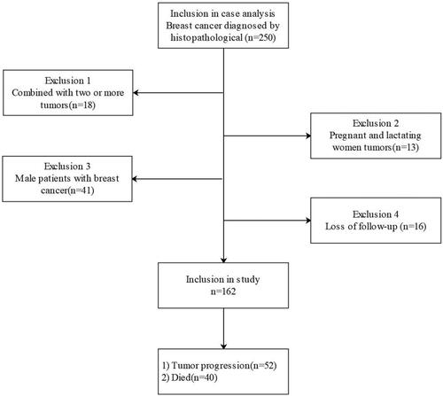
In this prospective study, we recorded data from breast caner patients between May 2014 and July 2017 at the First Affiliated Hospital of Wannan Medical College. Inclusion criteria: (1) All breast cancer patients were diagnosed by histopathological examination after surgery; (2) Accurate imaging evaluation should be taken before operation. Exclusion criteria: (1) Combined with two or more tumours (18 cases); (2) Pregnant and lactating women (13 cases); (3) Male patients with breast cancer (41 cases); (4) Loss of follow-up (16 cases). According to the exclusion criteria, 162 breast cancer patients were finally included in the study, as shown in .
Figure 1. Patients flow chart. According to the exclusion criteria, 162 breast cancer patients were finally included in the study.
Data extraction
A total of 16 clinical data, including basic clinical data, tumour diameter, pathological characteristics, metastasis, hormone receptor and prognosis were collected. According to the expression of human epidermal growth factor receptor 2 (HER2) and hormone receptor (HR) in tumour tissue, patients was divided into three molecular types: HR+/HER2−, HER2+ and triple negative breast cancer (TNBC). HER2 detection referred to China’s breast cancer HER2 Detection Guidelines (2019), and HR detection referred to the guidelines of American Society of Clinical Oncology/American Society of Pathologists in 2020 (Allison et al. Citation2020).
Definition
All subjects were followed up once a month via phone or outpatient service for 5 years, and the follow up endpoint was tumour progression (in any aspect) or death. Progression-free survival (PFS) was defined as the time from inclusion in the study to tumour progression (in any aspect) or death (due to any reason). Overall survival (OS) was defined as the time from inclusion in the study to death in any reason.
Cell transfection
Human breast cancer (MDA-MB-231 and MCF-7) cells in logarithmic growth phase (Purchased from the cell bank of Shanghai Chinese Academy of Sciences) were selected and plated in DMEM medium without serum, penicillin and streptomycin in normal air atmosphere at 37 °C. Human breast epithelial cells (MCF-10A) was cultured in DMEM/F12 (1:1) (Hyclone, USA) and supplemented with 5% horse serum, 10 mg/mL insulin, 20 ng/mL EGF, 100 ng/mL cholera toxin and 0.5 mg/mL hydrocortisone in 5% CO2 atmosphere at 37 °C. The cells (5 × 10⁵/mL) were plated in 6-well cell culture plate, and transfected when the cell reached 70–80% confluent. The transfection steps were strictly carried out according to the instructions of Lipo-fectamine 2000 transfection kit. The experiment was divided into miR-381 mimic group (transfected with miR-381 mimic), miR-NC group (transfected with miR-NC), and miR-381 inhibitor group (transfected with miR-381 inhibitor). The transfection sequence were: miR-NC were 5′-UCACAACCUCCUAGAAAGAGUAGA-3′; miR-381 mimic were 5′-UAUACAAGGGCAAGCUCUCUGU-3′; miR-381 inhibitor were 5′-ACAGAGAGCUUGCCCUUGUAUA-3′. The above sequences were synthesised by Shanghai Jima Pharmaceutical Technology Co., Ltd.
Cell proliferation assay
Breast cancer cells in logarithmic growth phase were adjusted to 5 × 10⁴/mL with the culture medium, and then plated in 96-well culture plates. Add 20 μL of MTT in each well, continue to culture for 4 h, and then add 250 μL of dimethyl sulfoxide, low speed oscillation for 10 min. The absorbance value was measured by Thermo ScientificTM microplate reader (490 nm) at 24, 48, 72h for three times to ensure the average value.
Cell migration and invasion assay
1 × 10⁴ cells were seeded into Transwell cells and placed in a 24 well cell culture plate, and then 600 μL of complete culture medium was added to each well. After incubation in an incubator under 37 °C and 5% CO2 condition for 24 h, cotton swabs were used to gently wipe off excess matrix glue from Transwell chamber and remove upper chamber cells. The cell migrated or invaded to the lower surface of the membrane were fixed with 4% paraformaldehyde for 30 min and stained in 10% crystal violet for 10 min. The stained cells were counted in 5 randomly selected fields for each chamber by high magnification field microscope, and the cell counts in each visual field was recorded.
Quantitative real-time PCR
The total of breast cells’ RNA was extracted according to the instructions of TaqMan miRNA kit, and the expression of miR-381 and CCNA2 in serum was detected by qRT-PCR. Total RNA was reverse transcribed into cDNA by M-MLV reverse transcriptase (Promega, USA). qPCR analysis was performed by SsoFastTM EvaGreen Supermix (Bio-Rad Company, USA) and StepOnePlusTM real-time fluorescence quantitative PCR (Thermo Fisher Company, USA). The PCR reaction system: cDNA 2 μL, upstream primer 1 μL, downstream primer 1 μL, PremixExTaqDNA Polymerase 25 μL and ddH2O2 21 μL. PCR reaction conditions: 95 °C pre-denaturation for 3 min, 98 °C 2s, 67 °C 15s, 72 °C 20s, 30 cycles. 2−ΔΔCt method was used to calculate the relative expression of miR-381 and CCNA2 (ΔΔCt = ΔCt experimental group − ΔCt control group, ΔCt = CT target gene − CT reference gene). Primer pairs used in this study were: miR-381 were 5′-TAATCTGA CTATACAAGGGCAAGC-3′ (upstream) and 5′-TATGGTT GTTCTGCTCTCTGTCTC-3′ (downstream); CCNA2 were 5′-CGCTGGCGGTACTGAAGTC-3′ (upstream) and 5′-GAGGAACGGTGACATGCTCAT-3′ (downstream); GAPDH (internal reference gene) were5′-GAAGGTGAAGGTCGGAGTC-3′ (upstream) and 5′-GAAGATGGTGATGGGATTT-3′ (downstream).
Western blot
The treated breast cancer cells were lysed on ice for 30 minutes. 10 μL sample was loaded into each well for 12% SDS-PAGE and then the proteins were transferred to a PVDF membrane; Soaked the membrane in 50 g/L skim milk for 2 hours; Added rabbit anti-human CCNA2 polyclonal primary antibody (diluted at 1:200) and incubated overnight with PBS at 4 °C; Then, the membranes were incubated with horseradish peroxidase labelled sheep anti immune IgG secondary antibodies for another 2 h at 37 °C. After chemiluminescence substrate development, the protein imprinting bands were analysed using Quantity One software.
Fluorescent reporter assay
In the detection of luciferase reporter assay, the wild type 3′ UTRs of CCNA2 containing predicted miR-381 target sites were amplifified by PCR from MDA-MB-231 and MCF-7 cell, and mutant 3′ UTRs were obtained by overlapping extension PCR method. MDA-MB-231 and MCF-7 cells were transfected with liposome 2000, and the mixture contained 5 pmol of miR-381 and 100 ng of firefly luciferase reporter plasmids. pRL-TK was also transfected as normalisation control. After 48 hours of transfection, the Dual-Luciferase Reporter Assay System was used to detect luciferase activity. The activity of firefly luciferase was normalised to Renin luciferase activity.
Data analysis
SPSS 20.0 software was used for statistical analysis. The categorical variables were expressed by n (%), and the rate between the two groups was compared by chi-squared test. The continuous variables of normal distribution were represented by mean ± SD, and the difference between two groups and more than two groups were analysed by unpaired Student’s t test and variance analysis, respectively. The predictive value was analysed by receiver operating curve (ROC), and the difference was compared by Medcalc software through Z test. The univariate and multivariate Cox regression analysis was used to find the the independent influencing factors for PFS and OS in breast cancer patients. Kaplan-Meier (K-M) analysis was used to analysis the PFS and OS between different groups, and the differences were tested by log-rank. P values less than 0.05 was considered statistically significant.
Ethics statement
The study was approved by the ethics committee (NO:20160501004) in accordance with the Declaration of Helsinki, and all research included agreed to participate in the study.
Results
miR-381 was down-regulated and CCNA2 was up-regulated in breast cancer patients and cell lines
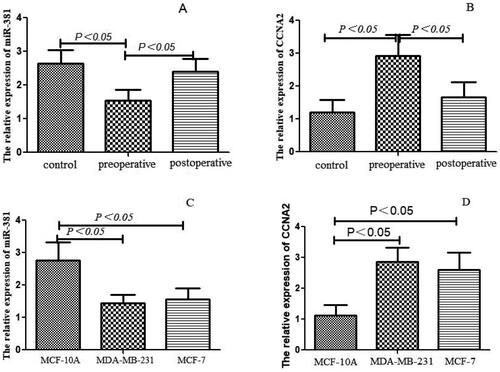
The expression of serum miR-381 in breast cancer group was significantly lower than that in the control group (), while the results of CCNA2 were opposite (). The expression of serum miR-381 in breast cancer patients after operation was significantly higher than that before operation (), while the results of CCNA2 were opposite (). We further analysed miR-381 expression in two breast cancer cells lines (MDA-MB-231, MCF-7) and breast epithelial cell line (MCF-10A). Compared with normal breast epithelial cells, lower expression of miR-381 and higher expression of CCNA2 were observed in breast cancer cells ( and ).
Figure 2. The expression of miR-381 and CCNA2 (detected by qRT-PCR) in breast cancer patient and cell lines. (A) The expression level of serum miR-381 in the control group and postoperative breast cancer patients were higher than preoperative breast cancer patients (all P < 0.05). (B) The expression level of serum CCNA2 in the control group and postoperative breast cancer patients were lower than preoperative breast cancer patients (all P < 0.05). (C) The expression of miR-381 in MCF-10A was higher than MDA-MB-231 and MCF-7 (all P < 0.05). (D) The expression of CCNA2 in MCF-10A was lower than MDA-MB-231 and MCF-7 (all P < 0.05). qRT-PCR: quantitative real-time polymerase chain reaction. MDA-MB-231 and MCF-7: human cancer cell lines. MCF-10A: human breast epithelial cell. CCNA2: Cyclin A2.
miR-381 reduced breast cancer cells proliferation and migration
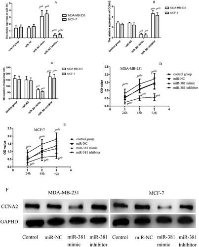
To reveal the potential function of miR-381 in breast cancer, we transfected MDA-MB-231 and MCF-7 cells with miR-381 mimics or miR-381 inhibitors to over express or silence miR-381 expression. After transfected with miR-381 mimic, the expression of miR-381 was effectively up-regulated () and the CCNA2 was effectively down-regulated (), while the opposite results were observed both in MDA-MB-231 and MCF-7 cells which transfected with miR-381 inhibitor. Migration and invasion were key determinants of malignant progression and metastasis. Compared with the control group, the migration ability of MCF-7 and MDA-MB-231 cells transfected with miR-381mimics was significantly reduced, while the opposite results were observed in cells transfected with miR-381 inhibitors (). We used the MTT assay to reveal the effect of miR-381 mimic and inhibitor on tumour cell proliferation at different time. After transfected with miR-381 mimic, the proliferation was significantly decreased both in MDA-MB-231 and MCF-7 cells, while the opposite results were observed in cells transfected with miR-381 inhibitor ( and ). After transfection with miR-381 mimic, the protein of CCNA2 was significantly down-regulated both in MDA-MB-231 and MCF-7 cells, while the opposite results were observed in cells transfected with miR-381 inhibitor according to the results of Western blot (). In other words, miR-381 can effectively down-regulate the expression of CCNA2 to inhibit the tumour cell proliferation and invasion.
Figure 3. miR-381 could inhibit breast cancer cells proliferation and migration. (A) MCF-7 and MDA-MB-231transfected with miR-381 mimic or miR-381 inhibitor both had significantly increased or decreased miR-381 expression (detected by qRT-PCR) compared with miR-NC and control groups; (B) MCF-7 and MDA-MB-231transfected with miR-381 mimic or miR-381 inhibitor both had significantly decreased or increased CCNA2 expression (detected by qRT-PCR) compared with miR-NC and control groups; (C) MCF-7 and MDA-MB-231 transfected with miR-381 mimics or miR-381 inhibitors both had significantly decreased or increased migrating cells compared with miR-NC and control groups; (D and E) MCF-7 and MDA-MB-231transfected with miR-381 mimics or miR-381 inhibitors both had significantly decreased or increased proliferation rate compared with miR-NC and control groups; (F) MCF-7 and MDA-MB-231 transfected with miR-381 mimics had obviously decreased the expression of CCNA2, while the cells transfected with miR-381 inhibitor had obviously increased the expression of CCNA2 (detected by Western blot). GAPHD: a reference gene. qRT-PCR: quantitative real-time polymerase chain reaction. MDA-MB-231 and MCF-7: human cancer cell lines. MCF-10A: human breast epithelial cell. CCNA2: Cyclin A2. Compared with control group, Pa < 0.05; Compared with miRNA-NC group, Pb < 0.05; Compared with miR-381 mimic group, Pc < 0.05.
CCNA2 is the direct target of miR-381
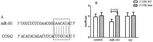
In order to explore the regulatory mechanisms of miR-381, we used target scan bioinformatics prediction to analyse the miR-381 target. It revealed that CCNA2 might be a potential target of miR-381 according to the assuming target sequence of CCNA2 3′ UTR (). The target sequence of CCNA2 3′ UTR or mutant sequence was cloned into luciferase vector. It showed that miR-381 significantly reduced the luciferase activity of CCNA2 3′ UTR, but did not reduce the mutant sequence ().
Figure 4. CCNA2 was a direct target of miR-381 in breast cancer cells. (A)The prediction of the binding between miR-381 and CCNA2 by TargetScan; (B) Luciferase reporter assays were used to verify the binding of miR-381 in 3′-UTR of CCNA2. CCNA2: Cyclin A2. NC: negative control.
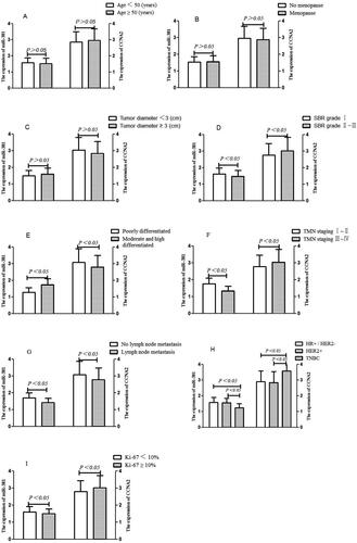
The expression of miR-381 and CCNA2 in breast cancer with different pathological parameters
The expression of serum miR-381 and CCNA2 were significantly different in breast cancer patients with different pathological grades (), TMN stages (), Scarff-Boom-Richardson (SBR) grades (), molecular pathology (), Ki-67 () and lymph node metastasis () (all P < 0.05), while the expression of serum miR-381 and CCNA2 were not significantly different in breast cancer patients with different age, menopause and tumour diameter (all P > 0.05), as shown in .
Figure 5. The expression of miR-381 and CCNA2 in breast cancer patients with different pathological characteristics. (A) Age, (B) Menopause, (C) Tumour diameter, (D) SBD grade, (E) Differentiated, (F) TMN staging, (G) Lymph node metastasis, (H) Molecular pathology, (I) Ki-67. SBR: Scarff-Bloom Richardson; TMN: Tumour Node Metastasis; HR: Hormone receptor; HER2: Human epidermal growth factor receptor-2; TNBC: Triple negative breast cancer; CCNA2: Cyclin A2.
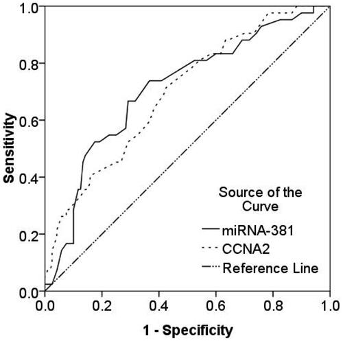
Predictive value of miR-381 and CCNA2 for PFS in breast cancer
52 of 162 patients with breast cancer had tumour progression during follow-up. The AUCs of miR-381 and CCNA2 for predicting PFS in breast cancer were 0.711 (0.619–0.802) and 0.695 (0.605–0.785) respectively (). According to the analysis of Medcalc, the predictive value between miR-381 and CCNA2 was not statistically significant (Z = 0.351, P = 0.755). The optima cut-off value, predictive sensitivity and specificity of miR-381 for predicting PFS were 1.65 2−ΔΔCt, 73.08% and 63.64%, respectively. The optima cut-off value, predictive sensitivity and specificity of CCNA2 for predicting PFS were 2.95 2−ΔΔCt, 71.16% and 57.27%, respectively.
Figure 6. ROC analysis of miR-381 and CCNA2 for predicting PFS in breast cancer. The AUC of miR-381 and CCNA2 for predicting PFS in breast cancer were 0.711 and 0.695, respectively. ROC: Receiver operating characteristic; AUC: Area under curve; CCNA2: Cyclin A2; PFS: Progression free survival.
Univariate cox regression analysis for risk of PFS in patients with breast cancer
High incidence of PFS was observed in patients with TMN staging III–IV, TNBC, miR-381 < 1.65 2−ΔΔCt and CCNA2 ≥ 2.95 2−ΔΔCt compared to patients with the opposite clinical characteristics, and the difference were statistically significant (all P < 0.05). The multivariate Cox regression analysis showed that TMN staging III–IV, TNBC and CCNA2 ≥ 2.95 2−ΔΔCt were positive correlated with PFS, while miR-381 ≥ 1.65 2−ΔΔCt were negative correlated with PFS, the hazard ratio (HR) values were 1.827, 2.192, 2.075 and 0.553 respectively (all P < 0.05) ().
Table 1. Influencing factors of PFS in breast cancer patients.
K-M analysis of PFS in breast cancer patients
The K-M survival curve analysis showed patients with TNBC, high TMN staging (III–IV) and CCNA2 (CCNA2 ≥ 2.95 2−ΔΔCt), and low miR-381 (miR-381 < 1.65 2−ΔΔCt) had significantly shorter PFS time (P < 0.05) than patients with non-TNBC, low TMN staging (I–II) and CCNA2 (CCNA2 < 2.95 2−ΔΔCt) and high miR-381 (miR-381 ≥ 1.65 2−ΔΔCt), the differences were statistically significant (χ2 = 4.762–8.552, all P < 0.05, Figure S1).
Predictive value of miR-381 and CCNA2 for OS in breast cancer
Forty of 162 patients with breast cancer died during follow-up. The AUCs of miR-381 and CCNA2 for predicting OS in breast cancer were 0.694 (0.602–0.786) and 0.675 (0.578–0.772) respectively (Figure S2). According to the analysis of Medcalc, the predictive value between miR-381 and CCNA2 wasn’t statistically significant (Z = 0.472, P = 0.711). The optima cut-off value, predictive sensitivity and specificity of miR-381 for predicting OS were 1.50 2−ΔΔCt, 65.00% and 69.70%, respectively.
Univariate cox regression analysis for risk of OS in patients with breast cancer
High incidence of OS was observed in patients with lymph node metastasis, TNBC, miR-381 < 1.50 2−ΔΔCt and CCNA2 ≥ 3.15 2−ΔΔCt compared to patients with the opposite clinical characteristics, and the difference were statistically significant (all P < 0.05). Which indicated that lymph node metastasis, molecular pathology, miR-381 and CCNA2 might be potential influencing factors of OS in breast cancer. The multivariate Cox regression analysis showed that lymph node metastasis, TNBC and CCNA2 ≥ 3.15 2−ΔΔCt were positive correlated with OS, while miR-381 ≥ 1.50 2−ΔΔCt were negative correlated with OS, the HR values were 2.240, 2.826, 2.089 and 0.462 respectively (all P < 0.05), as shown in .
Table 2. Influencing factors of OS in breast cancer patients.
K-M analysis of OS in breast cancer patients
The K-M survival curve analysis showed patients with lymph node metastasis, TNBC, high CCNA2 (CCNA2 ≥ 3.15 2−ΔΔCt) and low miR-381 (miR-381 < 1.50 2−ΔΔCt) had significantly shorter OS time (all P < 0.05) than patients with non-lymph node metastasis, non-TNBC, low CCNA2 (CCNA2 < 3.15 2−ΔΔCt) and high miR-381 (miR-381 ≥ 1.50 2−ΔΔCt), the differences were statistically significant (χ2 = 3.592–9.021, all P < 0.05, Figure S3).
Discussion
Previous studies confirmed that miR-381 involved in the occurrence and progress of non-small cell lung cancer, gastric cancer, pancreatic cancer and other tumours (Sápi et al. Citation2016). As the binding and activating partner of CDK, cyclin is one of the key proteins in cell cycle regulating protein. There are two types of CCNA2 in mammalian cells: CCNA21 (specifically expressed in testis) and CCNA2 (generally expressed in tissue cells) (Silva et al. Citation2021). As CCNA2 is associated with cell proliferation, it is usually expressed at a high level in human tumours. Previous studies found that the expression of miR-381 and CCNA2 in serum were both correlated with the occurrence of gastric cancer (Xing et al. Citation2021, Yu et al. Citation2021), and the direct target of miR-381-3p was CCNA2. However, the mechanism of miR-381 regulating the expression of CCNA2 was still unclear.
Zhang et al. (Citation2018) revealed the expression of miR-381 was down-regulated in gastric cancer, which could regulate transforming growth factor - β signal transduction, inhibit cancer epithelial mesenchymal transition through targeting sex-determining region Y box 4 and TWIST related protein 1. This study confirmed that the expression of miR-381 and CCNA2 in serum was significantly correlated with the occurrence of breast cancer. In order to identify the relationship between miR-381 and CCNA2 in breast cancer, we transfected miR-381 into MDA-MB-231 and MCF-7, and found that the expression level of CCNA2 was significantly reduced. However, the opposite result was found after transfection with miR-381 inhibitor. We used the TargetScan bioinformatics predictions to predict another genes, and the binding site of CCNA2 3′UTR was obviously located at miR-381 seed region. We also demonstrated that miR-381 could down-regulate the expression of CCNA2 via the directly target of 3′ UTR in breast cancer cell. These results further verified that CCNA2 was a target gene regulated by miR-381, and the expression of CCNA2 in breast cancer patients was significantly related to the regulation of miR-381. This study also found that miR-381 could inhibit the proliferation of breast cancer cells in the breast cancer cell proliferation test. After transfection of miR-381 inhibitor, the proliferation activity of breast cancer cells was significantly enhanced, which indicated that miR-381 played a role of tumour suppressor gene in breast cancer. These findings were consistent with the results of Cao et al. (Citation2017).
This study showed that the expression levels of miR-381 and CCNA2 in breast cancer patients were related to lymph node metastasis, TNM stage, pathological grade, TMN staging, SBR grade, molecular pathology and Ki-67, which indicated that miR-381 and CCNA2 were related to the malignant biological behaviour of breast cancer. The mechanism might be as follows: (1) Nicotinamide phosphoribosyltransferase was a rate limiting enzyme involved in the nicotinamide adenine dinucleotide rescue pathway. The expression of nicotinamide phosphoribosyltransferase was up-regulated in breast cancer cells, which could improve the vitality of cancer cells and promote the proliferation of cancer cells. miR-381 could directly target the 3 ‘- untranslated region of nicotinamide phosphoribosyltransferase, negatively regulate the expression of nicotinamide phosphoribosyltransferase, and promote the apoptosis of breast cancer cells (Bolandghamat Pour et al. Citation2019). (2) CXC chemokine receptor 4 was specific to stromal cell-derived factor 1 α-Chemokine receptor and stromal cell-derived factor 1/CXC chemokine receptor 4 signalling pathway was involved in the survival, proliferation and migration of breast cancer cells. CXC chemokine receptor 4 was the target of miR-381, was closely related to postoperative recurrence of breast cancer. The down-regulation of miR-381 could increase the expression of CXC chemokine receptor 4, and promote epithelial mesenchymal transformation and breast cancer cell proliferation (Li et al. Citation2020).
At present, there have been many reports on the influencing factors of PFS and OS in breast cancer, which mainly focuse on pathological characteristics and treatment methods (Almagro et al. Citation2016, Arnould et al. Citation2019). This study also demonstrated that TMN stage and TNBC were the influencing factors of PFS, while lymph metastasis and TNBC were the influencing factors of OS. TNBC is a special kind of breast cancer, and the expression of HR and HER2 is negative. Héquet et al. (Citation2017) reported that different molecular types of breast cancer showed different metastatic tendencies. HR + breast cancer were prone to bone metastasis, HER2+ breast cancer were prone to liver metastasis, and TNBC were prone to lung and brain metastasis. However, visceral metastasis and the number of metastatic parts more than 3 increased the risk of death. This study confirmed that the AUCs of miR-381 and CCNA2 on PFS and OS of breast cancer were 0.711, 0.695, 0.694 and 0.675, the optimal cut-off values were 1.65 2−ΔΔCt, 2.95 2−ΔΔCt, 1.50 2−ΔΔCt and 3.15 2−ΔΔCt respectively. In this study, after the longest 5 years follow-up, the Cox regression and K-M analysis showed that miR-381 was the influencing factor of PFS and OS in breast cancer, high level of miR-381 was associated with a better prognosis, while the opposite results were found in CCNA2. These findings further confirmed that miR-381 and CCNA2 played the role of tumour suppressor gene and oncogene respectively in breast cancer patients, which consistent with the report of Li et al. (Citation2020) in gastric cancer.
In conclusion, this study aimed to investigate the mechanism of miR-381 down-regulating the expression of CCNA2 and the predictive values of them on PFS and OS in breast cancer. Our data indicated that CCNA2 was the target gene of miR-381 and could be down-regulated by miR-381 to affect the proliferation, migration of tumour cells and the prognosis of patients. Therefore, both of them had good predictive values for PFS and OS in breast cancer patients. In other words, miR-381 and CCNA2 played a role similar to that of tumour suppressor gene and oncogene, which may provide a new strategy for clinical treatment in future.
Authors’ contributions
All authors made contributions to the research. Ming-gang Cao conceived and designed the study. Chun-sheng Liu and Ming-gang Cao conducted most of the experiments. Yan Wang and Yang Wang analysed the data. Zhi-min Yang and Mei-qing Wang performed the literature search and data extraction. Shuai Zhuo and Yan Yang drafted the manuscript. All authors read and approved the manuscript.
Supplemental Material
Download Zip (224.9 KB)Disclosure statement
All authors declare that there is no conflict of interest
Data availability statement
All data generated or analysed during this study are included in this published article.
Additional information
Funding
References
- Allison, K.H., et al., 2020. Estrogen and progesterone receptor testing in breast cancer: ASCO/CAP Guideline Update. Journal of Clinical Oncology, 38 (12), 1346–1366.
- Almagro, E., et al., 2016. Prognostic factors of early breast cancer. Medicine Clinica (Barcelona), 146 (4), 167–171.
- Arnould, L., et al., 2019. Breast cancer in young women. Histological and prognostic specificities: how are they different from older women? Bull Cancer, 106 (12S1), S10–S18.
- Bolandghamat Pour Z., et al., 2019. Up-regulation of miR-381 inhibits NAD + salvage pathway and promotes apoptosis in breast cancer cells. EXCLI Journal, 18, 683–696.
- Cao, Q., et al., 2017. MicroRNA-381 inhibits the metastasis of gastric cancer by targeting TMEM16A expression. Journal of Experimental & Clinical Cancer Research, 36 (1), 29.
- Chen, G., et al., 2022. Construction and analysis of a survival-associated competing endogenous RNA network in breast cancer. Frontiers in Surgery, 9, 1021195.
- Héquet, D., et al., 2017. Androgen receptors in breast cancer: Expression, value and therapeutic prospects. Bull Cancer, 104 (4), 363–369.
- Jiang, D., et al., 2022. The mortalities of female-specific cancers in China and other countries with distinct socioeconomic statuses: A longitudinal study. Journal of Advanced Research, 18, 127–139.
- Li, Y., et al., 2020. Protective effect of microRNA-381 against inflammatory damage of endothelial cells during coronary heart disease by targeting CXCR4. Molecular Medicine Reports, 21 (3), 1439–1448.
- Liu, L., et al., 2018. miR-381-3p knockdown improves intestinal epithelial proliferation and barrier function after intestinal ischemia/reperfusion injury by targeting nurr1. Cell Death & Disease, 9 (3), 411.
- Liu, N.Q., et al., 2022. Cyclin genes as potential novel prognostic biomarkers and therapeutic targets in breast cancer. Oncology Letters, 24 (4), 374.
- Pourgholamali, B., et al., 2023. Bioinformatic analysis divulged novel prognostic circulating microRNAs and their potential target genes in breast cancer. Applied Biochemistry and Biotechnology, 195 (1), 283–297.
- Sápi, Z., et al., 2016. Epigenetic regulation of SMARCB1 By miR-206, -381 and -671-5p is evident in a variety of SMARCB1 immunonegative soft tissue sarcomas, while miR-765 appears specific for epithelioid sarcoma. A miRNA study of 223 soft tissue sarcomas. Genes Chromosomes Cancer, 55 (10), 786–802.
- Silva, C. H., et al., 2021. Cyclin A2 localises in the cytoplasm at the S/G2 transition to activate PLK1. Life Science Alliance, 4 (3), e202000980.
- Su, F., et al., 2022. Integrated tissue and blood miRNA expression profiles identify novel biomarkers for accurate non-invasive diagnosis of breast cancer: preliminary results and future clinical implications. Genes, 13 (11), 1931.
- Tang, H., et al., 2011. Interaction of hsa-miR-381 and glioma suppressor LRRC4 is involved in glioma growth. Brain Research, 1390, 21–32.
- Xing, Z., et al., 2021. Expression and prognostic value of CDK1, CCNA2, and CCNB1 gene clusters in human breast cancer. Journal of International Medical Research, 49 (4), 300060520980647.
- Yu, Y.Z., et al., 2021. miR-381-3p suppresses breast cancer progression by inhibition of epithelial-mesenchymal transition. World Journal of Surgical Oncology, 19 (1), 230.
- Zhang, Y., et al., 2018. Effects of miRNAs on functions of breast cancer stem cells and treatment of breast cancer. OncoTargets and Therapy, 11, 4263–4270.