

Abstract
Background
This study aimed to analyse the expression of microRNA-223 (miR-223) in embryo culture medium and its correlation with pregnancy outcomes.
Methods
Two hundred and two patients undergoing in vitro fertilisation/intracytoplasmic sperm injection (IVF/ICSI) were divided into clinical pregnancy group (n = 101) and non-pregnant group (n = 101). The baseline data, clinical indicators, and the expression level of miR-223 in the embryo medium were compared between the two groups. Logistic regression analysis was used to analyse the relationship between each index and the pregnancy outcome. Receiver operator characteristic curve was carried out to evaluate the differential ability of miR-223 in pregnancy status. Bioinformatics methods were used to identify the target genes of miR-223 and elucidate their functions.
Results
Compared with pregnancy group, the non-pregnancy group exhibited a reduction in miR-223 expression (p < 0.001). Multivariate analysis revealed that miR-223 reduction was an independent factor for pregnancy failure (p < 0.05). The ROC curve demonstrated the discriminative capability of miR-223 in distinguishing pregnancy and non-pregnancy. In addition, bioinformatics analysis indicated that the target genes of miR-223 were predominantly located in the endocytic vesicle membrane and were primarily enriched in adenosine monophosphate-activated protein kinase (AMPK) and mammalian target of rapamycin (mTOR) signalling pathways.
Conclusion
In this study, levels of miR-223 in the embryo culture medium predicted pregnancy outcomes in subjects undergoing IVF/ICSI. Low expression of miR-223 was a risk factor for adverse pregnancy outcomes in subjects.
PLAIN LANGUAGE SUMMARY
In this study, 202 patients who underwent IVF/ICSI were retrospectively analysed and categorised into pregnant and non-pregnant groups based on their pregnancy status. The examination of embryo culture medium samples from both groups revealed that the non-pregnant group exhibited lower miR-223 expression compared to the pregnant group. Subsequent ROC analysis demonstrated the clinical relevance of miR-223 in effectively distinguishing between pregnant and non-pregnant states. Multi-factor analysis further established that the diminished expression of miR-223 independently influenced the likelihood of successful pregnancy.
Introduction
As assisted reproductive technology continually advances, in vitro fertilisation (IVF) -embryo transfer (ET) technology has emerged as a primary approach for addressing infertility (Tan et al. Citation2021). Compared to conventional IVF, IVF/intracytoplasmic sperm injection (ICSI) is primarily employed for males with oligozoospermia and asthenozoospermia. This is due to the inability of sperm to naturally achieve sperm-oocyte binding, necessitating technology intervention to facilitate sperm injection into the oocyte (Jawed et al. Citation2016). Despite over 30 years of development, the clinical pregnancy rate of IVF/ICSI remains within the range of 30–50% (Foong et al. Citation2006, Xing et al. Citation2020). Clinical pregnancy is a complicated process involving many factors such as embryo, endometrial and endocrine. Although researchers have tried to use age, antral follicle counting (AFC), and anti-Mullerian hormone (AMH) to predict pregnancy outcomes with assisted reproductive technology, the results have been unsatisfactory. For example, a study by Zhang et al. reported that serum AMH levels predicted pregnancy outcomes in IVF/ICSI patients aged 30–39 years, but not in patients younger than 30 years or older than 39 years (Zhang et al. Citation2021b). Therefore, enhancing the pregnancy success rate in IVF/ICSI, identifying more predictive factors, and investigating the underlying mechanisms influencing its outcomes remain pivotal concerns in the field of reproductive medicine.
MicroRNAs (miRNAs/miRs) constitute a class of small, highly conserved functional RNAs comprising 25 nucleotides. They are widely expressed across diverse biological systems, regulating gene and protein expression by targeting messenger RNA (mRNA)(Montagner et al. Citation2014, Gholizadeh et al. Citation2020). MiRNAs play a wide-ranging role in physiological and pathological processes in organisms, with the ability to be secreted extracellularly and detected in blood or other body fluids, rendering them potential biomarkers. Certain miRNAs, such as miR-103 and miR-30b, are known to be crucial in oocyte growth and development, meiosis, and the regulation of post-meiotic differentiation of male germ cells (Murchison et al. Citation2007). In preimplantation embryo development, miRNAs, including development-related miR-125a, exhibit varying expression levels (Byrne and Warner, Citation2008). In our extensive literature review, miR-223 (also known as miR-223-3p) was singled out for investigation. While miR-223 has been observed in prior studies, its exploration within the reproductive system remains limited. Encoded by a highly conserved gene on the X chromosome, miR-223 serves roles in regulating haematopoietic cell differentiation, suppressing inflammation during infection, and platelet adhesion (Almuntashiri et al. Citation2022). A study by Sadegh et al. noted a non-significant reduction in the expression of miR-223 in the blastocyst medium of unsuccessfully implanted embryos compared to successfully implanted embryos (Eivazi et al. Citation2023). The expression of miR-223 increased in foetuses of acute chorioamnionitis group (Lee et al. Citation2018). Other studies have shown that abnormal expression of miR-223 is associated with polycystic ovary syndrome (Li et al. Citation2017, Zou et al. Citation2023). Interestingly, studies have identified the release of specific miRNAs by human embryos into the culture medium, influencing endometrial responses during implantation. This phenomenon might directly impact clinical pregnancy rates in IVF (Kong et al. Citation2021).
This study retrospectively analysed the data of 202 patients who underwent IVF/ICSI treatment, and compared the clinical indicators and differences in the expression level of miR-223 in patients with different pregnancy outcomes, aiming to provide valuable experimental basis for clinical search for effective predictive markers of pregnancy outcomes.
Methods
Inclusion of the study population
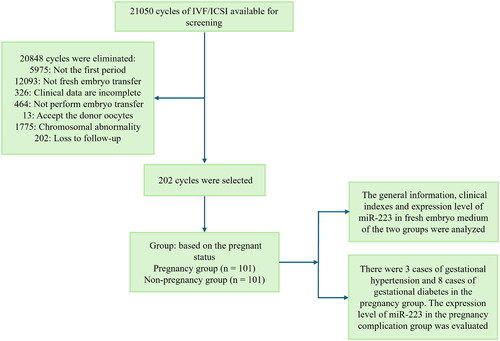
A retrospective analysis was conducted on 202 cases of infertility treated through assisted reproductive technology in Reproductive Medicine Centre of Maternity & Child Care Centre of Qinhuangdao Hospital. This study was approved by the Ethics Committee of Maternity & Child Care Centre of Qinhuangdao Hospital, and all participants provided informed consent (Ethical approval number: 2022019). The inclusion criteria were as follows: (1) infertility diagnosis, defined as no pregnancy for ≥ 12 months without contraception under normal sexual conditions; (2) indication for IVF-ICSI; (3) absence of prior use of drugs affecting pregnancy outcomes; (4) AMH levels ranging from 2 to 6.8 g/L; (5) patients with normal thyroid function. The exclusion criteria were as follows: (1) patients with conditions unsuitable for fertility; (2) patients with genital organ malformation, hydrosalpinx, intrauterine adhesions, and uterine leiomyoma (The flow chart of the experimental scheme is shown in ). The causes of infertility comprised 39 cases of pelvic and fallopian tube factors, 12 cases of ovarian dysfunction, 25 cases of polycystic ovary syndrome, 33 cases of endometriosis, 15 cases of immune infertility, and 78 cases of male oligozoospermia/asthenozoospermia/teratospermia. General information, such as age, duration of infertility, endometrial thickness, and basic sex hormone levels, was collected.
Figure 1. The flow chart of experimental scheme.
Basic procedure of IVF-ICSI
Ovarian function assessment: Before commencing the IVF cycle, a thorough evaluation of basic sex hormones, including follicle-stimulating hormone (FSH), luteinizing hormone (LH), oestradiol (E2), progesterone (P), and testosterone (T), was conducted on days 2–4 of menstruation. Additionally, the number of antral follicles in both ovaries, defined as follicles with a diameter of 2–8 mm, was measured using transvaginal ultrasound.
Protocol for controlled ovarian hyperstimulation: Tailored programs were developed based on the patient’s actual age and ovarian function. This included a long-acting regimen in early follicular phase and antagonist regimen. a) Long-acting regimen in the early follicular phase: a 3.7 mg dose of a long-acting gonadotropin-releasing hormone agonist (GnRH-a) was administered on days 2–4 of the menstrual period. Ovarian parameters reached the standard of reduced regulation (intima thickness < 5 mm, LH < 5 IU/L, P <3 nmol/L, and follicle diameter 4–6 mm) after 30–40 days of treatment. The choice of gonadotropin (Gn) type and the initial dose were based on the patient’s age, hormone profile, and other conditions, typically starting with 225–300 IU/day. Follicle growth was monitored 4–6 days after administration, and Gn dosage was adjusted accordingly. Human chorionic gonadotropin (hCG) 10000IU trigger was administrated when follicles ≥18 mm in diameter reached 40%–60% of the dominant follicle group (follicles ≥ 14mm in diameter). b) Antagonist regimen: exogenous Gn ovulation induction therapy began on the days 2–4 of the menstrual period. The Gn type and dose were selected based on patient specifics, typically at 75–300 IU/day. Monitoring of hormone levels, follicle size, and number occurs on the fourth day after medication. If the dominant follicle diameter exceeds 12 mm, GnRH-a should be introduced until the trigger date. In the presence of elevated LH levels, consideration is given to earlier addition. Considering E2 and P levels, when three dominant follicles reach a diameter of ≥17 mm or two dominant follicles reach a diameter of ≥18 mm, hCG or GnRH-a injection (or combined) was administrated, and oocyte retrieval was performed at 34–36 h based on LH levels.
Oocyte and sperm retrieval: Approximately 34–37 h after the trigger, guided by B-ultrasound, the oocyte retrieval needle was carefully inserted through the posterior fornix of the vagina, reaching the ovary via a puncture guide device. The oocytes were then aspirated using a negative pressure device. Immediately after retrieval, the oocytes were transferred to a petri dish containing embryo culture solution and cultured in an incubator at 37 °C. For males, ejaculation was required 2–3 days before oocyte retrieval and sperm retrieval was performed on the same day as oocyte retrieval. After semen liquefaction, the sperm was treated using density gradient centrifugation for subsequent use.
In vitro insemination: The routine insemination procedure in IVF involved placing the treated sperm and oocytes together in the same dish approximately 12–18 h after oocyte retrieval. The co-culture lasted for 12–18 h, and fertilisation was observed microscopically. In contrast, the ICSI protocol entails removing granulosa cells from the oocytes, injecting sperm into oocytes in metaphase II, and subsequently placing them in a petri dish for 12–18 h to monitor fertilisation. Normal zygotes are double pronuclei with regular morphology and intact zona pellucida, whereas abnormal fertilisation is indicated by a single pronucleus or multiple pronuclei.
Embryo assessment criteria: The grading criteria for embryos on day 3 were defined as follows: Grade I, characterised by uniform cell size, regular shape, intact zona pellucida, uniform and clear cytoplasm without granules, and cytoplasmic fragments < 5%; Grade II: displaying slightly uneven cell size, granular phenomenon in the cytoplasm, and fragments ranging from 6% to 20%; Grade III: with significantly uneven cell size, distinct granular phenomenon in the cytoplasm, and the fragments comprising 21% to 50%; Grade IV: exhibiting uneven cell size, marked granular phenomenon in the cytoplasm, and fragments exceeding 50%. In this study, high-quality embryos were selected based on three criteria: (a) the embryos originated from normal zygotes; (b) they featured 7–9 embryonic cells on the third day after fertilisation; (c) the embryos attained Grade I–II status at the cleavage stage.
Embryo transfer: Fresh cycle embryo transfer: embryo transfer, depending on the endometrium of the patient, can be performed 3–5 days after oocytes retrieval. The number of embryos transferred was 1 or 2, and the remaining transferable embryos were cryopreserved or frozen after blastocyst culture with the informed consent of the patient and her family.
Pregnancy assessment (Segura-Benitez et al. Citation2023): Urine hCG testing took place 14 days after embryo transfer, with additional blood hCG testing if the result was positive. A positive pregnancy test prompted a vaginal ultrasound 28 days after embryo transfer to confirm pregnancy. The pregnant group comprised cases where the test was positive, and the gestational sac and the original cardiac tube activity were visible under ultrasound. The non-pregnant group included cases with a negative pregnancy test 14 days after embryo transfer.
Detection of miR-223 expression level in embryo culture medium
Following the embryo transfer, the culture medium from the transferred embryos was promptly collected into a centrifuge tube. After centrifugation, the supernatant was retained, and the miR-223 expression in the supernatant was detected using quantitative reverse transcription polymerase chain reaction (qRT-PCR). In short, total RNA was extracted using TRIzol (Invitrogen), with subsequent determination of RNA concentration using an ultraviolet spectrophotometer and recording of the A260/A280 ratio (a qualified purity ratio fell within the range of 1.8–2.1). The reverse transcription reaction followed the instructions of the TaqMan MicroRNA Reverse Transcription kit (Applied Biosystems). Using the first cDNA strand as the template, the expression level of miR-223 was determined through PCR reaction. The reaction system adhered to the TaqMan MicroRNA assay kit (Applied Biosystems) instructions, with amplification procedures set on the quantitative system, and U6 served as the internal reference gene for standardisation. The sequence of primers required for the experiment is as follows: miR-223: (forward primer: 5′-CATTGTCAGTTTGTCAAATACC-3′), (reverse primer: 5′-CCATGAGAGATCCCTAGCG-3′). U6: (forward primer: 5′-CTCGCTTCGGCAGCACA-3′), (reverse primer: 5′-AACGCTTCACGAATTTGCGT-3′). The relative expression of miR-223 in the samples was calculated using the comparative cyclic threshold of 2-ΔΔCt. The experiment was conducted in triplicate.
Main observation index
The relationship between miR-223 levels and pregnancy complications and preterm birth in pregnant patients was investigated. Pregnancy complications included gestational hypertension and gestational diabetes. A preterm birth was defined as delivering occurring between 28 and less than 37 weeks of gestation.
Identification and analysis of miR-223 target genes
Four databases, including miRDB (http://www.mirdb.org/), TargetScan (http://www.targetscan.org/), miRWalk (http://mirwalk.umm.uni-heidelberg.de/) and ENCORI (https://www.ncbi.nlm.nih.gov/), were used to predict the downstream target genes of miR-223. Venn diagram of intersected candidate target genes of miR-223 was plotted via Online Venn diagram analysis (https://www.bioinformatics.com.cn).
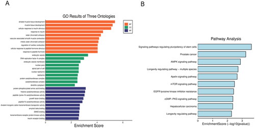
The intersecting target genes obtained above were analysed by Gene Ontology (GO) function analysis and Kyoto Encyclopaedia of Genes and Genomes (KEGG) pathway analysis (He et al. Citation2013). GO functional enrichment is a common method for functional enrichment analysis of genomic or transcriptome data, which is to annotate miR-223 target genes based on three levels of biological process, cell component and molecular function. KEGG is a knowledge database for the systematic analysis of gene function, linking genomic information to the high-level function and utility of biological systems, and its common terminology studies gene function and pathways from large datasets of high-throughput experiments.
Data analysis
Statistical analysis was performed using SPSS 27.0 software. The Kolmogorov-Smirnov test was used to evaluate the normality of data distribution. Measurement data exhibiting normal distribution and homogeneity of variance are expressed as mean ± standard deviation (SD), and inter-group comparisons were conducted using the independent sample t test. For categorical variables between groups, the Pearson’s Chi-square test or the Fisher’s exact test was used based on the validity of the assumption. The logistic regression model was employed, with odds ratio and their corresponding 95% confidence intervals (CIs) used to evaluate the correlation between each index and the outcome of embryo transfer. Additionally, a receiver operating characteristic curve was established to evaluate the predictive value of miR-223 expression level in embryo culture medium for clinical pregnancy after transfer. p < 0.05 indicates a statistically significant difference.
Results
Comparison of baseline indices between the clinical pregnant and non-pregnant groups
A total of 202 patients underwent fresh embryo transfer, providing clinical data for analysis, with 101 cases resulting in clinical pregnancies and 101 cases classified as clinical non-pregnancies. The relevant information and laboratory indicators for both groups are presented in . No significant differences were observed in age, body mass index (BMI), menstrual cycle, menstrual duration, disease course, and basic hormone levels between the two groups (p > 0.05). Additionally, it was also observed that the expression level of miR-223 in the embryo culture medium of patients in the clinically non-pregnant group was significantly lower compared to the clinically pregnant group (, p < 0.001).
Figure 2. MiR-223 expression decreased in the embryo culture medium of clinically non-pregnant patients. ***p < 0.001 vs. Pregnant group.
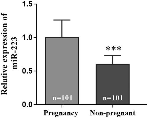
Table 1. Baseline data of the subjects.
Analysis of influencing factors on the outcomes of fresh embryo transfer
The indicators in were incorporated into the multi-factor logistic regression model. The findings in and indicate that reduced levels of miR-223 emerged as an independent factor significantly influencing non-pregnancy after embryo transfer (OR = 0.447, 95%CI = 0.248–0.805, p = 0.007).
Figure 3. Forest diagram of the multivariate logistic regression analysis.
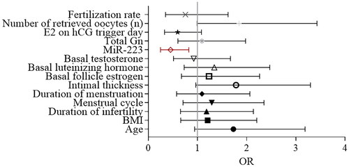
Table 2. Multi-factor logistics regression predicts independent influencing factors of pregnancy failure.
The predictive value of miR-223 levels in embryo culture medium for clinical pregnant
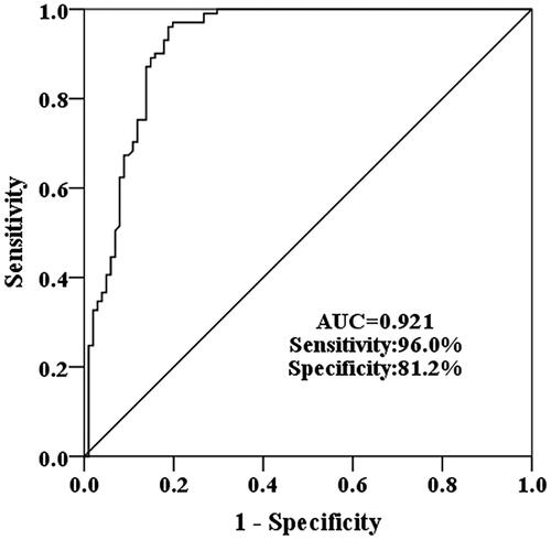
Using the level of miR-223 in embryo culture medium as the test variable and clinical pregnancy as the state variable, the ROC curve was drawn. The outcomes illustrated in revealed that the area under the curve (AUC) for miR-223 in predicting clinical pregnancy was 0.921, with a sensitivity and specificity were 96.0% and 81.2%, respectively. These results indicate that miR-223 holds a certain diagnostic value for distinguishing between clinical pregnancy and non-pregnancy.
Figure 4. Receiver operating characteristic (ROC) curves were used to evaluate the predictive value of miR-223 in clinical pregnancy after embryo transfer.
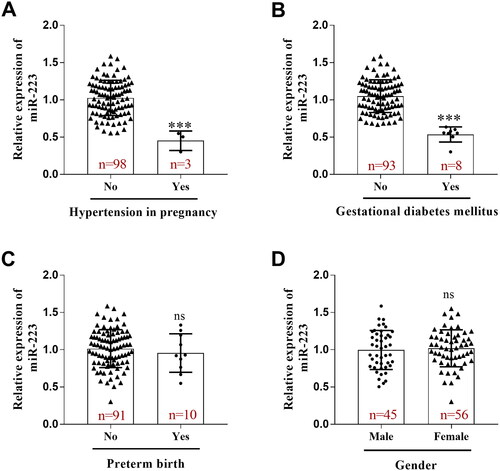
Expression of miR-223 in patients with different pregnancy complications
Upon analysing follow-up data from 101 patients with clinical pregnancy, it was noted that 3 patients developed gestational hypertension. In comparison to patients without gestational hypertension, those with gestational hypertension exhibited a down-regulation of miR-223 expression in the embryo culture medium (, p < 0.001). Additionally, 8 cases of gestational diabetes were observed, and miR-223 expression in the embryo culture medium of patients with gestational diabetes decreased compared to those without gestational diabetes (, p < 0.001). Among the 10 patients with preterm delivery, no different difference in miR-223 expression was observed compared to those with full-term delivery (, p > 0.05). Similarly, there was no significant difference in the expression level of miR-223 among groups with different infant sexes (, p > 0.05).
Figure 5. Expression of miR-223 in patients with varying pregnancy complications. (A) Decreased miR-223 expression in the embryo culture in patients with gestational hypertension. (B) Decreased miR-223 expression in the embryo culture in patients with gestational diabetes. (C) No difference in miR-223 expression was observed in preterm delivery compared to those with full-term delivery. (D) No significant difference was observed in the expression level of miR-223 in the embryo culture medium of the patients who delivered a female foetus compared to the patient group that delivered a male foetus. ***p < 0.001. ns, not significant.
Prediction and functional analysis of miR-223 target genes
As shown in , the miRDB database predicted 519 target genes of miR-223, TargetScan database identified 418, the miRWalk database identified 6326, and the ENCORI database identified 1108 target genes. Collectively, these four databases predicted 63 target genes. demonstrate that GO analysis highlighted the predominant localisation of miR-223 target genes at the endocytic vesicle membrane. These genes were involved in diverse biological processes, including striated muscle tissue development, vasoassociated smooth muscle contraction, cardiac conduction regulation, and cell response to insulin stimulation. The molecular functions associated with these genes included protein binding and regulation of enzyme activity. The KEGG pathway analysis indicated that the target genes of miR-223 were predominantly enriched in adenosine monophosphate-activated protein kinase (AMPK) signalling pathway, the regulating stem cell pluripotency signalling pathway, the Apelin signalling pathway, the mTOR signalling pathway, and cGMP-PKG signalling pathway ().
Figure 6. A Venn diagram of miR-223 target genes predicted by miRDB, TargetScan, miRWalk and ENCORI database. Abbreviations: ENCORI, Encyclopaedia of RNA Interactomes.
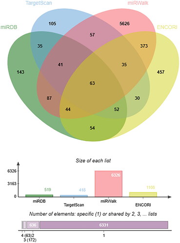
Figure 7. Bioinformatics analysis. (A) GO enrichment analysis for the predicted target genes of miR-223. (B) KEGG pathway analysis for the predicted target genes of miR-223. Abbreviations: GO, Gene Ontology; KEGG, Kyoto Encyclopaedia of Genes and Genomes.
Discussion
In this study, it was observed that the expression level of miR-223 in the embryo culture medium of patients with pregnancy failure after IVF/ICSI was significantly lower than that observed in the group with successful pregnancies. Through multivariate analysis, it was established that the reduction in miR-223 expression served as an independent factor associated with pregnancy failure after IVF/ICSI. Moreover, miR-223 exhibited significant diagnostic efficacy in distinguishing between successful pregnancies and pregnancy failures. Subsequent examinations revealed a significant reduction in the expression level of miR-223 in the embryo culture medium of individuals with gestational hypertension or gestational diabetes compared to patients without pregnancy complications. The above results revealed that reduced miR-223 expression was associated with pregnancy failure and complications after IVF/ICSI treatment.
Embryo implantation constitutes a pivotal stage in establishing pregnancy, encompassing early embryo development, the establishment of endometrial receptivity, mutual recognition, localisation, and adhesion between the embryo and endometrial epithelial cells, culminating in the penetration of the endometrial surface and implantation into the endometrial matrix (Toledo-Guardiola et al. Citation2023). Beyond the precise regulation by hormones and certain cytokines, this stage crucially relies on fundamental gene regulation. In the complex process of pre-implantation embryonic development in mammals, miRNA plays an important regulatory role. In this study, the expression level of miR-223 in the embryo culture medium of the pregnancy failure group was observed to be significantly lower than that of the successful pregnancy group. Notably, miR-223, well-established for its integral role in immune response regulation, governs multiple processes, including age differentiation in neutrophils, macrophages, and dendritic cells. Numerous studies have highlighted the dysregulated of miR-223 in inflammatory diseases, illustrating its ability to target multiple immune-related genes to mediate inflammation (Jiao et al. Citation2021, Yuan et al. Citation2021). Down-regulation of miR-223 has been observed in obesity, inflammatory diseases, cancer, and autoimmune diseases (Haneklaus et al. Citation2013, Dang and Leelahavanichkul, Citation2020, Nguyen et al. Citation2022). Several recent studies have reported the association between abnormal miR-223 and reproductive related diseases. For instance, in animal models of endometritis, such as horses and cows, serum miR-223 expression levels were found to be significantly elevated (Zhao et al. Citation2018, Asif et al. Citation2023). Another study showed that miR-223 was involved in the development of follicles in chickens (Shen et al. Citation2023). Besides, Dominguez et al. demonstrated an upregulation of miR-223 expression in patients with ectopic pregnancy (Dominguez et al. Citation2014). Conversely, this study revealed a down-regulation of miR-223 in the embryo culture from patients with pregnancy failure.
The successful transfer of high-quality embryos is pivotal for the efficacy of techniques such as IVF/ICSI, significantly enhancing clinical pregnancy rates. In previous studies, researchers have attempted to predict pregnancy outcomes in IVF patients by identifying biomarkers. Horka et al. reported that the concentration of MMP-9 in serum and follicular fluid of pregnant women after IVF assisted pregnancy was significantly higher than that of non-pregnant women, suggesting that the level of MMP-9 in serum or follicular fluid was related to the outcome of IVF assisted pregnancy (Horka et al. Citation2012). Shibahara et al. observed that the level of serum TIMP-1 in the pregnant group was significantly higher than that in the non-pregnant group 14 days after embryo transfer, and high levels of TIMP-1 predicted high-quality embryos (Shibahara et al. Citation2005). In this study, the observed dysregulation of miR-223 expression in the embryo culture medium might affect the quality of the embryo, potentially leading to pregnancy failure. In instances where there were no statistically significant difference in clinical features between patients with failed pregnancies and those with successful pregnancies, successful pregnancy patients showed higher miR-223 expression in the embryo culture medium. MiR-223 could play an important role in predicting pregnancy outcome. This suggests that, in the clinical selection of transferred embryos, especially when morphological scores are closely matched and the embryo quality are similar, emphasising the elevated expression level of miR-223 could be advantageous. The selection of embryos with highlighted miR-223 expression might enhance pregnancy outcomes and increase clinical pregnancy rate. Multivariate logistic regression analysis further corroborated the correlation between miR-223 and pregnancy outcomes, underscoring that changes in miR-223 levels in the embryo culture medium can objectively reflect the quality of embryos and sensitively predict pregnancy outcomes after IVF/ICSI.
Herein, the expression level of miR-223 in the embryo culture medium of patients with gestational diabetes mellitus or gestational hypertension was assessed, revealing a downward trend compared to patients without pregnancy complications. This suggests that the abnormal expression of miR-223 might be associated with the onset of pregnancy complications. Interestingly, certain related studies reported an abnormal up-regulation of miR-223 in the serum of patients with gestational diabetes (Yoffe et al. Citation2019, Dinesen et al. Citation2023). Another study identified a down-regulation of miR-223 expression in the placenta of patients with pregnancy-induced hypertension (Choi et al. Citation2013, Xu et al. Citation2014). The discrepancies in results might be attributed to variations in sample selection. The present study exclusively used pre-transplant embryo cultures as samples, warranting further investigation into miR-223 expression levels in the serum or placenta of the subjects.
Through bioinformatics analysis of miR-223, combined with existing literature studies, it was found that miR-223 can play an important role in cardiac conduction and hormone regulation through its target genes. The biological process results of GO function of miR-223 target genes also showed that miR-223 was involved in the regulation of peptide hormone stimulation. Studies have shown that upregulating miR-223 expression in the rat model of polycystic ovary can reduce insulin resistance in the rat model (Abuelezz et al. Citation2021). Other studies have shown that miR-223 can up-regulate the expression level of oestradiol, and play a protective role in the oxidative stress damage of Alzheimer’s disease mice (Pan et al. Citation2022). Through the KEGG pathway enrichment analysis of the intersection target genes of miR-223, this study found that most of these target genes were enriched in AMPK signalling pathway and mTOR signalling pathway. Overexpression of miR-223 may regulate oxidative stress damage in cardiovascular diseases by inhibiting AMPK signalling pathway (Zhang et al. Citation2021a). In addition, in a study on endometrial receptivity, increased phosphorylation of mTOR protein led to upregulation of miR-223 expression (Niknafs et al. Citation2021). In summary, miR-223 and its target genes may be related to the process of human growth and development, and are worthy of further study.
This study has certain limitations that cannot be ignored. First, this is a single-centre retrospective study, which not only has a small sample size, but also may inevitably introduce selection bias during sample inclusion. Second, the study did not incorporate data on perinatal death and placenta previa, potentially limiting the comprehensiveness of the results. However, given the relative rarity of such patients and the interpretability of the obtained results, the study is still presented. Third, this study only detected the expression level of miR-223 in the fresh embryo medium and did not detect the expression level of miR-223 in the blood of the subjects 14 days after embryo transfer. Therefore, the expression level of miR-223 in subjects’ peripheral blood after embryo transfer is still unknown. Taking these limitations into account, in future studies, it is necessary not only to continue to expand the sample size, but also to further study and evaluate the function of miR-223 on the basis of this study.
In summary, a reduction in miR-223 expression in the embryo culture medium of patients with pregnancy failure was observed compared to those with successful pregnancies. Multi-factor regression analysis showed that miR-223 independently influenced pregnancy failure. Furthermore, miR-223 demonstrated clinical value in predicting clinical pregnancy outcomes.
Authors contribution statement
This study was supervised by Hongzhi Shi. Qi Song and Jiajia Liu conceived and designed research. Chen Li and Rongrong Liu analysed data. Qi Song and Nan Zhang wrote the manuscript. All authors read and approved the manuscript.
Disclosure statement
Authors declare that they have no competing interests.
Data availability statement
The corresponding authors may provide data and materials upon reasonable request.
Additional information
Funding
References
- Abuelezz, N.Z., et al., 2021. Nanocurcumin modulates miR-223-3p and NF-kappaB levels in the pancreas of rat model of polycystic ovary syndrome to attenuate autophagy flare, insulin resistance and improve ss cell mass. Journal of Experimental Pharmacology, 13, 873–888.
- Almuntashiri, S., et al., 2022. Identification of circulating microvesicle-encapsulated miR-223 as a potential novel biomarker for ARDS. Physiological Reports, 10 (21), e15494.
- Asif, S., et al., 2023. MicroRNAs in equine Endometritis: a review of pathophysiology and molecular insights for diagnostic and therapeutic strategies. International Immunopharmacology, 124 (Pt B), 110949.
- Byrne, M.J. and Warner, C.M., 2008. MicroRNA expression in preimplantation mouse embryos from Ped gene positive compared to Ped gene negative mice. Journal of Assisted Reproduction and Genetics, 25 (5), 205–214.
- Choi, S.Y., et al., 2013. MicroRNA expression profiles in placenta with severe preeclampsia using a PNA-based microarray. Placenta, 34 (9), 799–804.
- Dang, C.P. and Leelahavanichkul, A., 2020. Over-expression of miR-223 induces M2 macrophage through glycolysis alteration and attenuates LPS-induced sepsis mouse model, the cell-based therapy in sepsis. PLoS One, 15 (7), e0236038.
- Dinesen, S., El-Faitarouni, A. and Dalgaard, L.T., 2023. Circulating microRNAs associated with gestational diabetes mellitus: useful biomarkers? The Journal of Endocrinology, 256 (1), e220170.
- Dominguez, F., et al., 2014. Embryonic miRNA profiles of normal and ectopic pregnancies. PLoS One, 9 (7), e102185.
- Eivazi, S., et al., 2023. MiRNAs secreted by human blastocysts could be potential gene expression regulators during implantation. Molecular Biology Reports, 50 (2), 1375–1383.
- Foong, S.C., et al., 2006. A prospective randomized trial of conventional in vitro fertilization versus intracytoplasmic sperm injection in unexplained infertility. Journal of Assisted Reproduction and Genetics, 23 (3), 137–140.
- Gholizadeh, M., et al., 2020. Identifying differentially expressed microRNAs, target genes, and key pathways deregulated in patients with liver diseases. International Journal of Molecular Sciences, 21 (19), 7368.
- Haneklaus, M., et al., 2013. miR-223: infection, inflammation and cancer. Journal of Internal Medicine, 274 (3), 215–226.
- He, X., et al., 2013. Host serum miR-223 is a potential new biomarker for Schistosoma japonicum infection and the response to chemotherapy. Parasites & Vectors, 6 (1), 272.
- Horka, P., et al., 2012. Matrix metalloproteinases in serum and the follicular fluid of women treated by in vitro fertilization. Journal of Assisted Reproduction and Genetics, 29 (11), 1207–1212.
- Jawed, S., et al., 2016. Fertilization rate and its determinants in intracytoplasmic sperm injection. Pakistan Journal of Medical Sciences, 32 (1), 3–7.
- Jiao, P., et al., 2021. miR-223: an effective regulator of immune cell differentiation and inflammation. International Journal of Biological Sciences, 17 (9), 2308–2322.
- Kong, C.S., et al., 2021. Embryo biosensing by uterine natural killer cells determines endometrial fate decisions at implantation. FASEB Journal, 35 (4), e21336.
- Lee, J., et al., 2018. Increased miR-223 expression in foetal organs is a signature of acute chorioamnionitis with systemic consequences. Journal of Cellular and Molecular Medicine, 22 (2), 1179–1189.
- Li, C., et al., 2017. Altered expression of miRNAs in the uterus from a letrozole-induced rat PCOS model. Gene, 598, 20–26.
- Montagner, S., Dehó, L. and Monticelli, S., 2014. MicroRNAs in hematopoietic development. BMC Immunology, 15 (1), 14.
- Murchison, E.P., et al., 2007. Critical roles for Dicer in the female germline. Genes & Development, 21 (6), 682–693.
- Nguyen, M.A., et al., 2022. miR-223 exerts translational control of proatherogenic genes in macrophages. Circulation Research, 131 (1), 42–58.
- Niknafs, B., Hesam Shariati, M.B. and Shokrzadeh, N., 2021. miR223-3p, HAND2, and LIF expression regulated by calcitonin in the ERK1/2-mTOR pathway during the implantation window in the endometrium of mice. American Journal of Reproductive Immunology, 85 (1), e13333.
- Pan, Q., Ma, J. and Guo, K., 2022. miR-223 enhances the neuroprotection of estradiol against oxidative stress injury by inhibiting the FOXO3/TXNIP axis. Neurochemical Research, 47 (7), 1865–1877.
- Segura-Benitez, M., et al., 2023. Human blastocysts uptake extracellular vesicles secreted by endometrial cells containing miRNAs related to implantation. Human Reproduction, 38 (8), 1547–1559.
- Shen, M., et al., 2023. microRNA transcriptome analysis of granulosa cells predicts that the Notch and insulin pathways affect follicular development in chickens. Theriogenology, 212, 140–147.
- Shibahara, H., et al., 2005. Serum matrix metalloproteinase and tissue inhibitor of metalloproteinase concentrations in infertile women achieved pregnancy following IVF-ET. American Journal of Reproductive Immunology, 54 (4), 186–192.
- Tan, X., et al., 2021. Development and validation of prediction model for high ovarian response in in vitro fertilization-embryo transfer: a longitudinal study. Computational and Mathematical Methods in Medicine, 2021, 7822119.
- Toledo-Guardiola, S.M., et al., 2023. Different seminal ejaculated fractions in artificial insemination condition the protein cargo of oviductal and uterine extracellular vesicles in pig. Frontiers in Cell and Developmental Biology, 11, 1231755.
- Xing, L., et al., 2020. Pregnancy outcome treated with stage-by-stage acupuncture and moxibustion therapy based on the chong channel being sea of blood theory in repeated IVF-ET failure patients: a randomized controlled trial. Medicine, 99 (47), e23234.
- Xu, P., et al., 2014. Variations of microRNAs in human placentas and plasma from preeclamptic pregnancy. Hypertension, 63 (6), 1276–1284.
- Yoffe, L., et al., 2019. Early diagnosis of gestational diabetes mellitus using circulating microRNAs. European Journal of Endocrinology, 181 (5), 565–577.
- Yuan, S., et al., 2021. miR-223: an immune regulator in infectious disorders. Frontiers in Immunology, 12, 781815.
- Zhang, D.M., et al., 2021a. MicroRNA-223-3p protect against radiation-induced cardiac toxicity by alleviating myocardial oxidative stress and programmed cell death via targeting the AMPK pathway. Frontiers in Cell and Developmental Biology, 9, 801661.
- Zhang, Y., et al., 2021b. Predictive value of anti-Mullerian hormone on pregnancy outcomes in in-vitro fertilization/intracytoplasmic single sperm injection patients at different ages. Archives of Gynecology and Obstetrics, 304 (6), 1611–1620.
- Zhao, G., et al., 2018. The potential therapeutic role of miR-223 in bovine endometritis by targeting the NLRP3 inflammasome. Frontiers in Immunology, 9, 1916.
- Zou, L., et al., 2023. Bioinformatics analysis of the common targets of miR-223-3p, miR-122-5p, and miR-93-5p in polycystic ovarian syndrome. Frontiers in Genetics, 14, 1097706.