Abstract
A 48-year-old male had been diagnosed with Behcet's disease two years previously. During the remission period a sudden decrease in visual acuity developed in the right eye. Visual acuity in the right eye was 1/10. Relative afferent pupillary defect and impaired colour vision was detected. The anterior segment was normal. Fundus examination revealed right optic disk swelling. Neurological examination and magnetic resonance imaging were normal. Mega dose steroid therapy was initiated. Visual acuity began to improve after the third day and was 9/10 at the end of the first month of the therapy.
KEYWORDS:
Dear Editor,
We report on a 48-year-old male, who developed acute optic neuropathy during the remission period of Behçet's disease. Two years previously, he had been diagnosed with Behçet's disease and was in remission with systemic azothiopurine therapy. When he first presented to our outpatient clinic, his best-corrected visual acuities (BCVAs) were 8/10 (Snellen Chart) in both eyes. There were ++ cells in the anterior chamber of the right eye and + vitritis in both eyes. There were no posterior synechiae. Fundus examination revealed intraretinal hemorrhages, and perivascular sheating and cuffing. Fundus fluorescein angiography (FFA) revealed multiple vascular leakage zones. Oral steroid (1 mg/kg/d) was added to the therapy and subsequently reduced at regular intervals. One month later, his BCVAs were 9/10 in both eyes. The cellular reaction in the anterior chamber and the vitritis had both resolved. At the end of the third month, both anterior segment findings and optic disc margins were normal. The patient remained in remission until the tenth month, when he presented with a sudden decrease in visual acuity in the right eye. BCVAs were 1/10 and 8/10 in the right and left eye, respectively. Intraocular pressure was normal in both eyes. Relative afferent pupillary defect (RAPD) was detected in the right eye and color vision in the right eye was impaired. Anterior segment examination revealed no uveal inflammation. On fundus examination, the right optic disc was swollen with blurred margins and peripapillary hemorrhages were also present (). Optic disc margins were normal in the left eye and there were no signs of vasculitis. FFA showed the right optic disc to be hyperfluorescent, although no macular edema was observed (). Neurological examination and magnetic resonance imaging (MRI) findings were normal. Megadose steroid (1000 mg/d intravenous methylprednisolone for 3 days) was administered with a diagnosis of right acute optic neuropathy. Following megadose steroid, oral corticosteroid therapy was initiated and the dose was reduced at regular intervals. BCVA was 4/10 after the third day. Visual field examination revealed peripheric field loss ().
FIGURE 1 The right eye fundus exhibiting a swollen optic disc with blurred margins.
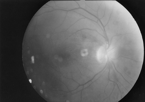
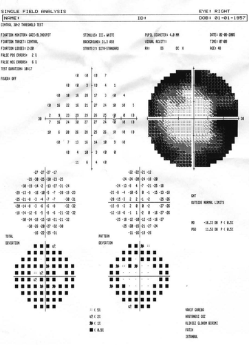
FIGURE 2 The right eye visual field showing peripheric field loss.
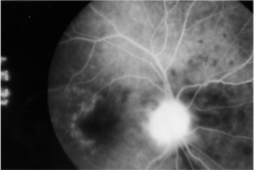
FIGURE 3 Right eye fundus fluorescein angiography revealing a hyperfluorescent optic disc on the first day.
BCVAs were 9/10 in both eyes after one month. Fundus examination revealed that the optic disc edema in the right eye had resolved and the disc margins were distinct. The optic disc hyperfluorescence, which was present in the FFA in the second month, had also disappeared and there was no leakage in the vessels ().
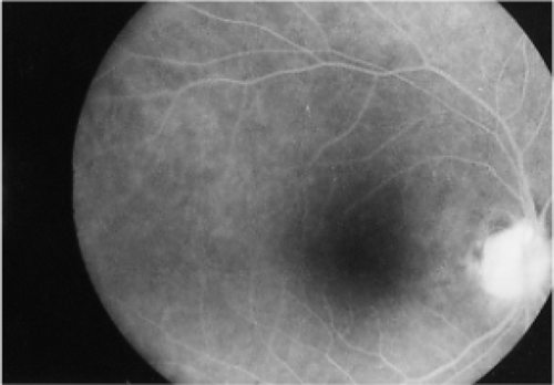
FIGURE 4 Right eye fundus fluorescein angiography after two months showing that the hyperfluorescence in the optic disc has disappeared.
Behçet's disease may involve the central nervous system and is then referred to as neuro-Behçet. Involvement of the optic nerve may appear as an isolated optic nerve inflammation or it may be related to inflammation of the entire central nervous system.Citation1, Citation2 Serdaroglu et al.Citation1 reported neurological involvement in Behçet's disease in Turkey to be 5.3%. In a study by Siva et al.,Citation2 the incidence of isolated optic neuropathy in Behçet's disease was determined to be 0.6%. In their series, Lamari et al.Citation3 determined that 148 out of 400 patients with Behçet's disease had optic nerve involvement over a period of eight years, 7% of which were in isolated form. Scouras and KoutroumanosCitation4 reported two patients with ischemic optic neuropathy in Behçet's disease, presumably on the basis of posterior ciliary arteritis. Kansu et al.Citation5 also reported three cases of optic neuropathy in Behçet's disease and suggested that the pathogenesis was the occlusion of the small vessels of the optic nerves and demyelinization on the basis of mild ischemia. Optic nerve involvement may occur during the course of Behçet's disease and can also occasionally be seen as the first sign of the disease without uveal or nervous system involvement.Citation6, Citation7 Yamauchi et al.Citation8 reported a woman with a one-year history of Behçet's disease who complained of sudden bilateral visual loss with concurrent anterior ischemic optic neuropathy.
Optic nerve damage in Behçet's disease is believed to be caused by various mechanisms, including uveopapillitis, vasculitis, or demyelinization. Inflammation of the optic nerve may precede that of the uvea.Citation4, Citation5 The acute inflammation in Behçet's disease is usually transient and self-limited.Citation9 In our case, the acute optic neuropathy resolved quickly with steroid therapy.
In conclusion, isolated optic disc involvement without neurological involvement or uveitis can occasionally be seen in Behçet's disease. It may be the first sign of the diseaseCitation6 or it may occur during the course of the disease and should be considered in all patients who experience visual loss.
Sincerely,
Muhittin Taskapili
Gokhan Gulkilik
Selim Kocabora
REFERENCES
- Serdaroglu P, Yazıcı H, Özdemir C, et al. Neurologic involvement in Behçet's syndrome. Arch Neurol. 1989; 46: 265–269
- Siva A, Kantarci O H, Saip S, et al. Behçet's disease: diagnostic and prognostic aspect of neurological involvement. J Neurol. 2001; 248(2)95–103
- Lamari H, Baha A T, Benhaddou M, et al. Involvement of the optic nerve in the course of Behçet's disease (presentation of 148 cases). Bull Soc Belge Ophtalmol. 2003; 289: 9–14
- Scouros J, Koutroumanos J. Ischaemic optic neuropathy in Behçet's syndrome. Ophthalmologica. 1976; 173(1)11–18
- Kansu T, Kırkali P, Kansu E, et al. Optic neuropathy in Behçet's disease. J Clin Neuro-Ophthalmol. 1989; 9: 277–280
- Yalcındag N, Yılmaz N, Tekeli O, Ozdemir O. Acute optic neuropathy in Behçet disease. Eur J Ophthalmol. 2004; 14: 578–580
- Salvi F, Mascalchi M, Malatesta R, et al. Optic neuropathy in Behçet's disease. Report of two cases. Italy J Neurol Sci. 1999; 20(3)183–186
- Yamauchi Y, Cruz J M, Kaplan H J, Goto H, Sakai J, Usui M. Suspected simultaneous bilateral anterior ischemic optic neuropathy in a patient with Behçet's disease. Ocul Immunol Inflamm. 2005; 13(4)317–325
- Nakamura T, Takahashi K, Kishi S. Optic nerve involvement in neuro-Behçet's disease. Jpn J Ophthalmol. 2002; 46: 100–102