
Abstract
Purpose: To evaluate the wetting ability and the microtensile bond strength of adhesive systems in various depths of dentin. Materials and Method: 48 extracted human molars cut in half in buccolingual direction. Buccal and lingual surfaces were used to obtain deep (n = 48) and superficial (n = 48) dentin. Groups were divided into 4 subgroups: Self-etch (CSE), etch&rinse (SB), multi-mode self-etch (SAU) and multimode etch&rinse (EAU) adhesive systems. 3 consecutive contact-angle measurements were obtained: T0- 3 μl drop of distilled water on dentin; T1-Droplet of the adhesive; T2- Distilled water after polymerization of the adhesive. After composite build-ups, microtensile measurements were performed. Contact angle data were analysed with analysis of variance for repeated measures. Bond strength data were analyzed by repeated measures analysis of variance, comparisons were made according to the logarithmic values (p < 0.05). Results: The difference between groups was not significant regardless of dentin depth for all measurements (p < 0.05). All groups except CSE enhanced the wetting ability of the adhesive but reduced the wetting ability of distilled water after application of the adhesive (p < 0.05). Regarding adhesive systems, the groups showed no significant difference between bond strengths to various depths of dentin except SAU (p > 0.05); in SAU, bond strength to deep dentine were significantly higher than superficial dentin (p < 0.05). Regarding adhesives’ bond strength, CSE showed significantly greater values than the other groups (p < 0.05). Conclusion: The cavity depth does not affect the bonding ability for all adhesive systems; self-etch adhesive systems might be a better choice since different adhesives may influence the wetting ability and microtensile bond strength of the dentin substrates.
Introduction
The intimate adaptation between the restorative material and the tooth structure is a crucial factor for the long-term prognosis of a resin-based restoration. Better interfacial proximity between the restorative material and dentin increases both the longevity and strength of restorations and improves fracture toughness [Citation1]. The integrity of the hybrid layer which is composed of the adhesive and tooth structure is the key factor that determines prognosis, as this layer is vulnerable to chemical and biological degradation by environmental conditions and inherited material tendency over time [Citation2]. The quality of the resin/dentin interfaces can be evaluated by microtensile bond strength (μTBS) testing, and wettability tests are used to evaluate resin/dentin interactions [Citation3].
Wettability is a measure of a liquid’s spreading tendency on a solid surface. In general, wettability determines interfacial proximity. It may be expressed as the contact angle between the applied agent and the dentin substrate [Citation4]. Contact angle is affected by the adherent’s viscosity, surface roughness, and heterogeneity of the substrate. Substrates with high wettability have a greater surface energy than the liquid’s surface tension [Citation5].
The procedure for applying the adhesive system not only affects the surface properties of the dentin substrate but also biomodifies the composition of the underlying dentin in the production of the hybrid layer. Contemporary adhesive systems condition enamel and dentin tissues using two distinct approaches. Etch&rinse technique removes the smear layer with acid etching while self-etch systems incorporate or modify this smear layer [Citation6].
Nowadays, dental manufacturers and clinicians seek to provide efficient adhesive ability using a simplified technique. Self-etch adhesive systems may comprise one- or two- steps, depending on how the manufacturers provide the acidic primer and the adhesive resin. One-step or so called all-in-one adhesives are very easy to use. However, there is evidence that they are inefficient at providing adequate etching of the enamel tissue [Citation7].
The more recent and versatile “universal” or “multi-mode” adhesive systems are designed to take advantage of the simplified all-in-one concept, while allowing clinicians to adopt a consistent adhesive approach; etch&rinse or self-etch [Citation8]. However, selecting the best adhesive protocol for multi-mode adhesive systems is difficult due to limited clinical evidence [Citation9Citation10Citation11].
Dentin is composed of collagen matrix and filled with apatite crystallites. Dentinal tubules extend from pulp to enamel which are surrounded with hyper mineralized, collagen-poor peritubular dentin. Intertubular dentin is located between dentin tubules and composed of mineralized collagen fibrils [Citation12]. Mineral density, tubular diameter and collagen composition of dentin differ with depth. Dentin tubules increase numerically and diametrically with depth, while the amount of intertubular dentin decreases from superficial to deep dentin. Therefore, superficial dentin has more intertubular matrix to form the hybrid layer [Citation12]. Since dentinal tubules make up most of the water content of dentin, dentin’s intrinsic wetness increases with depth [Citation13]. As the dentin substructures vary with respect to depth, the effectiveness of the adhesive systems also varies, even when the same adhesive system is employed [Citation14].
Thus, the aim of this in vitro research was to determine the wettability and the microtensile bond strength of different types of adhesive systems in superficial and deep dentin.
Material and methods
Specimen preparation
This study is funded by the Baskent University Medical and Health Sciences Research Committee (Project no: D-DA15/09). Forty-eight freshly extracted non-carious anonymized human molars were selected and stored in distilled water prior to the experiment. A total of 24 teeth were used for contact angle measurements, while the other 24 teeth were used for microtensile testing. All the teeth were mounted with occlusal surfaces facing upward with auto polymerizing resin (Steady-Resin; Scheu-Dental GmbH, Iserlohn, Germany).
Contact angle measurement
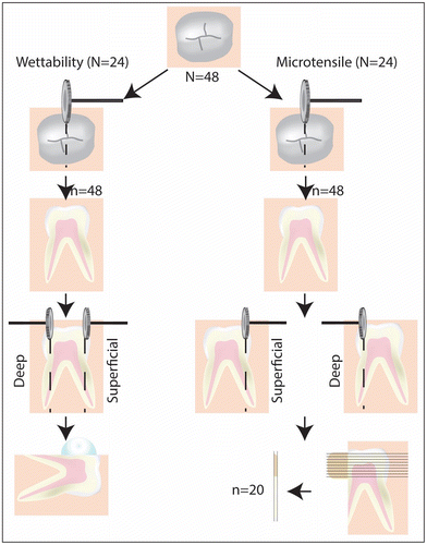
Twenty-four teeth were cut in half in the buccolingual direction using a diamond saw under running tap water and each half of the specimens was used separately (48 specimens) (Figure ). Each half of the specimens was further cut into half in a mesio-distal direction to obtain a quarter of a tooth (n = 96). To produce superficial dentin specimens (n = 48), buccal surfaces of the specimens were prepared with a standard-grit diamond rotary cutting instrument until the dentin surfaces were exposed. To produce deep dentin specimens (n = 48), lingual surfaces of the specimens were prepared up to 1 mm to the pulp. The thickness of each remaining dentin specimen was checked with a caliper. Dentin specimens were grounded wet with 600-grit silicon-carbide abrasive paper to obtain a standard smear layer.
Figure 1. Schematic representation of sequence of the experimental protocol.
Each group was further divided into 4 subgroups (n = 12) according to the following adhesive systems. Table shows the compositions, pH and application techniques of the adhesive systems.
| • | A two-step self-etch adhesive system (Clearfil SE Bond, Kuraray Medical Tokyo, Japan): CSE | ||||
| • | A two-step etch&rinse adhesive system (Single Bond2, 3 M ESPE, St. Paul, USA): SB | ||||
| • | A multi-mode adhesive system in self-etch mode (All Bond Universal (AU), Bisco, Schaumburg, IL, USA): SAU | ||||
| • | A multi-mode adhesive system in etch&rinse mode (All Bond Universal, Bisco, Schaumburg, IL, USA): EAU | ||||
Table 1. Materials used in the study.
For contact angle measurements, the Contact Angle System OCA (Dataphysics, Stuttgart, Germany) was employed. The technique enables measurement of the contact angle of sessile drops after reaching a flat profile (Figure ). Three consecutive contact angle measurements were obtained during the application of the adhesive systems at three stages. Prior to adhesive system application, a 3 μl drop of distilled water was placed on prepared dry dentin and the contact angle was measured (T0) (Figure ). The drop image was captured with a micro video system when the drop was in equilibrium. The contact angle values were calculated with the video signal transmitted to a computer. Prior to second measurements, specimens of SB and EAU groups were subjected to acid etching and specimens of the CSE group were subjected to primer application according to the respective manufacturers’ instructions. Then, a 3 μl drop of the adhesive system itself was placed on prepared dentin surfaces and the contact angle measurement was performed in the same way (T1) (Figure ). Finally, a 3 μl drop of distilled water was placed on the surface of the specimen after polymerization using a LED light curing unit at 1300 mW/cm2 power density (LEDMAX 550, Ankara, Turkey) of the adhesive system and contact angle measurement was done (T2) (Figure ). The contact angle measurements were performed under controlled temperature (22 °C) [Citation15].
Figure 2. The measurement of the contact angle of sessile drops after reaching a flat profile.
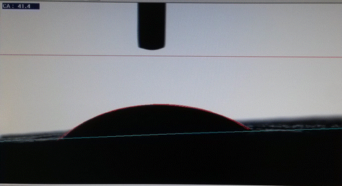
Figure 3. Replacement of 3 μl drop of distilled water on prepared dry dentin before adhesive system application.
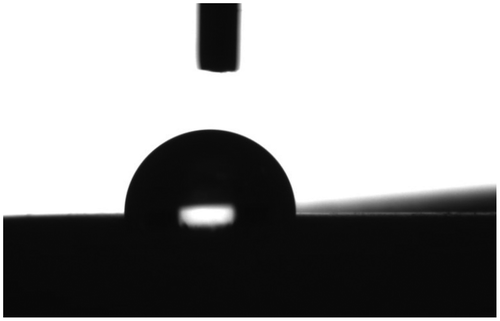
Figure 4. Replacement of 3 μl drop of the adhesive system on prepared dentin surface.
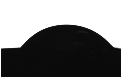
Figure 5. Replacement of 3 μl drop of distilled water on the adhesive system applied dentin surface.

Microtensile bond strength test
Twenty-four teeth were sectioned into two halves and both sides of the specimens used to obtain two groups: deep (n = 24) and superficial dentin (n = 24) (Figure ). They were further divided into 4 subgroups according to the adhesive systems utilized, as done in the contact angle measurement test (Group 1: CSE, Group 2: SB, Group 3: SAU and Group 4: EAU) (n = 6). The adhesives were applied on dentin surfaces according to manufacturers’ instructions. Resin composite measuring 4 mm wide (3 M™ ESPE™ Filtek™ Z250) was placed in two 2 mm layers. Each layer was polymerized for 20 s.
The resin–dentin blocks were sectioned perpendicular to the adhesive interface into approximately 1.0-mm thick slabs, using a water-cooled diamond disk in a sectioning machine (Metkon Micracut 201, Metkon Instruments Ltd., Turkey). A digital caliper was used to check the thickness of the disks and the measurements were recorded to calculate the bonded area. Each tooth yielded 3 or 4 beams for microtensile bond strength (μTBS) evaluation, for a total of 20 beams for each tooth structure/hybrid layer/resin composite (n = 20). The beams were placed on the jig of the microtensile testing machine (Micro Tensile Tester BİSCO Inc, Schaumburg, IL, USA) using cyanoacrylate glue (Pattex Transparent Contact Adhesive, Henkel KGaA, Düsseldorf). Each beam was subjected to μTBS test at a crosshead speed of 0.5 mm/min to create a fracture at the interface of composite and dentin. The values were recorded at the time of failure of the specimens. The bond strength for tensile forces was calculated by dividing the imposed force at the time of fracture by the bonded area. Values were reported in MPa.
Statistical analysis
Contact angle data from the groups were subjected to analysis of variance for repeated measures (p < 0.05) and multiple comparisons were done with Bonferroni test. The microtensile data were analyzed using analysis of variance for repeated measures.
Results
Contact angle measurements
The descriptive statistics and mean contact angle values obtained from all three measurements (T0, T1, T2) are given in Table . Superficial and deep dentin factors exerted no significant difference on dentin wettability for all measurements (T0, T1, T2) (p > 0.05). In T0, there was no significant difference among groups.
Table 2. The descriptive statistics and the mean°±SD° contact angle values.
In T1, all adhesive systems, except CSE, increased the wettability of the dentin surface (p < 0.05). CSE was significantly different from SAU (p = 0.001, p = 0.001) and EAU (p < 0.001, p = 0.001) in both dentin groups. The SAU and EAU groups increased the wettability of dentin for both superficial and deep dentin groups, respectively. There was no significant difference between the EAU and SAU groups (p = 1.000). However, there was a statistically significant difference between SB and EAU in superficial dentin only (p = 0.008). In superficial dentin, there was no significant difference between the SAU and SB groups (p = 0.182). However, there was a significant difference between the same groups in deep dentin (p = 0.049).
In T2, only CSE group was significantly different from other adhesive systems in both superficial and deep dentin groups (p < 0.05). Apart from CSE, all adhesive systems decreased the wettability of the surface (p < 0.05), reaching the first prepared state of the dentin in both superficial and deep dentin levels.
Only CSE showed significant difference between T0 and T2 in deep dentin (p = 0.008).
Microtensile bond strength measurements
The mean and standard deviation values of μTBS are shown in Table . Apart from SAU, the groups showed no significant difference at bond strength values for deep and superficial dentin (p > 0.05). In the SAU group, bond strength values for deep dentin were significantly higher than superficial dentin (p < 0.05). Additionally, apart from the CSE group, multiple comparison analysis of bond strength between groups with respect to dentin type revealed no significant difference between the tested adhesive systems.
Table 3. The descriptive statistics (mean values and the standard deviation) of microtensile bond strength measurements.
Discussion
The role of wettability in bonding stability between dentin and resin composite material is crucial. The quality and durability of the resin–dentin hybrid layer depends on dentin wettability and the depth of adhesive infiltration. Obtaining a successful adhesion is highly dependent on the adaptation and spreading of the adhesive system [Citation16]. Therefore, this study investigated the wettability of different multi-mode adhesive systems and the microtensile bond strength of a microhybrid composite resin. The tested adhesive systems vary with respect to their solvent type and pH: CSE contains water, SB contains ethanol and water, and AU contains ethanol as the solvent. (Table ) [Citation17Citation18].
Contact angle is generally affected by a solution’s viscosity, surface roughness, and heterogeneity [Citation5]. Ideal wettability may only be attained when the surface free energy of substrate is maximized, and the adhesive exhibits a lower contact angle [Citation19]. The substrates with high wettability have higher surface energy than the liquid’s surface tension [Citation5].
It is well established that the inorganic hydroxyapatite phase of dentin has high wettability, whereas the organic collagen phase of dentin has low wettability [Citation20Citation21]. As superficial and deep dentin have different characteristics, the effect of tubule density on roughness and wettability has been studied. Aguilera et al. found a significant increase in wettability on both superficial and deep dentins [Citation20]. Besides, on superficial dentin, mean observed contact angles were not statistically different [Citation22].
There are contradictory ideas about roughness and wettability interaction. Recent studies have suggested that lower dentin wettability is associated with higher surface roughness [Citation23Citation24]. These studies claimed that because of small air pockets inside them, open dentin tubules with larger diameters decrease dentin wettability. However, Liu et al. [Citation25] evaluated the correlation between roughness and wettability, and they concluded that the surface roughness reduced the contact angle and improved wettability.
According to a study by Pashley et al., the thickness of the smear layer has a greater effect on the wettability of the surface than surface roughness [Citation26]. The smear layer obstructs the dentinal tubules preventing the diffusion of oral fluids and restorative materials [Citation25]. Therefore, some researchers recommend acid etching of the dentin to eliminate the smear layer and develop perfect adhesion [Citation27]. Application of acid also removes or dissolves the superficial part of the dentin and increases the microporosity of the intertubular dentin. Therefore, both the morphology and wettability of the dentin surface are modified considerably [Citation28]. The smear layer is mainly hydrophobic. Functional monomers may dissolve the smear layer and modify the intact dentin surface underneath the smear layer [Citation29].
Acid etching not only modifies the adherent surface topology by increasing both the tubule diameters and the surface roughness, it also exposes collagen fibers and increases the organic/mineral ratio component resulting in a decreased surface energy. However, there is no consensus in literature on the effectiveness of wettability of dentin after acid etching. Some researchers found that dentin wettability increases after acid etching [Citation16Citation22Citation30Citation31], while others reported no significant alteration [Citation32] or decrease in dentin wettability [Citation23].
The pKa values (acid dissociation constant) of the acidic monomers of self-etching adhesives determine the etching potential. However, the application procedure, viscosity and solubility of the adhesives and monomer diffusion dynamics are other factors that may contribute to the aggressiveness of these adhesives. The pH of CSE, SB and AU is 2.1; 4.1 and 3.2, respectively [Citation33].
In the current study, the EAU and SB groups were etched with phosphoric acid prior to the measurement of the adhesive system’s contact angle. Acid etching removed the inorganic components and exposed the collagen network. However, in T1 and T2, no statistical difference was observed between the EAU and SAU groups. Recent studies have shown that the interaction energy between hydroxyapatite and water is greater than between collagen and water. Additionally, it has been shown that the interaction between hydroxyapatite and collagen occurs through water [Citation34]. CSE contained water as solvent and it is assumed that in the presence of water, the hydrated state of demineralized collagen can be preserved. In T2, CSE was significantly different from other adhesive systems in both dentin groups, and the contact angle values in the CSE group were lower than in other groups.
Hitmi et al. evaluated the effect of drying time and primer application on water spreading/infiltration on dentin and they concluded that air drying dentin after acid etching procedure significantly reduced the wettability of dentin [Citation35]. They also claimed that HEMA-based primers decreased the hydrophobicity of dentin, prevented collagen collapse and partially restored the water amount in air-dried dentin. These results are comparable with the results of our study in which a HEMA-based primer application increased both the wettability of dentin and the microtensile bond strength in superficial and deep dentin. However, Gregoire et al. found no correlation between water contact angles and the use of HEMA-based primers [Citation34].
Incorporating ethanol to temporarily alter the acid-etched dentin from a hydrophilic to a hydrophobic state was first proposed in 2007 [Citation36]. Recent in vitro studies have demonstrated reduced hydrophilicity of the hybrid layer and improved restoration interfaces when ethanol replaces water in the tissue [Citation37]. This technique is called “ethanol-wet bonding” and involves impregnation of hydrophobic monomers by acid-etched dentine to form a hybrid layer less susceptible to hydrolytic degradation over time [Citation36].
In this current study, one adhesive system contained only ethanol as the solvent (AU), and one adhesive system contained both ethanol and water as solvents (SB). Ethanol based adhesive systems showed increased wettability in T1. This may be due to better infiltration of resins into the interfibrillar spaces previously treated with ethanol [Citation16]. Li et al. evaluated the contact angles formed with the water and ethanol-wet bonding techniques and reported that ethanol saturated dentin provided lower contact angles compared with water saturated ones [Citation38]. However, the composition of the adhesive system may also have positive effects on ethanol-wet bonding, because the surface tension of the liquid plays a significant part in wettability [Citation38Citation39]. The solvents in SB; water and ethanol have a surface tension of 72.0 mN/m and 22.4 mN/m, respectively [Citation23]. Compared to purely ethanol-solvated adhesive systems, the higher surface tension of water limits penetration of the water-based adhesive systems into the substrate [Citation23]. Ideally, the solvent should be removed by air drying since residual solvent affects the mechanical characteristics of the adhesive system [Citation40]. Furthermore, acetone and ethanol based adhesive systems achieve greater solvent volatilization than water based adhesive systems, and consequently may increase the longevity of adhesive restorations [Citation26Citation41].
Of the three adhesive systems chosen for this investigation, all of them contained HEMA but CSE and AU also contained 10-methacryloyloxydecyl dihydrogen phosphate (10-MDP). 10-MDP containing adhesive agents bond chemically to dentin. Yoshida et al. compared adhesive-dentin interfaces chemically using different adhesive systems and showed that CSE extends into adhesive layer and that 10-MDP bonds ionically to hydroxyapatite [Citation42]. As a result, a strong adhesive interface can be formed. This layer might enhance the mechanical characteristics of adhesive layer. AU is a “ultra-mild” MDP-containing one-step self-etch adhesive (pH = 3.1) that is capable of chemically interacting with calcium in hydroxyapatite [Citation43]. However, AU does not have a polyalkenoic acid copolymer like Vitrebond copolymer. The significance of Vitrebond copolymer was evaluated by μTBS testing [Citation44]. The absence of the additional chemical bonding provided by Vitrebond copolymer may be responsible for the lower μTBS values obtained with AU compared to CSE.
Regarding the composition of AU and CSE, it was expected that the AU group and the CSE group would be comparable. However, this study did not show that. In terms of μTBS, there was a significant difference between the CSE and AU groups. This is probably because AU is a single-bottle adhesive and its 10-MDP concentration might be lower than the concentration in CSE. Muñoz et al. investigated μTBS for both etch-and-rinse and self-etch modes of universal adhesives [Citation45]. The results are comparable to this study and they claim that universal adhesive system used in both modes showed lower μTBS values compared to self-etch and etch-and-rinse adhesive systems.
In previous research studies, AU resulted in higher bond strength in ER mode than in SE mode [Citation17Citation46]. Toledano et al. reported that when the bond strengths of superficial and deep dentin were compared, 10-MDP containing, water-based, self-etch adhesives exhibited superior bond performance [Citation47]. Inoue et al. confirmed that the acetone solvent performed better when applied on deep dentin [Citation48]. Tagami et al. revealed controversial results about the adhesive’s performance and the remaining dentin thickness [Citation49]. All-in-one adhesive systems performed better. Generally, adhesive properties were better with superficial dentin than with deep dentin [Citation13Citation50].
Conclusion
Given the results of this study, the choice of adhesive system and application methods are not correlated with the depth of the cavity. Furthermore, self-etch adhesive systems might be a better choice for successful restorations. Clinicians should remember that the success of the restorations is dependent on the choice of adhesive system, as these systems affect the wettability and the microtensile bond strength of the dentin substrates.
Disclosure statement
No potential conflict of interest was reported by the authors.
Acknowledgement
The manuscript’s wettability parameters’ results were presented in Meeting Presentation: ConsEuro 2015 London, England and the abstract were printed in Clin Oral Invest 2015;19:1701–1754
References
- Neppelenbroek KH. The cilinical challenge of achieving marginal adaptation in direct and indirect restorations. J. Appl. Oral Sci. 2015;23:448–449.10.1590/1678-77572015ed005
- Shokati B, Tam LE, Santerre JP, et al. Effect of salivary esterase on the integrity and fracture toughness of the dentin-resin interface. J Biomed Mater Res B Appl Biomater. 2010;94:230–237.
- Sabatini C, Ortiz PA, Pashley DH. Preservation of resin-dentin interfaces treated with benzalkonium chloride adhesive blends. Eur J Oral Sci. 2015;123:108–115.10.1111/eos.2015.123.issue-2
- Leme A, Vidal CMP, Hassan LS, et al. Potential role of surface wettability on the long-term stability of dentin bonds after surface biomodification. J Biomech. 2015;48:2067–2071.10.1016/j.jbiomech.2015.03.016
- Marshall SJ, Bayne SC, Baier R, et al. A review of adhesion science. Dent Mater. 2010;26:e11–e16.10.1016/j.dental.2009.11.157
- Peumans M, Kanumilli P, Demunck J, et al. Clinical effectiveness of contemporary adhesives: A systematic review of current clinical trials. Dent Mater. 2005;21:864–881.10.1016/j.dental.2005.02.003
- Erickson RL, Barkmeier WW, Kimmes NS. Bond strength of self-etch adhesives to pre-etched enamel. Dent Mater. 2009;25(10):1187–1194.10.1016/j.dental.2009.04.004
- Rosa WL, Piva E, Silva AF. Bond strength of universal adhesives: A systematic review and meta-analysis. J Dent. 2015;43:765–776.10.1016/j.jdent.2015.04.003
- Perdigão J, Kose C, Mena-Serrano AP, et al. A new universal simplified adhesive: 18-month clinical evaluation. Oper Dent. 2014;39:113–127.10.2341/13-045-C
- Loguercio AD, de Paula EA, Hass V, et al. A new universal simplified adhesive: 36-Month randomized double-blind clinical trial. J Dent. 2015;43:1083–1092.10.1016/j.jdent.2015.07.005
- Lopes LS, Calazans FS, Hidalgo R, et al. Six-month follow-up of cervical composite restorations placed with a new universal adhesive system: a randomized clinical trial. Oper Dent. 2016;41:465–480.10.2341/15-309-C
- Fawzy AS. Variations in collagen fibrils network structure and surface dehydration of acid demineralized intertubular dentin: effect of dentin depth and air-exposure time. Dent Mater. 2010;26:35–43.10.1016/j.dental.2009.08.009
- Pegado REF, do Amaral FLB, Flório FM, et al. Effect of different bonding strategies on adhesion to deep and superficial permanent dentin. Eur J Dent. 2010;4:110–117.
- Ting S, Chowdhury AA, Pan F, et al. Effect of remaining dentin thickness on microtensile bond strength of current adhesive systems. Dent Mater J. 2015;34:181–188.10.4012/dmj.2014-130
- Halacoglu DM, Tuncer D, Salman ZC, The Arhun N. The wettability of different adhesive systems in superficial and deep dentine. Conseuro conference 2015, London, May 14–16. Clin Oral Investig. 2015;2015(19):1701–1754.
- Ricci HA, Scheffel DLS, de Souza Costa CA, et al. Wettability of chlorhexidine treated non-carious and caries-affected dentine. Aust Dent J. 2014;59:37–42.10.1111/adj.12150
- Luque-Martinez IV, Perdigão J, Muñoz M, et al. Effects of solvent evaporation time on immediate adhesive properties of universal adhesives to dentin. Dent Mater. 2014;30:1126–1135.10.1016/j.dental.2014.07.002
- Vogler EA. Structure and reactivity of water at biomaterial surfaces. Adv Colloid Interface Sci. 1998;74:69–117.10.1016/S0001-8686(97)00040-7
- Tsujimoto A, Iwasa M, Shimamura Y, et al. Enamel bonding of single-step self-etch adhesives: Influence of surface energy characteristics. J Dent. 2010;38:123–130.10.1016/j.jdent.2009.09.011
- Aguilera FS, Osorio R, Osorio E, et al. Wetting ability of an acetone/based etch rinse adhesive after NaOCl-treatment. Med Oral Patol Oral Cir Bucal. 2012;17:644–648.10.4317/medoral.17654
- Yassen GH, Sabrah AHA, Eckert GJ, et al. Effect of different endodontic regeneration protocols on wettability, roughness, and chemical composition of surface dentin. J Endod. 2015;41:956–960.10.1016/j.joen.2015.02.023
- Toledano M, Osorio R, Perdigao J, et al. Effect of acid etching and collagen removal on dentin wettability and roughness. J Biomed Mater Res. 1999;47:198–203.10.1002/(ISSN)1097-4636
- Farge P, Alderete L, Ramos SMM. Dentin wetting by three adhesive systems: Influence of etching time, temperature and relative humidity. J Dent. 2010;38:698–706.10.1016/j.jdent.2010.03.013
- Ramos SMM, Alderete L, Farge P. Dentinal tubules driven wetting of dentin: Cassie-Baxter modelling. Eur Phys J. 2009;30:187–195.
- Liu J, Lü P, Sun Y, et al. Wettability of dentin after Yb:KYW thin-disk femtosecond ablation. Lasers Med Sci. 2015;30:1689–1693.10.1007/s10103-014-1655-8
- Pashley EL, Agee KA, Pashley DH, et al. Effects of one versus two applications of an unfilled, all-in-one adhesive on dentine bonding. J Dent. 2002;30:83–90.10.1016/S0300-5712(02)00002-7
- Wagner A, Wendler M, Petschelt A, et al. Bonding performance of universal adhesives in different etching modes. J Dent. 2014;42:800–807.10.1016/j.jdent.2014.04.012
- Eldarrat AH, High AS, Kale GM. In vitro analysis of “smear layer” on human dentine using ac-impedance spectroscopy. J Dent. 2004;32:547–554.10.1016/j.jdent.2004.05.003
- Yoshida Y, Van Meerbeek B, Nakayama Y, et al. Evidence of chemical bonding at biomaterial-hard tissue interfaces. J Dent Res. 2000;79:709–714.10.1177/00220345000790020301
- Aguilar-Mendoza JA, Rosales-Leal JI, Rodríguez-Valverde MA, et al. Effect of acid etching on dentin wettability and roughness: self-etching primers versus phosphoric acid. J Biomed Mater Res B Appl Biomater. 2008;84:277–285.10.1002/(ISSN)1552-4981
- Rosales-Leal JI, Osorio R, Holgado-Terriza JA, et al. Dentin wetting by four adhesive systems. Dent Mater. 2001;17:526–532.10.1016/S0109-5641(01)00014-8
- Attal JP, Asmussen E, Degrange M. Effects of surface treatment on the free surface energy of dentin. Dent Mater. 1994;10:259–264.10.1016/0109-5641(94)90071-X
- Pucci CR, de Oliveira RS, Caneppele TMF, et al. Effects of surface treatment, hydration and application method on the bond strength of a silorane adhesive and resin system to dentine. J Dent. 2013;41:278–286.10.1016/j.jdent.2012.11.016
- Grégoire G, Dabsie F, Dieng-Sarr F, et al. Solvent composition of one-step self-etch adhesives and dentine wettability. J Dent. 2011;39:30–39.10.1016/j.jdent.2010.09.008
- Hitmi L, Bouter D, Degrange M. Influence of drying and HEMA treatment on dentin wettability. Dent Mater. 2002;18:503–511.10.1016/S0109-5641(01)00075-6
- Tay FR, Pashley DH, Kapur RR, et al. Bonding BisGMA to dentin – a proof of concept for hydrophobic dentin bonding. J Dent Res. 2007;86:1034–1039.10.1177/154405910708601103
- Pashley DH, Tay FR, Carvalho RM, et al. From dry bonding to water-wet bonding to ethanol-wet bonding. A review of the interactions between dentin matrix and solvated resins using a macromodel of the hybrid layer. Am J Dent. 2007;20:7–20.
- Li F, Liu XY, Zhang L, Kang J-J, et al. Ethanol-wet bonding technique may enhance the bonding performance of contemporary etch-and-rinse dental adhesives. J Adhes Dent. 2012;14:113–120.
- Hosaka K, Nishitani Y, Tagami J, et al. Durability of resin-dentin bonds to water- vs. ethanol-saturated dentin. J Dent Res. 2009;88:146–151.10.1177/0022034508328910
- Garcia FCP, Almeida JCF, Osorio R, et al. Influence of drying time and temperature on bond strength of contemporary adhesives to dentine. J Dent. 2009;37:315–320.10.1016/j.jdent.2008.12.007
- Thitthaweerat S, Nakajima M, Foxton RM, et al. Effect of solvent evaporation strategies on regional bond strength of one-step self-etch adhesives to root canal dentine. Int Endod J. 2013;46:1023–1031.
- Yoshida Y, Yoshihara K, Nagaoka N, et al. Self-assembled nano-layering at the adhesive interface. J Dent Res. 2012;91:376–381.10.1177/0022034512437375
- Yoshihara K, Yoshida Y, Hayakawa S, et al. Self-etch monomer-calcium salt deposition on dentin. J Dent Res. 2011;90:602–606.10.1177/0022034510397197
- Perdigão J, Sezinando A, Monteiro PC. Effect of substrate age and adhesive composition on dentin bonding. Oper Dent. 2013;38:267–274.10.2341/12-307-L
- Muñoz MA, Luque I, Hass V, et al. Immediate bonding properties of universal adhesives to dentine. J Dent. 2013;41:404–411.
- Sezinando A, Luque-Martinez I, Muñoz MA, et al. Influence of a hydrophobic resin coating on the immediate and 6-month dentin bonding of three universal adhesives. Dent Mater. 2015;31:e236–e246.10.1016/j.dental.2015.07.002
- Toledano M, Osorio R, Ceballos L, et al. Microtensile bond strength of several adhesive systems to different dentin depths. Am J Dent. 2003;16:292–298.
- Inoue S, Vargas MA, Abe Y, et al. Microtensile bond strength of eleven contemporary adhesives to dentin. J Adhes Dent. 2001;3:237–245.
- Tagami J, Tao L, Pashley DH. Correlation among dentin depth, permeability, and bond strength of adhesive resins. Dent Mater. 1990;6:45–50.10.1016/0109-5641(90)90044-F
- Yoshikawa T, Wattanawongpitak N, Cho E, et al. Effect of remaining dentin thickness on bond strength of various adhesive systems to dentin. Dent Mater J. 2012;31:1033–1038.10.4012/dmj.2012-143