Abstract
A variety of neoplasms with rhabdoid differentiation have been reported in many sites. The authors describe a case of gastrointestinal stromal tumor (GIST) of the stomach that exhibited prominent rhabdoid features. Immunohistochemically, the tumor cells displayed positive staining for vimentin, c-kit, CD34, and alpha smooth muscle actin. Ultrastructural examination of the rhabdoid tumor cells revealed paranuclear whorls of intermediate filaments, which were immunoreactive for vimentin by both light microscopic immunohistochemical and protein A gold immunoelectron microscopic techniques. On H&E light microscopic examination alone, such a tumor could be mistaken for a variety of epithelial, mesenchymal, or other neoplasms that may show rhabdoid features. One report of GIST with a rhabdoid histologic phenotype has been described. This is the second known report of such a case with immunophenotypic and ultrastructural evaluation, and the first case with immunoelectron microscopic examination.
Malignant rhabdoid tumors have been reported arising in the kidney as well as in extrarenal locations [Citation[1–3]]. Rhabdoid morphologic features have also been described as a component in multiple tumors, including epithelial neoplasma such as renal cell carcinoma, transitional cell carcinoma, adenocarcinoma of the small intestine, adenocarcinoma of the pancreas, colorectal adenocarcinoma, Merkel cell carcinoma, vulvar carcinoma, and malignant mixed mullerian tumor [Citation[4–7]]. Mesenchymal tumors that have been shown to display rhabdoid differentiation include leiomysarcoma, endometrial stromal sarcoma, pleomaorphic fibrosarcoma with rhabdoid cells, myxoid chondrosarcoma, cutaneous epithelial malignant nerve sheath tumor, and synovial sarcoma [Citation[7–10]]. In addition, rhabdoid phenotype has also been described in melanoma, mesothelioma, meningioma, lymphoma, and desmoplastic small round blue cell tumor [Citation[7]]. This is the second report of gastrointestinal stromal tumor (GIST) with a rhabdoid histologic phenotype, and the first to be examined by immunoelectron microscopy [Citation[11]].
CASE HISTORY
A 68-year-old male presented with nausea and mid-epigastric discomfort. Upper endoscopy studies revealed a submucosal mass in the greater curvature of the stomach. The patient underwent partial gastrectomy. The postoperative course was complicated by pneumonia and respiratory failure, and the patient died 3 weeks following surgery. Postmortem examination revealed no evidence of residual or metastatic GIST.
MATERIALS AND METHODS
Multiple sections from the tumor were fixed in 10% neutral buffered formalin and processed for routine light microscopy and avidin–biotin-based immunohistochemistry. Antibodies directed against vimentin (V9, Dako, Carpinteria, CA, USA), c-kit (polyclonal CD117, Dako), CD34 (My10, Becton Dickinson Immunocyutometry Systems, San Jose, CA, USA), alpha smooth muscle actin (SMA) (1A4, Sigma Chemical, St. Louis, MO, USA), CD31 (JC/70A, Dako), CD99 (HBA71, 013, Signet Laboratories, Dedham, MA, USA), desmin (33, Biogenex Laboratories, San Ramon, CA, USA), S-100 (polyclonal, Dako), keratin (AE1/AE3, Serologicals, Norcross, GA, USA), GFAP (polyclonal, Dako), and neurofilament (2F11, Dako) were applied to sections of the tumor using a polymer-based Envision + detection system (Dako). Appropriate positive and negative isotype matched IgG1 tissue controls were used.
Formalin-fixed tissue for conventional electron microscopy was retrieved from a paraffin-embedded block, deparaffinized, rehydrated, and fixed in Karnovsky's fixative (2.5% glutaraldehyde, 1% paraformaldehyde). Following rines with Millonig's buffer, the specimen was postfixed with 1% OsO4, and again rinsed with buffer. After being osmicated, the tissue was dehydrated through a graduated series of ethanols, and embedded in Spurr's resin in the conventional manner. Then 100-nm-thick sections were cut to include rhabdoid tumor cells, contrasted with uranyl acetate and lead citrate, and viewed with a JEOL 1210 electron microscope.
Formalin-fixed tumor tissue for immuno-electron microscopic study was retrieved from a paraffin block and embedded in Lowicryl K4M as previously described [Citation[12]]. Polymerization was achieved by exposing the blocks to UV light for 24 h at − 35°C, followed by UV light for 3 days at room temperature. Then 100-nm sections were cut and placed on parlodion-coated, carbon nickel grids. Immunoelectron microscopic localization of vimentin was performed with the postembedding protein A-gold technique as described by Taatjes et al. using monoclonal anti-vimentin at dilutions of 1:2, 1:5, and 1:10 [Citation[13]]. Immunoelectron microscopic examination for SMA and c-kit was performed using a technique similar to that described above.
RESULTS
Gross Description
The partial gastrectomy specimen measured 8.0 × 5.5 × 4.5 cm. Beneath a raised, ulcerated area of mucosa was a well-circumscribed submucosal mass that measured 5.0 × 4.5 × 4.0 cm. On the cut surface the mass was soft, tan-yellow to tan-white, focally hemorrhagic, and focally lobulated. The mass abutted but did not penetrate the underlying serosa, and did not invade the overlying mucosa.
Light Microscopy
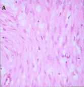
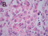
H&E sections showed a tumor composed of mixed patterns, the most common being atypical spindle cells arranged in intersecting fascicles. These spindled cells had round to ovoid nuclei, prominent nucleoli, abundant eosinophilic cytoplasm, and poorly defined cell borders (. Cytoplasmic vacuoles and nuclear palisading were not observed. In addition, areas showing epithelioid morphology, including round to polygonal cells with large eccentric nuclei and prominent nucleoli, were present. Many of the cells in these foci demonstrated rhabdoid morphologic features exemplified by a moderate to large amount of densely eosinophilic cytoplasm with eccentric nuclei (. Mitotic rate was 2 per 10 high-power fields. Necrosis was absent.
Immunohistochemistry
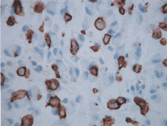
Both spindled and rhabdoid tumor cells showed strong positive cytoplasmic staining for vimentin, c-kit (), CD34, SMA, and CD99. The rhabdoid cells demonstrated strong paranuclear cytoplasmic staining with vimentin consistent with the cytoplasmic paranuclear location of the intermediate filaments identified ultrastructurally. Tumor cells were negative for antibodies directed against desmin, keratin AE1/AE3, S-100, CD31, GFAP, and neurofilament.
Electron Microscopy
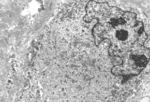
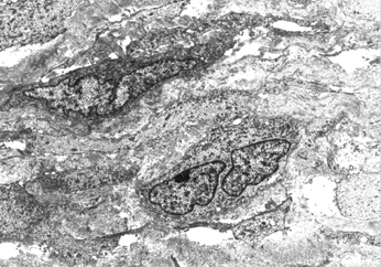
The tumor cells with rhabdoid histologic phenotype contained abundant cytoplasm and irregularly shaped nuclei, many with marginated chromatin. Nucleoli, when present, were eccentrically placed. Intercellular junctions were not identified. The most distinctive ultrastructural feature in the rhabdoid cells was the presence of whorled arrays of intermediate filaments that occupied a paranuclear position (). Other cytoplasmic organelles included mitochondria and rough endoplasmic reticulum and in some cells these structures were embedded into the filament whorls. The nonrhabdoid spindle tumor cells were composed of cells with oval elongated nuclei and long cytoplasmic processes (). No diagnostic structures were identified, although preservation was suboptimal due to processing to tissue retrieved from formalin-fixed, paraffin-embedded specimen.
Immunoelectron Microscopy
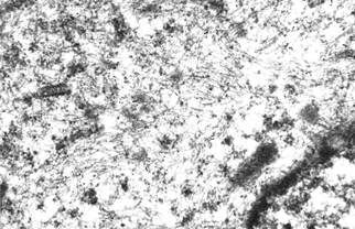
Collodial gold particles indicative of vimentin antigenicity were concentrated over whorls of paranuclear intermediate filaments (). Immunoelectron microscopy study was attempted using antibodies directed against c-kit and SMA, but no specific staining was achieved.
DISCUSSION
GISTs consists of a heterogeneous class of stromal neoplasms that occur throughout the gastrointestinal tract, most commonly arising in the stomach [Citation[14], Citation[15]]. The cell of origin resembles the interstitial cells of Cajal, which are normally located within the myenteric plexus, and are presumably involved in peristaltic pacemaker activity, although the actual role of the interstial cells of Cajal remains contraversial [Citation[16]]. The mixed immunogenicity of GIST tumors, along with some cases occurring outside the GI tract, suggests an origin of multipotential cells that can differentiate into Cajal and smooth muscle cells [Citation[14]]. Ultrastructurally, the tumor cells often show neural, neuronal, and indistinct myogenic features, correlating with the variable immunophenotype of these tumors. Although the gastrointestinal autonomic nerve tumor (GANT) can be separated from the GIST by differing ultrastructural features [Citation[17]], because of their similar immunohistochemical staining profile, GANTs are included in the GIST category.
A recent study performed immunohistochemical staining on 109 GISTs in the duodenum. In this series, 100% of cases were c-kit positive, 54% were CD34 positive, 39% of cases were SMA positive, and 20% of cases were S-100 positive [Citation[18]]. It is well documented that GISTs typically stain positively for vimentin and negatively for desmin and keratin AE1/AE3 [Citation[19–21]]. Additionally, a smaller series has shown CD99 to be positive in a majority of GISTs (89%) [Citation[19]]. Our case displayed an immunoreactive pattern that was similar to the immunohistochemical profile reported in the literature, showing positive staining for vimentin, c-kit, CD34, SMA, and CD99, and negative staining for keratin AE1-AE3, CD31, and desmin.
There is a wide differential to consider when faced with a tumor showing a rhabdoid histologic phenotype, including a variety of epithelial and mesenchymal tumors that could present as primary or metastatic tumors. The epithelial neoplasms to consider include renal cell carcinoma, transitional cell carcinoma, adenocarcinoma of the gastrointestinal tract, Merkel cell carcinoma, malignant mixed mullerian tumor, and vulvar carcinoma [Citation[4–7]]. Perhaps even more relevant in this case, given the spindle cell component, are the mesenchymal tumors that can show a rhabdoid histologic phenotype, including rhabdomyosarcoma, leiomyosarcoma, endometrial stromal sarcoma, pleomorphic fibrosarcoma with rhabdoid cells, myxoid chondrosarcoma, and synovial sarcoma [Citation[7–9]]. In addition, melanoma, mesothelioma, lymphoma, and desmoplastic small round blue cell tumor may also show a rhabdoid histologic phenotype. In children, one should also consider the true rhabdoid tumors, both renal and extrarenal [Citation[7]]. Histologic distinction between such tumors on H&E alone may be difficult. However, if one employs immunohistochemistry, the true rhabdoid tumors are typically positive for cytokeratins, which are negative in GIST. Another entity that we considered in this case was signet ring cell carcinoma, which was easily excluded by keratin negativity.
GISTs classically display three histologic patterns, namely spindle cell, epithelial cell, or mixed type. In addition, GIST with prominent signet-ring cell features has been described [Citation[22], Citation[23]] as well as a case of signet ring epithelioid stromal tumor of the small intestine in which the signet ring morphology was found by ultrastructural examination to reflect retraction artifact [Citation[24]]. One case of GANT, a variant of GIST, has been reported to show a rhabdoid histologic phenotype [Citation[11]]. In addition to prominent cytoplasmic inclusions on H&E, this case report included electron microscopic and immunohistochemical features consistent with GANT. Other authors have shown that electron microscopy can be helpful in the diagnostic evaluation and subclassification of GIST [Citation[25], Citation[26]].
In our case, foci of rhabdoid cells, in addition to spindled and epithelioid cells, were identified by light microscopy. When studied ultrastructurally, the rhabdoid cells contained abundant whorls of paranuclear intermediate filaments that were vimentin positive by immunoelectron microscopy. The lack of specific immunoelectron microscopic staining for c-kit and SMA may reflect susceptibility of these antigens to degeneration during the processing required for protein A gold immunoelectron microscopy. Immunoreactivity was achieved for both antigents by light microscopy.
Similar electron and immunoelectron microscopic features have been used to confirm rhabdoid differentiation in other tumors, such as malignant mixed mullerian tumor and malignant rhabdoid tumor of the kidney [Citation[3], Citation[27]]. However, the marked accumulation of paranuclear intermediate filaments, characteristic of rhabdoid differentiation, may reflect a nonspecific degenerative change. Paranuclear filamentous inclusions have been identified in colchicine-treated cells, in which depolymerization of microtubules may result in a general collapse of cytoskeletal elements into a paranuclear cytoplasmic location [Citation[28]]. Furthermore, Ghadially has speculated that the formation of globular filamentous bodies may be a result of cytoskeletal derangement related to aging or injury [Citation[29]].
The rhabdoid histologic morphology seen in our case, together with the immunohistochemical and electron microscopic findings, supports that diagnosis of GIST with a rhabdoid histologic phenotype. Recognizing that GISTs may demonstrate a rhabdoid appearance may be useful when faced with a tumor in the gastrointestinal tract displaying prominent rhabdoid features.
REFERENCES
- Blatt J, Russo P, Taylor S.. Extrarenal rhabdoid sarcoma. Med Pediatr Oncol. 1986; 14: 221–226. [PUBMED], [INFOTRIEVE]
- Cho K, Rosenshein NB, Epstein JI.. Malignant rhabdoid tumor of the uterus. Int J Gynecol Pathol. 1989; 8: 381–387. [PUBMED], [INFOTRIEVE]
- Haas JE, Palmer NF, Weinberg AG, Beckwith JB.. Ultrastructure of malignant rhabdoid tumor of the kidney: a distinctive renal tumor of children. Hum Pathol. 1981; 12: 646–657. [PUBMED], [INFOTRIEVE]
- Al-Nafussi A, O'Donnell M.. Poorly differentiated adenocarcinoma with extensive rhabdoid differentiation: clinicopathologic features of two cases arising in the gastrointestinal tract. Pathol Int. 1999; 49: 160–163. [PUBMED], [INFOTRIEVE], [CSA], [CROSSREF]
- Baschinsky DY, Niemann TH, Eaton LA, Frankel WL. Malignant mixed Mullerian tumor with rhabdoid features: a report of two cases and a review of the literature. Gynecol Oncol. 1999; 73: 145–150. [PUBMED], [INFOTRIEVE], [CSA], [CROSSREF]
- Gokden N, Nappi O, Swanson PE. Renal cell carcinoma with rhabdoid features.. Am J Surg Pathol. 2000; 24: 1329–1338. [PUBMED], [INFOTRIEVE], [CSA]
- Wen P, Prasad ML. Synovial sarcoma with rhabdoid features. Arch Pathol Lab Med. 2003; 127: 1391–1392. [PUBMED], [INFOTRIEVE]
- Kim YH, Cho H, Kyeom-Kim H, Kim I. Uterine endometrial stromal sarcoma with rhabdoid and smooth muscle differentiation. J Korean Med Sci. 1996; 11: 88–93. [PUBMED], [INFOTRIEVE], [CSA]
- McCluggage WG, Date A, Bharucha H, Toner PG. Endometrial stromal sarcoma with sex cord-like areas and focal rhabdoid differentiation. Histopathology. 1996; 29: 369–374. [PUBMED], [INFOTRIEVE], [CSA]
- Morgan MB, Stevens L, Patterson J, Tannenbaum M. Cutaneous epithelioid malignant nerve sheath tumor with rhabdoid features: a histologic, immunohistochemical, and ultrastructural study of three cases. J Cutan Pathol. 2000; 27: 529–534. [PUBMED], [INFOTRIEVE], [CSA], [CROSSREF]
- Lee JR, Joshi V, Griffin JW, Lasota J, Miettinen M.. Gastrointestinal autonomic nerve tumor: immunohistochemical and molecular identity with gastrointestinal stromal tumor. Am J Surg Pathol. 2001; 25: 979–987. [PUBMED], [INFOTRIEVE], [CSA], [CROSSREF]
- Mount SL, Taatjes DJ, von Turkovich M, Tindle BH, Trainer TD. Diagnostic immunoelectron microscopy in surgical pathology: assessment of various tissue fixation and processing protocols. Ultrastruct Pathol. 1993; 17: 547–556. [PUBMED], [INFOTRIEVE]
- Taatjes DJ, Arendash-Durand B, von Turkovich M, Trainer TD. HMB-45 antibody demonstrates melanosome specificity by immunoelectron microscopy. Arch Pathol Lab Med. 1993; 117: 264–68. [PUBMED], [INFOTRIEVE]
- Miettinen M, Sarlomo-Rikala M, LaSota J.. Gastrointestinal stromal tumors: recent advances in understanding of their biology. Hum Pathol. 1999; 30: 1213–1220. [PUBMED], [INFOTRIEVE], [CSA], [CROSSREF]
- Trupiano JK, Stewart RE, Misick C, Appelman HD, Goldblum JR.. Gastric stromal tumors: a clinicopathologic study of 77 cases with correlation of features with nonaggressive and aggressive clinical behaviors. Am J Surg Pathol. 2002; 26: 705–714. [PUBMED], [INFOTRIEVE], [CSA], [CROSSREF]
- Min KW, Seo IS.. Intestitial cells of Cajal in the human small intestine: immunochemical and ultrastructural study. Ultrastruct Pathol. 2003; 27: 67–78. [PUBMED], [INFOTRIEVE], [CSA], [CROSSREF]
- Eyden B, Chorneyko KA, Shanks JH, Menasce LP, Banerjee SS.. Contribution of electron microscopy to understanding cellular differentiation in mesenchymal tumors of the gastrointestinal tract: a study of 82 tumors. Ultrastruct Pathol. 2002; 26: 269–285. [PUBMED], [INFOTRIEVE], [CSA], [CROSSREF]
- Miettinen M, Kopczynski J, Makhlouf HR. et al.. Gastrointestinal stromal tumors, intramural leiomyomas, and leiomyosarcomas in the duodenum: a clinicopathologic, immunohistochemical, and molecular genetic study of 167 cases. Am J Surg Pathol. 2003; 27: 625–641. [PUBMED], [INFOTRIEVE], [CSA], [CROSSREF]
- Shidham VB, Chivukula M, Gupta D, Rao RN, Komorowski R.. Immunohistochemical comparison of gastrointestinal stromal tumor and solitary fibrous tumor. Arch Pathol Lab Med. 2002; 126: 1189–1192. [PUBMED], [INFOTRIEVE], [CSA]
- Tworek JA, Appelman HD, Singleton TP, Greenson JK.. Stromal tumors of the jejunum and ileum. Mod Pathol. 1997; 10: 200–209. [PUBMED], [CSA]
- Tworek JA, Goldblum JR, Weiss SW, Greenson JK, Appelman HD.. Stromal tumors of the abdominal colon: a clinicopathologic study of 20 cases. Am J Surg Pathol. 1999; 23: 937–945. [PUBMED], [INFOTRIEVE], [CSA], [CROSSREF]
- Schned AR.. Correspondence Re: Suster S. Fletcher DM. Gastrointestinal stromal tumors with prominent signet-ring cell features. Mod Pathol. 1996; 9: 1089, [PUBMED], [INFOTRIEVE], [CSA]
- Suster S, Fletcher CD.. Gastrointestinal stromal tumors with prominent signet-ring cell features. Mod Pathol. 1996; 9: 609–613. [PUBMED], [INFOTRIEVE], [CSA]
- Ferrer MD, Corominas JM, Ribalta T, Iglesias M, Serrano S.. Signet ring epithelioid stromal tumor of the small intestine. Ultrastruct Pathol. 1999; 23: 45–50. [PUBMED], [INFOTRIEVE], [CSA], [CROSSREF]
- Erlandson RA, Woodruff JM.. Role of electron microscopy in the evaluation of soft tissue neoplasms, with emphasis on spindle cell and pleomorphic tumors. Hum Pathol. 1998; 29: 1372–1381. [PUBMED], [INFOTRIEVE], [CSA], [CROSSREF]
- Erlandson RA, klimstra DS, Woodruff JM. Subclassification of gastrointestinal stromal tumors based on evaluation by electron microscopy and immunohistochemistry. Ultrastruct Pathol. 1997; 21: 311–313. [CSA]
- Mount SL, Lee KR, Taatjes DJ.. Carcinosarcoma (malignant mixed mullerian tumor) of the uterus with a rhabdoid tumor component: an immunohistochemical, ultrastructural, and immunoelectron microscopic case study. Am J Clin Pathol. 1995; 103: 235–239. [PUBMED], [INFOTRIEVE], [CSA]
- Lazarides E.. Intermediate filaments as mechanical integrators of cellular space. Nature. 1980; 283: 249–256. [PUBMED], [INFOTRIEVE], [CROSSREF]
- Ghadially FN.. Ultrastructural Pathology of the Cell and Matrix. 2, , London, Butterworths. 1988
