ABSTRACT
The nanopathological diagnostics (ND) is an ultra-specialized branch of pathological anatomy aimed to identify the nanoparticles of metallic, semimetallic, or nonmetallic elements in the inorganic particulate matter present inside pathological tissues, even on the nanometer scale. ND exploits an environmental scanning electron microscope, connected to an X-ray microprobe mounted on an energy-dispersive spectrometer. The searching of nanoparticles can be performed on paraffin-embedded material, omitting emissions of black overlay and plating procedures. The technique is highly sensitive and specific, reproducible and rapid, covering an entire operating cycle in few hours. Nowadays, ND finds many applications: (I) intratumor detection of heavy metals and endocrine metal disruptors; (II) identification of pathogenic nanoparticles in medical or veterinary drugs and devices, cosmetics, household products, and foodstuffs; (III) differential diagnosis of sarcoid-type granulomas (berylliosis, baritosis) and foreign body granulomas (prosthetic, iatrogenic); (IV) attestation of occupational disease correlating the datum with the occupational risk (anthracosis, asbestosis, bauxite fibrosis, byssinosis, chalicosis, siderosis, silicosis, stannosis, talcosis); and (V) forensic investigations to ascertain a causal link between disease and environmental, military, or work exposure. In addition to filling a knowledge gap, ND offers to the pathologist a current research field, with particular reference to the impact of occupational and environmental pollution on the human health and cancer.
Declaration of interest
The authors report no conflicts of interest. The authors alone are responsible for the content and writing of this article.
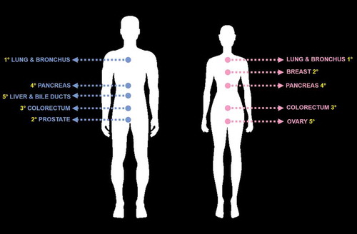
Figure 1. Today, the top five leading sites of cancer death in American men (left picture) are lung and bronchus (1°), prostate (2°), colorectum (3°), pancreas (4°), liver and intrahepatic bile ducts (5°). For American women (right picture), they are in descending order: lung and bronchus (1°), breast (2°), colorectum (3°), pancreas (4°), and ovary (5°).