Abstract
Nanotechnology is developing rapidly and, in the future, it is expected that increasingly more products will contain some sort of nanomaterial. However, to date, little is known about the occurrence, fate and toxicity of nanoparticles. The limitations in our knowledge are partly due to the lack of methodology for the detection and characterisation of engineered nanoparticles in complex matrices, i.e. water, soil or food. This review provides an overview of the characteristics of nanoparticles that could affect their behaviour and toxicity, as well as techniques available for their determination. Important properties include size, shape, surface properties, aggregation state, solubility, structure and chemical composition. Methods have been developed for natural or engineered nanomaterials in simple matrices, which could be optimized to provide the necessary information, including microscopy, chromatography, spectroscopy, centrifugation, as well as filtration and related techniques. A combination of these is often required. A number of challenges will arise when analysing environmental and food materials, including extraction challenges, the presence of analytical artifacts caused by sample preparation, problems of distinction between natural and engineered nanoparticles and lack of reference materials. Future work should focus on addressing these challenges.
Introduction and background
Nanomaterials are commonly regarded as materials with at least one dimension below 100 nm (Borm et al. Citation2006), although there is no official definition. They include nanofilms and coatings (<100 nm in one dimension), nanotubes and wires (<100 nm in two dimensions) and nanoparticles (<100 nm in three dimensions) (Hochella Citation2002). Nanoparticles can occur naturally (e.g. in ashes, as soil particles or biomolecules), be produced unintentionally (e.g. in diesel exhaust) or be intentionally engineered. This review will mainly focus on engineered or manufactured nanoparticles (ENPs).
As a consequence of their size, nanoparticles show different physico-chemical properties compared to their respective bulk material. These include changes in optical properties, which can cause changes in colour (e.g. gold colloids appear as deep red), thermal behaviour, material strength, solubility, conductivity and (photo) catalytic activity (Hochella Citation2002; Kamat Citation2002; Burleson et al. Citation2004). Nanoparticles are effectively a bridge between atomic or molecular structures and bulk materials (Henglein Citation1993). For example, nanoparticles made of semi-conducting materials and with a size between ∼1 and 10 nm (corresponding to the diameter of ∼10–50 atoms) are small enough to show quantum effects (quantization of electronic energy levels) and are typically called quantum dots (Rao et al. Citation2002). Probably the most significant influence on the behaviour of nanoparticles, however, is the change in surface-to-volume ratio (Banfield and Zhang Citation2001). Volume decreases with size but the proportion of atoms at the particle surface increases, and, therefore, the surface properties can dominate the properties of the bulk material (Waychunas Citation2001). Furthermore, the structure and properties of the surfaces of nanoparticles are substantially modified compared to the surfaces of the same materials in bulk form owing to the proportionally high curvature of the nanoparticle surfaces, more surface defects and edges, as well as the presence of highly catalytically active sites (Madden and Hochella Citation2005). Additionally, targeted change in surface properties of ENPs can be achieved by coating or functionalisation of nanoparticles.
The potential benefits of engineered nanomaterials have been long recognized but not until recently has the step from research to manufacture and use been made. Engineered nanomaterials are now being manufactured in ever increasing quantities and finding application in a wide range of products and sectors, including medicines, cosmetics, clothing, engineering, electronics and environmental protection (Ponder et al. Citation2001; Obare and Meyer Citation2004). Current applications range from antibacterial wound dressings and clothing to reinforced tennis rackets to advanced, transparent sun protection.
In the food sector, the uses of nanotechnology-derived food ingredients, additives, supplements and contact materials are expected to grow rapidly. Chaudhry et al. (2007) claim that, worldwide, over 200 companies are conducting R&D into the use of nanotechnology in either agriculture, engineering, processing, packaging or delivery of food and nutritional supplements. Food safety will also potentially benefit with the introduction of nano-based detectors, sensors and labelling (Weiss et al. Citation2006). In some countries, nanomaterials are already used in food supplements and food packaging, with nanoclays as diffusion barriers and nano-silver as antimicrobial agents (Sanguansri and Augustin Citation2006; Chaudhry et al. Citation2008; Corporate Watch Citation2007) ().
Table 1. Examples of nanomaterial application in consumer products.
The proliferation of nanotechnology has prompted discussions over the safety of these materials to human health and the environment. It is almost inevitable that humans will be exposed to engineered nanoparticles; for example, due to migration of nanoparticles from food packaging into food, as well as from the application of creams directly to the skin. In addition, unintended (e.g. waste, wastewater, sludge) and intended (e.g. groundwater remediation) release of nanoparticles into the environment may lead to indirect human exposure (e.g. via drinking water, food chain, etc.).
The pulmonary toxicity of airborne particles (mostly referred to as ultrafine particles <10 μm) has been well studied and it is known that toxicity is strongly related to particle size (Brown et al. Citation2001; Hasegawa et al. Citation2004; Geiser et al. Citation2005; Frampton et al. Citation2006). However, the toxicity of engineered nanoparticles and their effects on human health, as well as their environmental fate and impact in water and soil, is still widely unknown (Burleson et al. Citation2004), although some studies suggest (eco-) toxicity. It has been reported that different types of nanoparticles can cause cytotoxicity and cross cellular layers (Shiohara et al. Citation2004; Koch et al. Citation2005; Chen and von Mikecz Citation2005; Hardman Citation2006; Brunner et al. Citation2006), as well as accumulate in tissue (Bullard-Dillard et al. Citation1996). Further toxicity of fullerenes and TiO2 nanoparticles to Daphnia, large mouth bass and other aquatic species has been reported (Oberdorster Citation2004; Oberdorster et al. Citation2006; Lovern and Klaper Citation2006), whereas Yang and Watts (Citation2005) recorded phytotoxicity of alumina nanoparticles (Yang and Watts Citation2005). Fullerenes, silver and other nanoparticles have also shown antibacterial behaviour, e.g. in healthcare applications and in aquatic environments (Sondi and Salopek-Sondi Citation2004; Oberdorster et al. Citation2006; Lyon et al. Citation2006) ().
Table 2. Examples for nanoparticle (eco-) toxicity and other effects.
Even in cases where nanoparticles do not show any acute toxicity, questions of long-term effects, bioaccumulation and the impact on food webs remain unanswered. Engineered nanoparticles may also affect the toxicity of other substances, since natural nanomaterials are known to act as nanovectors for contaminants (McCarthy and Zachara Citation1989; Kersting et al. Citation1999; Lyven et al. Citation2003; Lamelas and Slaveykova Citation2007). For example, a study on carp showed enhanced cadmium bioaccumulation in the presence of TiO2 nanoparticles (Zhang et al. Citation2007).
Therefore, it is crucial that we begin to understand the behaviour of engineered nanoparticles in food materials, consumer products and environmental matrices, as well as their toxicity to humans and the environment. To accomplish this, access to robust analytical methodologies is essential for detecting and characterizing engineered nanoparticles in a range of matrix types.
This paper provides an overview of the different analytical techniques available for the detection as well as physical and chemical characterization of engineered nanoparticles in product formulations, environmental matrices and food materials. As limited work has been done to date on the detection and characterization of engineered nanoparticles in food, the review draws heavily on studies reporting characterization of nanoparticles in raw products and environmental matrices where much more information is available (e.g. Walther Citation2003; Lead and Wilkinson Citation2006; Wigginton et al. Citation2007a). Possible future directions of ENP analysis and characterization in biological, environmental or food samples are identified and areas of further research are recommended.
Nanoparticle properties and their analysis
The potential toxicity and behaviour of nanoparticles will be affected by a wide range of factors including particle number and mass concentration, surface area, charge, chemistry and reactivity, size and size distribution, state of aggregation, elemental composition, as well as structure and shape (Borm et al. Citation2006; Chau et al. Citation2007) (). Therefore, when analysing nanoparticles in different matrices, it is not only the composition and concentration that will need to be determined but also the physical and chemical properties of the engineered nanoparticles within the sample and the chemical characteristics of any capping/functional layer on the particle surface.
Table 3. Nanoparticle properties and their importance for measurement.
The analytical techniques should be sensitive enough to measure low concentrations, as small particles normally represent only a small part of the total mass. The techniques should also minimise sample disturbance to ensure that laboratory analyses reflect the unperturbed environmental state (Chen and Buffle Citation1996; Gimbert et al. Citation2007b). A range of analytical techniques is available for providing information on concentration and properties, including microscopy approaches, chromatography, centrifugation and filtration, spectroscopic and related techniques (). In the following sections, a selection of these methods will be discussed that are potentially suitable for nanoparticle characterization and literature examples will be used to demonstrate the application of different techniques to complex media.
Table 4. Nanoparticle properties and examples of analytical methods potentially suitable for their measurement.
Overview of analytical methods applicable to nanoparticle analysis
A wide range of methods is available for the detection and characterization of nanoparticles; a choice of different approaches are described below and a summary of the information generated by different techniques and their application to complex media is given in and , respectively.
Table 5. Overview of discussed analytical methods suitable for nanoparticle characterization (in alphabetical order) with literature examples for their application in complex media.
Microscopy and microscopy-related techniques
Microscopy-based methods include optical approaches, i.e. confocal microscopy, as well as electron and scanning probe microscopy.
The typical dimensions of nanoparticles are below the diffraction limit of visible light, so that they are outside of the range for optical microscopy. However, near-field scanning optical microscopy (NSOM)–a scanning probe microscopy (SPM) technique–can obtain a spatial resolution of ∼50–100 nm, much better than conventional optical microscopes. This is achieved through the use of a sub-wavelength diameter aperture. NSOM may, therefore, be suitable for optical imaging of nanoparticle aggregates (Maynard Citation2000).
The diffraction of light is also the limiting factor for confocal microscopy. However, using confocal laser scanning microscopy (CLSM), resolutions of up to 200 nm can be achieved and tiny fluorescent objects can often be located more precisely than the resolution limit. Another feature of a CLSM is the high-resolution optical imaging of thick specimens (optical sectioning). Naturally fluorescent samples or samples treated with fluorescent dyes are detectable. Confocal microscopy has only recently been applied in colloid characterization and has been combined with fluorescence correlation spectroscopy (FCS) to characterize fluorescent species in complex systems (Lead et al. Citation2000b; Prasad et al. Citation2007).
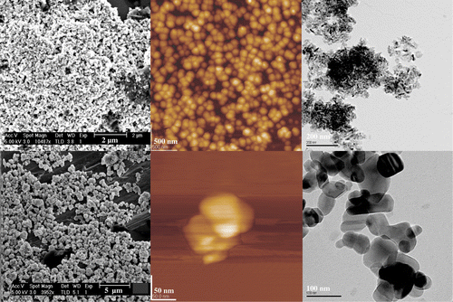
The most popular tools for the visualization of engineered nanoparticles are electron and scanning probe microscopes. Depending on the technique, resolutions down to the sub-nanometer range can be achieved. Using atomic force microscopy (AFM), scanning electron (SEM) and transmission electron microscopy (TEM), nanoparticles can not only be visualized, but also properties such as the state of aggregation, dispersion, sorption, size, structure and shape can be observed (Mavrocordatos et al. Citation2004). For comparison, shows TiO2 and ZnO nanoparticles imaged by SEM, TEM and AFM.
Figure 1. ZnO (1st row) and TiO2 (2nd row) nanoparticles suspended in distilled water, allowed to dry and imaged in order from left to right by SEM, AFM and TEM. Initial sizes as stated by the manufacturer (Sigma-Aldrich UK): 50–70 nm for ZnO particles and 5–10 nm for TiO2 particles.
In TEM, electrons are transmitted through a specimen (therefore, the specimen has to be very thin) to obtain an image; in SEM, scattered electrons are detected at the sample interface for imaging. In general, imaging of lighter atoms in an electron microscope is more difficult as they scatter electrons less efficiently.
Analytical (mostly spectroscopic) tools can be coupled to electron microscopes for additional elemental composition analysis, generally known as analytical electron microscopy (AEM). For example, energy dispersive X-ray spectroscopy (EDS) can be combined with SEM and TEM permitting a clear determination of the composition of elements heavier than oxygen, quantitative analysis, however, leads to a ∼ 20% uncertainty (Mavrocordatos et al. Citation2004).
Electron energy loss spectroscopy (EELS) is based on the loss of energy of the incident electron through the specimen; thus, elements can be discriminated. This technique can only be used with TEM and quantitative analysis has uncertainties as low as 10% (Mavrocordatos et al. Citation2004). Selected area electron diffraction (SAED) can also be combined with TEM and provides information on crystalline properties of particles (Mavrocordatos et al. Citation2004).
Electron microscopy is usually a destructive method, meaning that the same sample cannot be analyzed twice or by another method for validation. Other disadvantages of electron microscopes are charging effects caused by accumulation of static electric fields at the specimen due to the electron irradiation required during imaging. This can normally be overcome by using a sample coating made of a conducting material, but this can result in a loss of information. Also, biological samples often need treatment, such as heavy metal staining, for improved contrast.
For biological samples, a scanning transmission electron microscope (STEM) belonging to the group of TEMs can be of use. Dark-field microscopy with a STEM allows high contrasts and, therefore, imaging of biological samples without staining. In combination with diffraction and spectroscopic techniques, STEMs can also provide images and chemical data for nanomaterials with a sub-nanometer spatial resolution (Liu Citation2005). Utsunomiya and Ewing (Citation2003) successfully applied high-angle annular dark-field scanning transmission electron microscopy, scanning transmission electron microscopy–energy dispersive X-ray spectrometry and energy-filtered transmission electron microscopy to the characterization of heavy metals on airborne particulates (Utsunomiya and Ewing Citation2003).
X-ray microscopy (XRM) can provide spatial resolution (down to ∼30 nm, limited by the X-ray beam focusing optics) imaging of a specimen in the aqueous state without the need for sample preparation, e.g. fixation, staining or sectioning (Jearanaikoon and Braham-Peskir Citation2005; Thieme et al. Citation2007). X-ray microscopy can also be combined with computer tomography to enable 3D imaging (Thieme et al. Citation2003). A variation of the XRM is the scanning transmission X-ray microscopy (STXM), which has been used, for example, to characterize metallic Fe particles for remediation purposes (Nurmi et al. Citation2005).
The major limitation of conventional electron microscopes, such as transmission electron and scanning electron microscopes, is that they have to be operated under vacuum conditions. This means no liquid samples can be introduced to the sample chamber and sample preparation (dehydration, cryo-fixation or embedding) is necessary, which usually leads to sample alteration and dehydration artifacts (Mavrocordatos et al. Citation2007).
To limit artifacts, efforts have been made to improve sample preparation techniques for electron microscope imaging. For example, Lonsdale et al. (Citation1999) applied high pressure freezing and freeze substitution to image barley aleurone protoplasts by transmission electron microscopy (TEM). This method preserves the cellular fine structure and antigenicity of proteins better than conventional chemical fixation and dehydration techniques. Another possibility is the use of a cryo-TEM, which enables imaging of frozen samples on a cold specimen-stage and microscope. This has the advantage of preserving and visualizing structures that would be lost or altered by other sample preparation methods. Wang et al. (Citation2004) employed this method to image Fe(III)-doped TiO2 nanoparticles (2–4 nm) in an aqueous environment with a special sample holder. Mavrocordatos and Perret (Citation1998) embedded iron-rich particles (30–200 nm) in resin and then sectioned these samples for visualization by TEM and EELS (Mavrocordatos and Perret Citation1998).
However, none of these preparative techniques can fully avoid artifacts caused by sample drying or preparation. As imaging of nanoparticles in their original state is crucial for nanoparticle research, other methods are required. One possibility to image nanoparticles under more natural conditions is to use an environmental scanning electron microscope (ESEM). In an ESEM, the gun and lenses of the microscope are under vacuum conditions as in a conventional SEM, but, due to a detector that is able to operate under higher pressure and multiple pressure limiting apertures to separate the sample chamber from the column, the sample chamber itself can be operated at around 10–50 Torr. Therefore, samples can theoretically be imaged in their natural state without modification or preparation under variable pressure and humidity, theoretically up to 100%. Additionally, the gas ionization in the ESEM sample chamber eliminates the charging artifacts and, therefore, materials no longer have to be coated with a conducting material. Other advantages of an ESEM are that the detector is insensitive to light and fluorescence or cathodoluminescence does not disturb imaging. ESEM still allows X-ray data, e.g. from EDS, to be obtained. However, an ESEM cannot achieve real atmospheric pressure and only the top surface of a specimen can be imaged, which, in the case of a liquid sample, is the water surface. The contrast is increasingly poor with increasing humidity and there is the possibility of specimen drifting. Also, a loss in resolution from ∼10 nm up to ∼100 nm is unavoidable.
Doucet et al. (Citation2005) compared the performance of an environmental and a conventional scanning electron microscope (ESEM and SEM, respectively) for the imaging of natural aquatic particles and colloids. Analyzing river estuary samples they found that the conventional SEM provides sharper images and lower resolution limits, but produces more imaging artifacts due to drying of the sample. To some extent, ESEM samples retain their morphological structures without the need of sample preparation, but image interpretation and imaging itself is more complex. Also, it has been stated that the maximum relative humidity at which imaging could be performed was 75%, as, at 100%, layers of free water over the sample made colloid visualization impossible. Sizing of colloids revealed technique-dependent differences; hence, Doucet et al. (Citation2005a) suggest that ESEM and SEM should be used as complementary techniques, but are in favor of the ESEM for imaging colloids and colloid aggregation. Redwood et al. (Citation2005) applied an ESEM to analyze and quantify humic substances (Suwannee river humic acid, 100 mg l–1) as a function of humidity and pH (3.3–9.8). They concluded that ESEM is an important complementary technique to other analytical methods for probing changes in colloid structure as a function of hydration state; however, they also concluded that at present non-perturbed samples cannot be imaged (Redwood et al. Citation2005).
The technique of WetSTEM allows transmission observations of wet samples in an ESEM under annular dark-field imaging conditions down to a few tens of nm. Combining elements of TEM and ESEM, samples that are fully submerged can be imaged. The imaging is achieved by placing a TEM grid with the sample on a TEM sample holder. This holder is placed in the ESEM chamber allowing transmission imaging under non-vacuum conditions (Bogner et al. Citation2005).
An alternative to the ESEM methods described above is the use of a WetSEM™ capsule as a specimen holder, in which the sample is added and the holder is then sealed. These capsules have been developed by QuantomiX (Rohovot, Israel) for imaging of samples in a conventional SEM under hydrated conditions. There are two different types of WetSEM capsules on the market suitable for conventional SEM with a back-scattered electron detector: one for imaging in liquids and another for imaging of solid but wet materials (e.g. biological samples, food or soil). With this technique, in situ imaging of nanoparticles in natural media is possible. The capsule separates the sample from the vacuum chamber of the microscope and a membrane in the capsule allows electrons to pass into the sample; thus, enabling imaging under atmospheric pressure. It is possible to conduct semi-quantitative and qualitative elemental analysis with these capsules provided the microscope is equipped with an energy dispersive X-ray spectrometer (Thiberge et al. Citation2004a, Citationb; Joy and Joy Citation2006; Timp et al. Citation2007). Limitations are a loss of resolution and the sensitivity of the membrane to radiation damage. Also, objects have to be close to the membrane to be visible. Thiberge et al. (Citation2004) describe in detail the theory, characteristics, limitations and possible applications of WetSEM capsules using a conventional SEM and an ESEM (Thiberge et al. Citation2004a, Citationb).
Imaging under fully liquid conditions is also possible using atomic force microscopy (AFM). The AFM belongs to the family of scanning probe microscopes (SPMs) (Balnois et al. Citation2007). An oscillating cantilever is scanning over the specimen surface and electrostatic forces (down to 10–12 N) are measured between the tip and the surface. An AFM can achieve 3D surface profiles from these force measurements with height resolutions of ∼0.5 nm. The main advantage of an AFM is that it images sub-nanometer structures under wet or moist conditions. Although under liquid conditions particles not fixed to a substrate will float around and eventually stick to the cantilever, which leads to imaging artifacts, both as smearing effects and changes in the cantilever oscillation properties, as the tip gains weight. This smearing effect could be minimized by using a non-contact scanning mode where the tip is not touching the particles but only feel its forces (Balnois et al. Citation2007).
The main limitation of AFM for nanoparticle visualization is that the geometry of the tip is often larger than the particles being probed and this leads to errors in the onset and offset of particle topography on a scan, resulting in severe overestimations of the lateral dimensions of the nanoparticles. Therefore, accurate size measurements should only be taken on the height (z-axis) of the particles and the lateral dimensions only used with great caution. Furthermore, AFM for environmental or food related samples is limited in the ability to obtain qualitative or quantitative information of the sample composition. Nevertheless, the force patterns that emerge can also help in identifying the nature of individual atoms via a technique called chemical force microscopy (CFM) (Sugimoto et al. Citation2007; Shluger and Trevethan Citation2007). This recent development could lead to progress in AFM application to more complex samples. Scanning tunneling microscopy (STM) is another type of scanning probe microscopy and is based on the quantum mechanical nature of electrons on the sub-nanometre scale. A conducting tip is brought into proximity of a metallic or semiconducting surface such that when the gap between the surface and the tip is ∼<1 nm, and a small voltage applied to the tip (or surface), electrons can “tunnel” through this gap creating a very small current, the magnitude of which is very sensitive to the tip-surface separation. By scanning the tip across the surface and adjusting the height of the tip to maintain a constant tunnelling current, the surface can be imaged with a resolution of ∼1 nm or better. STM has been applied to environmental samples to image redox properties of microbial enzymes (Wigginton et al. Citation2007b).
AFM has been used to characterize natural colloidal matter. For example, Lead et al. (Citation2005) analyzed natural aquatic colloids by AFM and their structure was found to vary as a function of pH. Mica slides were dipped for 30 min into filtered samples rinsed with distilled water and allowed to dry prior to imaging in tapping mode. It has been stated that it is not known whether imaging under ambient humidity or liquid water produces better results. A priori, imaging under liquid water appears to provide ideal experimental conditions. However, atmospheric humidity retains colloid-bound water, helping to maintain structure, and AFM tips exposed to organic matter in solution soon become coated in the organic matter, potentially affecting the veracity of the images. This is also a possibility in imaging after air-drying. Comparing TEM and AFM using different sample preparation methods indicated similar morphologies (Lead et al. Citation2005). Balnois et al. (Citation1999) employed tapping mode AFM for the analysis of humic acid on mica. They found that aggregation might be related to the hydrophobicity of the sample. No aggregates were observed for relatively hydrophilic humic acids (Suwannee river) at pH 3–10, but aggregates were seen for peat humic acid at low pH and high ionic strength. A comparison between AFM, Fluorescence Correlation Spectroscopy, Field-Flow Fractionation and Pulsed Field Gradient-NMR on a reference fulvic acid sample (Lead et al. Citation2000a) consistently showed that AFM resulted in smaller particle sizes measurements than the other techniques, even though AFM is a number-average method whereas the others are mass-average methods. This underestimation of the size of the fulvic acid was thought to be due to drying or other substrate effects during the AFM procedure.
Although an AFM is operated under ambient conditions, samples still have to be applied to a specimen holder, which can cause alterations; thus, sample application has to be done carefully. A range of sample preparation techniques have been reported by Balnois and Wilkinson (Citation2002), including drop deposition, adsorption, ultracentrifugation, which have successfully been applied in the characterization of environmental biopolymers (e.g. humic substances, polysaccharides) by AFM. Bickmore et al. (Citation1999) developed methods (including electrostatic attraction and adhesion based) to fix clay minerals to a substrate thus allowing imaging in aqueous suspensions by AFM. Further applications of AFM to environmental colloids have been reviewed by Maurice (Citation1996). He describes the AFM as powerful tool to image environmental colloids and surfaces in air or immersed in water at sub-nanometer-scale resolution with examples of applications and limitations (Maurice Citation1996). Very recently a review has also been published relating the application of AFM to nanotechnology in food science (Yang et al. Citation2007).
From the above, it is clear that, using a combination of microscopic techniques, we can not only visualize nanoparticles but also generate useful data on the size, size distribution and other measurable properties (Jose-Yacaman et al. Citation2001; Biberthaler et al. Citation2003; Rabinski and Thomas Citation2004; Chuklanov et al. Citation2006; Baatz et al. Citation2006). However, it needs to be recognized that the image analysis of the microscope outputs is as crucial as imaging itself. Only small amounts of samples can be analyzed by microscopic techniques and this has an impact on the statistical significance of the results. The average particle size is a number average, and size distribution obtained by image analysis depends on the number of particles measured. Since there are often fewer larger particles, it is important to count and measure enough particles to obtain good counting statistics on these size fractions. The same issues need to be considered when measuring ENPs in food or environmental samples in the presence of high concentrations of natural nanomaterials. It may, therefore, be necessary to measure thousands of particles to generate reliable data. Therefore, it is essential to develop automation and image analysis procedures. Image contrast can have an influence on the visible size of the particles or light element particle coatings may be invisible, leading to controversial or incomparable results.
Chromatography and related techniques
Techniques based on or related to chromatography can be used for the separation of nanoparticles in samples. These techniques are rapid, sensitive (detector-dependent) and non-destructive, so that samples are available for further analysis. Although some chromatographic tools allow a range of solvents to be used, samples usually cannot be run in their original media, which can cause sample alteration and sample solvent interaction. By attaching traditional analytical tools (e.g. ICP–MS, DLS) as detectors to size separation techniques, it is not only possible to quantify different nanoparticles in food, water, biota and soil, but also to characterise or elementally analyse them.
The best known technique for size separation is size exclusion chromatography (SEC). A size exclusion column is packed with porous beads as the stationary phase. The pores of the column retain particles, depending on their size and shape. This method has been applied to the size characterization of quantum dots, single-walled carbon nanotubes and polystyrene nanoparticles (e.g. Krueger et al. Citation2005; Ziegler et al. Citation2005; Huang et al. Citation2005). Size exclusion chromatography has good separation efficiency, but major disadvantages include possible interactions of the solute with the solid phase (Lead and Wilkinson Citation2006) or the limited size separation range of the columns, which may not cover the size range of both the primary nanoparticles and their aggregates. Methods employed to overcome the problem of solid-phase interactions include the addition of capping agents to the mobile phase and the recycling of the analyte. SEC has been successfully combined with a range of detection techniques to not only monitor the size fractionation of the particles but also to characterize them. For example, Song et al. (Citation2004) used voltammetric detection for gold nanoparticles separation and Helfrich et al. (Citation2006) employed ICP–MS as multi-element detection method, whereas Porsch et al. (Citation2005) worked with multi-angle laser light-scattering (MALLS).
Unlike SEC, in capillary electrophoresis (CE) there are no solid phase interactions. CE allows the separation of particles in different solutions based on the charge and size distribution of the components. However, as separation is not based on size alone, data interpretation is more complex. Also, mobile phase interactions cannot be excluded. Lin et al. (Citation2007) used CE for the sizing of engineered Au and Au/Ag nanoparticles and Schmitt-Kopplin and Junkers (Citation2003) have used CE in the characterization of humic substances and other natural organic matter.
Hydrodynamic chromatography (HDC) separates particles based on their hydrodynamic radius. A HDC column is packed with non-porous beads building up flow channels in which particles are separated by flow velocity and the velocity gradient across the particle. Therefore, larger particles elute faster from the column than smaller ones (McGowan and Langhorst Citation1982). The non-porous beads considerably reduce the risk of solid-phase interactions compared to the porous packaging in a SEC column. Available HDC columns show size separation ranges from 5 to 1200 nm depending on the column length, whereas the size-separation range of a SEC column is dominated by its pore size distribution. The wider particle size-separation range of HDC allows a whole range of nanoparticles to be sized in different media and is particularly helpful in allowing a better understanding of formation of aggregates. HDC has been connected to the most common UV–Vis detector for the size characterization of (fluorescent) nanoparticles, colloidal suspensions and biomolecules (Williams et al. Citation2002; Chmela et al. Citation2002; Blom et al. Citation2003), but also to dynamic light scattering (DLS) for sizing separate lipid nanocapsules (Yegin and Lamprecht Citation2006). A major limitation of HDC is poor peak resolution.
A highly promising technique for the size separation of ENPs in complex natural samples is field-flow fractionation (FFF) (Giddings Citation1993; Beckett and Hart Citation1993; Schimpf et al. Citation2000; Hassellöv et al. Citation2007). It is similar to chromatographic techniques, but separation is solely based on physical separation in an open channel without relying on a stationary phase. The particles are separated based on how they are affected by an applied field. The field controls the particle transport velocity by positioning them in different average laminar flow vectors in a thin channel. The field can be a centrifugal force (sedimentation FFF) or a hydrodynamic flow perpendicular to the separation flow (flow FFF). FFF is able to fractionate particles in a range from 1 nm to 1 µm in Brownian mode.
FFF instruments can be coupled to online or offline detection and characterization, which in addition to size distributions, allows analysis and visualisation of the fractionated samples by electron microscopy (Baalousha et al. Citation2005a). FFF can also be coupled to a range of sensitive and multi-element techniques, such as multi-angle laser light-scattering (MALLS) and ICP–MS (Hassellöv et al. Citation1999b; von der Kammer et al. Citation2005a). FFF coupling techniques have been successfully applied in geochemistry and natural colloid research as well as studies into the behaviour of engineered nanoparticles. Applications range from colloids in fresh and marine water to size separation of soil suspensions (Ranville et al. Citation1999; Hassellöv et al. Citation1999a, Citationb; Chen and Beckett Citation2001; Lyven et al. Citation2003; Siepmann et al. Citation2004; von der Kammer et al. Citation2004, Citation2005a, Citationb; Stolpe et al. Citation2005; Baalousha et al. Citation2005a; Graff and Frazier Citation2006; Lead and Wilkinson Citation2006; Gimbert et al. Citation2006; Peng et al. Citation2006; Baalousha et al. Citation2006a, Citationb; Baalousha and Lead Citation2007). Also, single-walled carbon nanotubes have been length-separated by dielectrophoresis FFF (Peng et al. Citation2006) and many engineered nanoparticles, such as SiO2, metals, metal oxides, carbon black, etc. have been analysed by FFF (Schimpf et al. Citation2000).
The limitations of FFF techniques are membrane or accumulation wall interactions, the continuous re-equilibration in the channel (for trace constituent studies) and the need (in some circumstances) of pre-concentration, additional concentration of sample during equilibration and an increasing possibility of aggregation in the channel (Beckett and Hart Citation1993; Hassellöv et al. Citation2007).
In theory, any aqueous or non-aqueous phase of any ionic strength and a pH between 2 and 11 can be used as a carrier. This gives versatility in terms of selecting the carrier composition to favor colloidal stability, thus minimizing wall and membrane interactions and particle–particle interactions.
Stegeman et al. (Citation1994) compared the resolving power and separation time in thermal field-flow fractionation (TFFF), hydrodynamic chromatography and size exclusion chromatography for the size separation of polymers, and concluded that TFFF theoretically has the best separation potential due to high selectivity, but this may not be exploitable in practice owing to the technical requirements. On the other hand, SEC was found to be the fastest method for low molecular masses (Stegeman et al. Citation1994). In general, FFF and HDC have a wider dynamic size range than SEC, while SEC has higher separation efficiency (less peak broadening). SEC also suffers from more sample perturbations than FFF or HDC.
Centrifugation and filtration techniques
Centrifugation and filtration techniques are well-established tools for the preparative, size fractionation of samples. These are low-cost, high speed and high volume techniques. Ultracentrifugation (UC), for example, is a centrifuge system capable of very high spinning speeds for accelerations up to 1,000,000 g. There are two different types of ultracentrifugation: analytical and preparative UC. In an analytical ultracentrifuge (ANUC), a sample can be monitored in real time through an optical detection system using ultraviolet light absorption and/or interference optical refractive index sensitive systems. This allows the operator to observe the evolution of the sample concentration versus the axis of rotation profile as a result of the applied centrifugal field, and is valuable for sedimentation velocity and sedimentation equilibrium experiments (gross shape of macromolecules, conformational changes in macromolecules and size distribution). Preparative ultracentrifugation has been used for pelleting of fine particulate fractions, for gradient separations (Bootz et al. Citation2004) and for harvesting aquatic colloids and nanoparticles on TEM and AFM substrates (Mavrocordatos et al. Citation2007; Balnois et al. Citation2007).
Traditional membrane filtration allows the fractionation of particle sizes between 0.2 and 1 µm (Lead and Wilkinson Citation2006). Comparative data obtained for soil suspensions, for filtration and sedimentation FFF indicates that membrane filtration can both over- and under-estimate smaller size fractions due to clogging as well as electrostatic interactions (Gimbert et al. Citation2005). Microfiltration with pore sizes >0.1 µm is a simple and common method, although exhibiting many artifacts caused by, for example, filter-cake formation and concentration polarization (Morrison and Benoit Citation2001). Ultrafiltration is applicable for large sample volumes; however, with decreasing pore sizes, common filtration artifacts are even more likely. For the separation of nanoparticles and ions, nanofiltration with pore sizes of 0.5 or 1 nm can be used.
Cross flow filtration (CFF) or tangential filtration recirculates the samples and, therefore, reduces clogging, concentration polarization and other artifacts caused by traditional dead-end filtration (Lead and Wilkinson Citation2006). It has become the standard method for separating colloids and particles and its efficacy has been evaluated against AFM by Liu and Lead (Citation2006). The method has been applied to fluorescence investigations of colloidal organic matter and dissolved organic matter in lake and river water (Liu et al. Citation2007) as well as in seawater (Guo et al. Citation2000). Electrically assisted cross-flow filtration has also been used for the separation of nanoparticles (Sung et al. Citation2007). Doucet et al. (Citation2004) evaluated cross-flow ultrafiltration (CFUF) for the size fractionation of freshwater colloids and particles (1 nm–1 µm) by AFM and SEM, and concluded that CFUF is not fully quantitative and separation is not always based on size alone. Amounts of large colloids might be overestimated and fractionation is not always consistent with the nominal pore size of the membranes. These conclusions have to be treated with some caution as the validation techniques used (i.e. AFM and SEM) also have their limitations (Doucet et al. Citation2004).
Spectroscopic and related techniques
A wide range of spectroscopic methods is available for nanoparticle analysis and characterization. Scattering techniques useful for nanoparticle characterization include light scattering methods, such as static (SLS) and dynamic light-scattering (DLS), or neutron scattering, such as small-angle neutron scattering (SANS).
DLS or photon correlation spectroscopy (PCS) is particularly useful for sizing nanoparticles and determining their state of aggregation in suspensions. DLS provides fast in situ and real-time sizing (Ledin et al. Citation1994), but also has considerable limitations. For example, interferences can be caused by a range of possible artifact sources, such as dust particles, which will influence the scattering intensity compared to smaller particles and, therefore, on the sizing result. Also, data obtained from samples containing particles with heterogeneous size distributions are difficult to interpret. DLS is solely quantitative and unless the sample content is known or pure, size fractions cannot be related to particles of a specific composition. (e.g. Bootz et al. Citation2004).
Static light scattering, also known as multi-angle (laser) light-scattering (MAL(L)S), gives information on particle structure and, in combination with dynamic light-scattering or FFF, particle shape can be determined.
SANS can be used on solid or liquid samples. For example, Diallo et al. (Citation2005) have applied SANS for the characterization of Suwannee River fulvic acid aggregates in aqueous solutions.
Small angle X-ray scattering (SAXS) is an analytical X-ray application technique for investigating the structural characterization of solid and fluid materials in the nanometer range. Monodisperse and polydisperse systems can be studied. In monodisperse systems, size, shape and structure determination is possible, whereas, in polydisperse systems, only the size distribution can be calculated.
Laser-induced breakdown detection (LIBD) is a laser-based technique featuring extremely low detection limits, which is capable of analyzing the size and concentration of colloids, depending on the measured breakdown probability (BP). LIBD is, therefore, a highly promising tool for nanoparticle characterization, although it cannot distinguish between different types of particles and requires particle-specific size calibration (Bundschuh et al. Citation2001a,b).
Other laser-based techniques include Raman spectroscopy and laser-induced fluorescence (LIF). Instruments are now available combining these techniques, allowing the atomic, molecular and structural characterization of a specimen, as well as a better understanding of physical properties.
UV–Vis and infrared spectroscopy offer the possibility of characterizing nanoparticles, especially quantum dots and organic-based nanoparticles, such as fullerenes and carbon nanotubes. Fourier transformation infrared (FTIR) and UV–Vis spectroscopy have been used to compare aqueous colloidal suspensions of C60 (Andrievsky et al. Citation2002). Pesika et al. (Citation2003) also used UV spectroscopy to study the relationship between absorbance spectra and particle-size distributions for quantum-sized nanocrystals.
Nuclear magnetic resonance (NMR) is a powerful technique providing information on the dynamics and three-dimensional structure of a solid compound or a suspension. Carter et al. (Citation2005) characterized air- and water-stable silica nanoparticles by NMR. Diffusion NMR spectroscopy has also been used for the characterization of the size and interactions of colloidal matter (Valentini et al. Citation2004; Carter et al. Citation2005). Lead et al. (Citation2000a) used pulsed field gradient NMR to measure the diffusion coefficients of fulvic acids.
X-ray spectroscopy comprises X-ray photoelectron (XPS), X-ray fluorescence (XRF) as well as X-ray absorption spectroscopy (XAS) and X-ray diffraction (XRD). XPS is highly surface-specific due to the short range of the photoelectrons that are excited from the solid sample and, therefore, XPS could be useful to characterize nanoparticle surfaces and coatings. X-ray diffraction is non-destructive and can reveal information about the crystallographic structure or elemental composition of natural and manufactured materials. Nurmi et al. (Citation2005) used this technique, as well as XPS, for the characterization of zero-valent Fe nanoparticles for use in remediation. X-ray fluorescence (XRF) spectroscopy is also non-destructive and can be used to identify and determine the concentrations of elements present in solid, powdered or liquid samples. XRF can be subdivided into wavelength separation (WDXRF) and energy dispersive XRF (EDXRF).
X-ray absorption (XAS) and emission spectroscopy is used in chemistry and material sciences to determine elemental composition and chemical bonding.
Other potentially suitable spectroscopic techniques for nanoparticle characterization include electron paramagnetic resonance (EPR), Mössbauer, Auger electron (AES) and 3D fluorescence excitation–emission matrix spectroscopy (EEM). Mössbauer spectroscopy provides information about chemical, physical and magnetic properties by analyzing the resonant absorption of characteristic energy gamma-rays, known as the Mössbauer effect. Liu et al. (Citation2007) and Lead et al. (Citation2006) applied 3D fluorescence excitation–emission matrix (EEM) spectrophotometry for the fluorescence investigation of colloidal organic matter and dissolved organic matter in lake and river water. EPR spectroscopy can be applied for particle surface reactivity analysis, and is a sensitive, specific method for studying organic and inorganic radicals formed in chemical reactions or the reactions themselves, similar to NMR. Auger electron spectroscopy is also commonly used in the surface characterization of nanostructures. Quantitative bulk analysis by AES has been described by Powell and Seah (1980).
Mass spectrometry
Mass spectrometers consist of an ion source, a mass analyzer and a detector system. Two ionization techniques often used with liquid and solid biological samples include electrospray ionization (ESI) and matrix-assisted laser desorption/ionization (MALDI). Inductively coupled plasma (ICP) sources are mainly used for metal analysis. Mass analyzers (e.g. ion trap, quadrupole or time-of-flight) cover different mass-to-charge ranges, differ in mass accuracy and achievable resolution. Most of the available analyzers are compatible with electrospray ionization, whereas MALDI is not usually coupled to a quadrupole analyzer.
Mass spectrometry (MS) approaches, such as MALDI, laser-induced fluorescence (LIF) or ion trap (IT) mass spectrometry, have been applied for the analysis of fluorescently labeled nanoparticles (Peng et al. Citation2003; Cai et al. Citation2003).
In the case of ICP–MS, samples cannot only be injected directly into the ion source but via a combined technique, such as HPLC. An increasingly popular combination in this respect is FFF–ICP–MS, which allows the size separation of the sample with quantitative and elemental analysis of the obtained size fractions. This development is highly promising for nanoparticle analysis, as particles can be simultaneously sized and analyzed in their original environment (Ranville et al. Citation1999; Hassellöv et al. Citation1999a,b; Lyven et al. Citation2003; von der Kammer et al. Citation2004; Bolea et al. Citation2006; Baalousha et al. Citation2006a).
Whereas conventional mass spectrometry (MS) is applicable for identifying unknown compounds and their mass concentrations, as well as their isotopic composition, single particle mass spectrometry (SPMS) has also the ability to size single particles. MS techniques have also been used in aerosol characterization, including aerosol time-of-flight mass spectrometer (ATOF-MS). An ATOF-MS consists of an aerosol introduction interface; a light-scattering region for sizing and a TOF–MS. Suess and Prather (Citation1999) published a review on the topic of mass spectrometry of aerosols, describing tools for offline MS of aerosols, such as LAMMS, SIMS and ICP-MS, tools for online MS, such as surface/thermal ionization MS (SIMP, DIMS, CAART, PAMS), and laser desorption/ionization MS (ATOFMS, PALMS, RSMS, LAMPAS). More applied examples are described by Janzen et al. (Citation2002), who compared the sizing of nanoparticles with SPMS and TEM. Lee et al. (Citation2005) used SPMS to characterize the size and composition of polydisperse aerosol nanoparticles. They estimated particle size with a laser ablation/ionization time-of-flight single-particle mass spectrometer and validated their results by differential mobility analysis (DMA). In situ characterization of size and elemental composition of individual aerosol particles in real time was performed by Prather et al. (Citation1994) with the help of an ATOF–MS. For the sizing and analysis of aerosol nanoparticles, a DMA has also been coupled to an ICP–MS (Okada et al. Citation2002).
Other techniques
Particle counters for number concentrations
The electrical sensing zone method counts and sizes particles by detecting changes in electrical conductance as particles suspended in a weak electrolyte solution are drawn through a small aperture. The technique has been successfully applied to the size and surface charge characterization of nanoparticles using a carbon nanotube-based Coulter counter (Ito et al. Citation2003). Condensation particle counter (CPC) measurements can also provide data on the number and concentration of individual particles by growing the particles through a condensing process using various operating liquids, such as alcohol and water.
DMA for sizing aerosols
A differential mobility analyzer (DMA) can be used to determine the size distribution of sub-micrometer aerosol particles. Particles are firstly charged and then their electrical mobility is measured as a function of their charge and size. After sizing, the particles are still suspended in air and are ready for further analysis (McMurry et al. Citation1996; Weber et al. Citation1996; Okada et al. Citation2002).
SMPS for sizing and number concentration determination
A scanning mobility particle sizer (SMPS) consists of a DMA and a CPC. First, particles are separated by their electrical mobility in the DMA; then, the size fractionations enter a CPC, which determines the particle concentration at that size.
BET method for surface area determination
The very common Brunauer–Emmett–Teller (BET) method enables the determination of the specific surface area of solids and, thus, nanoparticles by gas adsorption (Brunauer et al. Citation1938).
Thermogravimetry and differential thermo analysis (TG–DTA)
DTA can be applied for phase changes and other thermal processes, such as the determination of melting point. In combination, TG–DTA is useful for investigating the thermal stability and decomposition, dehydration oxidation, as well as the determination of volatile content and other compositional analysis. Thermogravimetry in combination with a mass spectrometer can be used for surface analysis. Surface molecules are removed by heating and afterwards analysed by MS.
Electrophoretic mobility and the zeta potential
Electrophoresis is used for studying properties of dispersed particles, in particular, for measuring the zeta potential. The zeta potential is a measure of the overall charge a particle acquires in a specific medium and gives an indication of the potential stability of a colloidal system. If all the particles have a large negative or positive zeta potential, they will repel each other, which leads to higher stability than if the particle charge is nearly neutral. The zeta potential is a measure of the net charge and there may be significant charge heterogeneities that can still lead to aggregation, even though the net zeta potential suggests otherwise. Information about the aggregation state of a nanoparticle dispersion is highly valuable for nanoparticle fate and behaviour studies. As an example, the electrophoretic mobility of silica spheres suspended in water at different concentrations and salinities has been studied by Reiber et al. (Citation2007).
Nanomaterial analysis in food and biological samples
As previously discussed, when measuring nanoparticles in different media, it is not only necessary to generate data on concentrations but information will also be required on the size distribution and properties of the particles. No single technique can provide all this information, so a range of analytical techniques is required. Moreover, while a range of methods have been shown to be applicable to the analysis of nanoparticles, the current methods do not fulfill all data requirements.
As shown in the previous section, many analytical tools are theoretically suitable for the characterization of nanoparticles, ranging from electron microscopy to dynamic light scattering to field flow fractionation techniques, but only a few of these are applicable to the analysis of more complex samples. Requirements for analysis of engineered nanoparticles in natural and food related samples will differ greatly from their analysis in pure or neutral media (e.g. air, distilled water). In complex media, it is essential to analyze samples of diverse elemental compositions and samples containing more than one type of nanoparticle. Many techniques are destructive or, if not, application of some sample preparation methods can lead to artifacts. In addition, natural samples will be hetero-dispersed and, for measuring size distributions, instruments providing a wide size-separation range from, ideally, 1 nm to up to several µm, are needed. There are many methods available for the sizing of particles, but very few, if any, is applicable to the entire size range. In the next section, some of these challenges are discussed in more detail.
Bulk versus single particle analysis
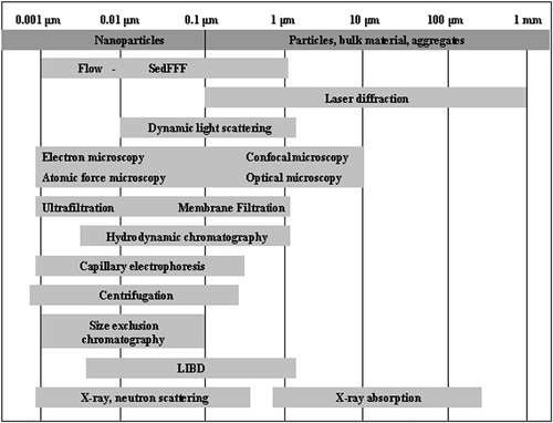
One problem with some methods, as discussed in the previous chapter, is their application range. Existing techniques have to be divided between tools suitable for analysing individual particles (depending on particle size) or the bulk material. Classic composition- and mass-based tools are readily applicable for the bulk material; however, elemental analysis of single particles in a dilute environment has only recently become available (e.g. aerosol mass spectrometry). Whereas, standard tools for elemental composition and mass concentration are restricted by their limit of detection (LOD), techniques capable of characterizing individual particles face spatial limitations. Especially, particle sizing techniques are restricted by their size separation range. illustrates the size range of selected methods for particle sizing.
Figure 2. Sizing methods and their size ranges for nanoparticle measurements Adapted from Lead and Wilkinson (Citation2006) and Gimbert et al. (Citation2007b).
Sizing artifacts and the lack of reference materials
The limitations of each analytical method for nanoparticle characterization can lead to inconsistent results and, therefore, to inaccurate predictions of material properties and structure (Carter et al. Citation2005). For example, it is still almost impossible to determine the absolute size of particles. Correct size measurements are difficult, which often leads to artifacts, depending on the applied tool and the medium the particles are analyzed in. For example, organic coatings that are not visible in the electron microscope (due to light elements, such as carbon) can lead to errors in sizing, especially when compared to sizing tools that measure the hydrodynamic radius of particles, such as FFF or DLS. It has been reported that the average size and size distribution of nanoparticles can significantly vary when comparing results from different techniques, such as electron microscopy, dynamic light scattering, CFF or ultracentrifugation (Bootz et al. Citation2004).
The lack of consistent reference materials and standards further exacerbates this problem (Lead and Wilkinson Citation2006). Nanoparticle sizing standards, as well as standardized methods for sampling and measurement, are, therefore, urgently required to overcome the problem of inconsistent data (Borm et al. Citation2006). To the best of our knowledge, standardized nanoparticles are not yet available and researchers have to rely on commercially available, often not well-characterized, nanoparticles.
Sample preparation
Depending on the technique, to analyse natural samples, sample preparation and/or digestion is often required. As nanoparticles can and do change structure and composition in response to their environment, results obtained for pre-treated or digested samples can often differ from the situation where the particles are characterised in situ (Burleson et al. Citation2004). These artifacts in analysis can be avoided by using techniques that do not require or reduce sample preparation to a minimum. The complexity of data obtained for some techniques (e.g. NMR, CE) for samples in their original state can make analysis and interpretation difficult.
If sample preparation cannot be avoided, a careful record of sampling and preparation steps is essential to track artifacts. The nature of nanoparticles can also change over time; for example, aggregation can increase or decrease and particles could dissolve. A lot of effort has been put into the development of sample preparation methods that improve the conservation of the original state of the sample. Especially in the field of microscopy, advances have been made in sample preparation ranging from gel-trapping techniques for imaging emulsions under the SEM (Paunov et al. Citation2007) to high-pressure freezing and freeze-drying for imaging biological specimen under the TEM (Lonsdale et al. Citation1999; Bootz et al. Citation2004). Fixation methods for imaging clay minerals and particles in aqueous solutions under the AFM have also been developed (Bickmore et al. Citation1999).
Natural versus engineered nanoparticles
At the moment, it is very difficult to distinguish between particles of engineered origin and particles from a natural or other source (Burleson et al. Citation2004). A way has to be found to differentiate between natural occurring and engineered nanoparticles. As the number of engineered nanoparticles actually reaching the environment or their bioavailability is unknown, this will allow concentrations in consumer products and the environment to be determined. Therefore, selective detection methods need to be developed. Another solution to this problem could be nanomaterial labeling, with suggestions ranging from fluorescent- and radioactive-labeling for carbon-based nanoparticles to isotopic enrichment or depletion of metal-based nanoparticles. Also, special particle coatings or entrapment of rare elements in nanotubes or fullerenes could be used to enable the detection of these distinctive chemical characteristics after an experimental study. Gulson and Wong (Citation2006) reviewed the possibilities of isotopic labeling and tracking of metal and metal oxide nanoparticles for nanotechnology research. Isotopic labelling of carbon nanotubes and fullerenes has already been performed; for example, 13C isotope carbon nanotubes are available and 14C C60s have been synthesized, with subsequent uptake and toxicity studies (Scrivens et al. Citation1994b; Bullard-Dillard et al. Citation1996).
Conclusions and recommendations for future work
Analytical methods are required to reliably detect and characterise nanoparticles and their properties in matrices to which humans and ecosystems are exposed, including air, soil and water as well as food and consumer products. These methods must also be applicable for nanoparticle characterisation in toxicological and ecotoxicological testing; only then can an appropriate risk assessment be performed and nanoparticle properties of risk identified and regulated or used in standard testing (SCENIHR Citation2005).
These techniques have to be able (a) to deal with heterogeneous samples, (b) minimize sample alteration to avoid artifacts and (c) provide as much information as possible, because most characterization techniques are destructive and, therefore, samples often cannot be analyzed twice or by more than one technique. An ideal analytical instrument should allow simultaneous determination of all physico-chemical properties of a nanoparticle and, as many nanoparticles are transient in nature, obtain them by real-time sampling (Prather et al. Citation1994). While a wide range of tools is available, the existing tools do not fulfil all desirable criteria and have limitations when considering their application for food and natural samples. Therefore, until new tools have been developed, existing tools have to be used and combined in such a way that data can be validated. Analysis of the unperturbed sample or further analysis of the size fractionations is preferred. Complementary analytical tools should be applied and care taken with sample preparation.
As this review demonstrates, promising developments have been made in nanoparticle analysis; however, further advances are essential to overcome these deficiencies. Especially, in situ analysis, as well as routine and reliable techniques, to improve size determination, size distribution of particles and other nanoparticle properties, are important.
Nanotoxicology and nanoecotoxicology are still in their infancy and risk assessments are practically non-existent, especially in the food sector. Therefore, progress in nanoparticle testing (in vivo and in vitro) is urgently needed to guarantee consumer safety, including the development of standard testing materials and testing guidelines. In addition to toxicity studies, various uptake paths have to be studied, including dermal, oral and intestinal, as well as nanoparticle accumulation and potential long-term effects. Other effects of nanoparticle uptake could be the interaction with other (toxic) substances and their mobilisation or dislocation, not only in the human body, but also in consumer products. The environmental fate, behaviour and bioavailability of nanoparticles is unknown and, thereby, their potential impact on food webs and persistence. Their effect on other substances also needs examination; for example, whether contaminant transport in the environment could be facilitated through adsorption to nanoparticles, whether nanoparticles enhance contaminant uptake or have a negative impact on bacteria useful for natural remediation. Furthermore, data on environmental and exposure concentrations are unavailable. Developments in the above-mentioned analytical fields will be crucial to further our knowledge of nanoparticle and related issues.
Acknowledgements
Karen Tiede thanks Unilever UK for funding this work and Prof. John Gilbert for his support.
Related Research Data
References
- Akthakul , A , Hochbaum , AI , Stellacci , F and Mayes , AM . 2005 . Size fractionation of metal nanoparticles by membrane filtration . Adv Mater , 17 : 532
- Andrievsky , GV , Klochkov , VK , Bordyuh , AB and Dovbeshko , GI . 2002 . Comparative analysis of two aqueous-colloidal solutions of C-60 fullerene with help of FTIR reflectance and UV–Vis spectroscopy . Chem Phys Lett , 364 : 8 – 17 .
- Angelino , S , Suess , DT and Prather , KA . 2001 . Formation of aerosol particles from reactions of secondary and tertiary alkylamines: Characterization by aerosol time-of-flight mass spectrometry . Environ Sci Technol , 35 : 3130 – 3138 .
- Arcon , I , Mozetic , M and Kodre , A . 2005 . XAS study of oxygen plasma-treated micronized iron oxide pigments . Vacuum , 80 : 178 – 183 .
- Baalousha , M and Lead , JR . 2007 . Characterization of natural aquatic colloids (<5 nm) by flow-field flow fractionation and atomic force microscopy . Environ Sci Technol , 41 : 1111 – 1117 .
- Baalousha , M , Kammer , F von der , Motelica-Heino , M and Le Coustumer , P . 2005a . Natural sample fractionation by F1FFF–MALLS–TEM: Sample stabilization, preparation, pre-concentration and fractionation . J Chromatogr A , 1093 : 156 – 166 .
- Baalousha , M , Kammer , F von der , Motelica-Heino , M and Le Coustumer , P . 2005b . 3D characterization of natural colloids by FIFFF–MALLS–TEM . Anal Bioanal Chem , 383 : 549 – 556 .
- Baalousha , M , Kammer , F von der , Motelica-Heino , M , Baborowski , M , Hofmeister , C and Le Coustumer , P . 2006a . Size-based speciation of natural colloidal particles by flow field flow fractionation, inductively coupled plasma-mass spectroscopy, and transmission electron microscopy/X-ray energy dispersive spectroscopy: Colloids-trace element interaction . Environ Sci Technol , 40 : 2156 – 2162 .
- Baalousha , M , Kammer , F von der , Motelica-Heino , M , Hilal , HS and Le Coustumer , P . 2006b . Size fractionation and characterization of natural colloids by flow-field flow fractionation coupled to multi-angle laser light scattering . J Chromatogr A , 1104 : 272 – 281 .
- Baatz , M , Arini , N , Schape , A , Binnig , G and Linssen , B . 2006 . Object-oriented image analysis for high content screening: Detailed quantification of cells and sub cellular structures with the cellenger software . Cytometry A , 69 : 652 – 658 .
- Balnois , E and Wilkinson , KJ . 2002 . Sample preparation techniques for the observation of environmental biopolymers by atomic force microscopy . Colloids Surf A , 207 : 229 – 242 .
- Balnois , E , Papastavrou , G and Wilkinson , KJ . 2007 . “ Force microscopy and force measurements of environmental colloids ” . In Environmental Colloids and Particles: Behaviour, Structure and Characterization , Edited by: Wilkinson , KJ and Lead , JR . 405 – 468 . Chichester : Wiley .
- Balnois , E , Wilkinson , KJ , Lead , JR and Buffle , J . 1999a . Atomic force microscopy of humic substances: Effects of pH and ionic strength. Environ . Sci Technol , 33 : 3911 – 3917 .
- Balnois , E , Wilkinson , KJ , Lead , JR and Buffle , J . 1999b . Atomic force microscopy of humic substances: Effects of pH and ionic strength. Environ . Sci Technol , 33 : 3911 – 3917 .
- Banfield , JF and Zhang , HZ . 2001 . Nanoparticles in the environment . Nanoparticles Environ , 44 : 1 – 58 .
- Bauer , F , Ernst , H , Hirsch , D , Naumov , S , Pelzing , M , Sauerland , V and Mehnert , R . 2004 . Preparation of scratch and abrasion resistant polymeric nanocomposites by monomer grafting onto nanoparticles. 5(a) Application of mass Spectroscopy and atomic force microscopy to the characterization of silane-modified silica surface . Macromol Chem Phys , 205 : 1587 – 1593 .
- Beckett , R and Hart , BT . 1993 . “ Use of field-flow fractionation techniques to characterise aquatic particles, colloids, and macromolecules ” . In Environmental Particles , Edited by: Buffle , J and van Leeuwen , HP . 165 – 205 . Boca Raton, FL : Lewis Publishers .
- Biberthaler , P , Athelogou , M , Langer , S , Luchting , B , Leiderer , R and Messmer , K . 2003 . Evaluation of murine liver transmission electron micrographs by an innovative object-based quantitative image analysis system (Cellenger (R)) . Eur J Med Res , 8 : 275 – 282 .
- Bickmore , BR , Hochella , MF , Bosbach , D and Charlet , L . 1999 . Methods for performing atomic force microscopy imaging of clay minerals in aqueous solutions . Clays Clay Miner , 47 : 573 – 581 .
- Blom , MT , Chmela , E , Oosterbroek , RE , Tijssen , R and van den Berg , A . 2003 . On-chip hydrodynamic chromatography separation and detection of nanoparticles and biomolecules . Anal Chem , 75 : 6761 – 6768 .
- Bogner , A , Thollet , G , Basset , D , Jouneau , PH and Gauthier , C . 2005 . Wet STEM: A new development in environmental SEM for imaging nano-objects included in a liquid phase . Ultramicroscopy , 104 : 290 – 301 .
- Bolea , E , Gorriz , MP , Bouby , M , Laborda , F , Castillo , JR and Geckeis , H . 2006 . Multielement characterization of metal-humic substances complexation by size exclusion chromatography, asymmetrical flow field-flow fractionation, ultrafiltration and inductively coupled plasma-mass spectrometry detection: A comparative approach . J Chromatogr A , 1129 : 236 – 246 .
- Bootz , A , Vogel , V , Schubert , D and Kreuter , J . 2004 . Comparison of scanning electron microscopy, dynamic light scattering and analytical ultracentrifugation for the sizing of poly(butyl cyanoacrylate) nanoparticles . Eur J Pharm Biopharm , 57 : 369 – 375 .
- Borm , PJA , Robbins , D , Haubold , S , Kuhlbusch , T , Fissan , H , Donaldson , K , Schins , R , Stone , V , Kreyling , W Lademann , J . 2006 . The potential risks of nanomaterials: a review carried out for ECETOC . Particle Fibre Toxicol , 3
- Brant , J , Lecoanet , H and Wiesner , MR . 2005a . Aggregation and deposition characteristics of fullerene nanoparticles in aqueous systems . J Nanoparticle Res , 7 : 545 – 553 .
- Brant , J , Lecoanet , H , Hotze , M and Wiesner , MR . 2005b . Comparison of electrokinetic properties of colloidal fullerenes (n-C-60) formed using two procedures . Environ Sci. Technol , 39 : 6343 – 6351 .
- Brown , DM , Wilson , MR , MacNee , W , Stone , V and Donaldson , K . 2001 . Size-dependent proinflammatory effects of ultrafine polystyrene particles: A role for surface area and oxidative stress in the enhanced activity of ultrafines . Toxicol Appl Pharmacol , 175 : 191 – 199 .
- Brunauer , S , Emmett , PH and Teller , E . 1938 . Adsorption of gases in multimolecular layers . J Am Chem Soc , 60 : 309 – 319 .
- Brunner , TJ , Wick , P , Manser , P , Spohn , P , Grass , RN , Limbach , LK , Bruinink , A and Stark , WJ . 2006 . In vitro cytotoxicity of oxide nanoparticles: Comparison to asbestos, silica, and the effect of particle solubility . Environ Sci Technol , 40 : 4374 – 4381 .
- Bullard-Dillard , R , Creek , KE , Scrivens , WA and Tour , JM . 1996 . Tissue sites of uptake of C-14-labeled C-60 . Bioorg Chem , 24 : 376 – 385 .
- Bundschuh , T , Knopp , R and Kim , JI . 2001a . Laser-induced breakdown detection (LIBD) of aquatic colloids with different laser systems . Colloids Surf A , 177 : 47 – 55 .
- Bundschuh , T , Yun , JI and Knopp , R . 2001b . Determination of size, concentration and elemental composition of colloids with laser-induced breakdown detection/spectroscopy (LIBD/S) . Fresenius J Anal Chem , 371 : 1063 – 1069 .
- Burleson , DJ , Driessen , MD and Penn , RL . 2004 . On the characterization of environmental nanoparticles . J Environ Sci Health A , 39 : 2707 – 2753 .
- Cai , Y , Peng , WP and Chang , HC . 2003 . Ion trap mass spectrometry of fluorescently labeled nanoparticles . Anal Chem , 75 : 1805 – 1811 .
- Cai , Y , Peng , WP , Kuo , SJ , Lee , YT and Chang , HC . 2002 . Single-particle mass spectrometry of polystyrene microspheres and diamond nanocrystals . Anal Chem , 74 : 232 – 238 .
- Carter , RS , Harley , SJ , Power , PP and Augustine , MP . 2005 . Use of NMR spectroscopy in the synthesis and characterization of air- and water-stable silicon nanoparticles from porous silicon . Chem Mater , 17 : 2932 – 2939 .
- Cass , GR , Hughes , LA , Bhave , P , Kleeman , MJ , Allen , JO and Salmon , LG . 2000 . The chemical composition of atmospheric ultrafine particles . Philos Trans R Soc Lond A , 358 : 2581 – 2592 .
- Chan , KC , Patri , AK , Veenstra , TD , Mcneil , SE and Issaq , HJ . 2007 . Analysis of fullerene-based nanomaterial in serum matrix by CE . Electrophoresis , 28 : 1518 – 1524 .
- Chau , CF , Wu , SH and Yen , GC . 2007 . The development of regulations for food nanotechnology . Trends Food Sci Technol , 18 : 269 – 280 .
- Chaudhry , Q , Aitken , R , Scotter , R , Blackburn , J , Ross , B , Boxall , A , Castle , L and Watkins , R . 2008 . Applications and implications of nanotechnologies for the food sector. . Food Addit Contam. , (in press)
- Chen , BL and Beckett , R . 2001 . Development of SdFFF-ETAAS for characterising soil and sediment colloids . Analyst , 126 : 1588 – 1593 .
- Chen , KL and Elimelech , M . 2007 . Influence of humic acid on the aggregation kinetics of fullerene (C-60) nanoparticles in monovalent and divalent electrolyte solutions . J Colloid Interface Sci , 309 : 126 – 134 .
- Chen , M and von Mikecz , A . 2005 . Formation of nucleoplasmic protein aggregates impairs nuclear function in response to SiO2 nanoparticles . Exp Cell Res , 305 : 51 – 62 .
- Chen , YW and Buffle , J . 1996 . Physicochemical and microbial preservation of colloid characteristics of natural water samples.1. Experimental conditions . Water Res , 30 : 2178 – 2184 .
- Chmela , E , Tijssen , R , Blom , M , Gardeniers , HJGE and van den Berg , A . 2002 . A chip system for size separation of macromolecules and particles by hydrodynamic chromatography . Anal Chem , 74 : 3470 – 3475 .
- Chuklanov , AP , Ziganshina , SA and Bukharaev , AA . 2006 . Computer program for the grain analysis of AFM images of nanoparticles placed on a rough surface . Surf Interface Anal , 38 : 679 – 681 .
- Corporate Watch . 2007 . Engineered nanomaterials in UK consumer products. . Available: http://www2.cst.gov.uk/cst/business/files/nano_review_evidence.pdf. Accessed: 16 November 2007
- De Momi , A and Lead , JR . 2006 . Size Fractionation and characterisation of fresh water colloids and particles: Split-flow thin-cell and electron microscopy analyses . Environ Sci Technol , 40 : 6738 – 6743 .
- Diallo , MS , Glinka , CJ , Goddard , WA and Johnson , JH . 2005 . Characterization of nanoparticles and colloids in aquatic systems 1. Small angle neutron scattering investigations of Suwannee River fulvic acid aggregates in aqueous solutions . J Nanoparticle Res , 7 : 435 – 448 .
- Doucet , FJ , Lead , JR , Maguire , L , Achterberg , EP and Millward , GE . 2005a . Visualisation of natural aquatic colloids and particles–a comparison of conventional high vacuum and environmental scanning electron microscopy . J Environ Monit , 7 : 115 – 121 .
- Doucet , FJ , Maguire , L and Lead , JR . 2005b . Assessment of cross-flow filtration for the size fractionation of freshwater colloids and particles . Talanta , 67 : 144 – 154 .
- Doucet , FJ , Maguire , L and Lead , JR . 2004 . Size fractionation of aquatic colloids and particles by cross-flow filtration: analysis by scanning electron and atomic force microscopy . Anal Chim Acta , 522 : 59 – 71 .
- Flagan , R and Ginley , D . 2006 . “ Nanoscale Processes in the Environment ” . In Nanotechnology research directions: IWGN workshop report. , Edited by: Roco , MC , Williams , RS and Alivisatos , P . 143 – 152 . Dordrecht, Boston, London : Kluwer Academic Publishers .
- Frampton , MW , Stewart , JC , Oberdorster , G , Morrow , PE , Chalupa , D , Pietropaoli , AP , Frasier , LM , Speers , DM , Cox , C Huang , LS . 2006 . Inhalation of ultrafine particles alters blood leukocyte expression of adhesion molecules in humans . Environ Health Perspect , 114 : 51 – 58 .
- Friedbacher , G , Grasserbauer , M , Meslmani , Y , Klaus , N and Higatsberger , MJ . 1995 . Investigation of Environmental Aerosol by Atomic-Force Microscopy . Anal Chem , 67 : 1749 – 1754 .
- Geiser , M , Rothen-Rutishauser , B , Kapp , N , Schurch , S , Kreyling , W , Schulz , H , Semmler , M , Hof , VI , Heyder , J and Gehr , P . 2005 . Ultrafine particles cross cellular membranes by nonphagocytic mechanisms in lungs and in cultured cells . Environ Health Perspect , 113 : 1555 – 1560 .
- Giddings , JC . 1993 . Field-Flow Fractionation–Analysis of Macromolecular, Colloidal, and Particulate Materials . Science , 260 : 1456 – 1465 .
- Gilbert , B , Zhang , HZ , Huang , F , Banfield , JF , Ren , Y , Haskel , D , Lang , JC , Srajer , G , Jurgensen , A and Waychunas , GA . 2004 . Analysis and simulation of the structure of nanoparticles that undergo a surface-driven structural transformation . J Chem Phys , 120 : 11785 – 11795 .
- Gimbert , LJ , Andrew , KN , Haygarth , PM and Worsfold , PJ . 2003 . Environmental applications of flow field-flow fractionation (FIFFF) . Trends Anal Chem , 22 : 615 – 633 .
- Gimbert , LJ , Haygarth , PM , Beckett , R and Worsfold , PJ . 2005 . Comparison of centrifugation and filtration techniques for the size fractionation of colloidal material in soil suspensions using sedimentation field-flow fractionation . Environ Sci Technol , 39 : 1731 – 1735 .
- Gimbert , LJ , Haygarth , PM , Beckett , R and Worsfold , PJ . 2006 . The influence of sample preparation on observed particle size distributions for contrasting soil suspensions using flow field-flow fractionation . Environ Chem , 3 : 184 – 191 .
- Gimbert , LJ , Hamon , RE , Casey , PS and Worsfold , PJ . 2007a . Partitioning and stability of engineered ZnO nanoparticles in soil suspensions using flow field-flow fractionation . Environ Chem , 4 : 8 – 10 .
- Gimbert , LJ , Worsfold , PJ and Haygarth , PM . 2007b . Processes affecting transfer of sediment and colloids, with associated phosphorus, from intensively farmed grasslands: colloid and sediment characterization methods . Hydrol Processes , 21 : 275 – 279 .
- Giusti , P , Schaumloffel , D , Encinar , JR and Szpunar , J . 2005 . Interfacing reversed-phase nanoHPLC with ICP-MS and on-line isotope dilution analysis for the accurate quantification of selenium-containing peptides in protein tryptic digests . J Anal Atomic Spectrom , 20 : 1101 – 1107 .
- Graff , M and Frazier , AB . 2006 . Resonance light scattering (RLS) detection of nanoparticle separations in a microelectrical field-flow fractionation system . IEEE Trans Nanotechnol , 5 : 8 – 13 .
- Gulson , B and Wong , H . 2006 . Stable isotopic tracing–A way forward for nanotechnology . Environ Health Perspect , 114 : 1486 – 8 .
- Guo , LD , Wen , LS , Tang , DG and Santschi , PH . 2000 . Re-examination of cross-flow ultrafiltration for sampling aquatic colloids: evidence from molecular probes . Mar Chem , 69 : 75 – 90 .
- Guzman , KAD , Finnegan , MP and Banfield , JF . 2006 . Influence of surface potential on aggregation and transport of titania nanoparticles . Environ Sci Technol , 40 : 7688 – 7693 .
- Hardman , R . 2006 . A toxicologic review of quantum dots: Toxicity depends on physicochemical and environmental factors . Environ Health Perspect , 114 : 165 – 172 .
- Hasegawa , S , Hirabayashi , M , Kobayashi , S , Moriguchi , Y , Kondo , Y , Tanabe , K and Wakamatsu , S . 2004 . Size distribution and characterization of ultrafine particles in roadside atmosphere . J Environ Sci Health A , 39 : 2671 – 2690 .
- Hassellöv , M , Kammer , F von der and Beckett , R . 2007 . “ Characterisation of Aquatic Colloids and Macromolecules by Field-flow Fractionation ” . In Environmental Colloids and Particles: Behaviour, Structure and Characterization , Edited by: Wilkinson , KJ and Lead , JR . 223 – 276 . Chichester : Wiley .
- Hassellöv , M , Lyven , B and Beckett , R . 1999a . Sedimentation field-flow fractionation coupled online to inductively coupled plasma mass spectrometry–New possibilities for studies of trace metal adsorption onto natural colloids . Environ Sci Technol , 33 : 4528 – 4531 .
- Hassellöv , M , Lyven , B , Haraldsson , C and Sirinawin , W . 1999b . Determination of continuous size and trace element distribution of colloidal material in natural water by on-line coupling of flow field-flow fractionation with ICPMS . Anal Chem , 71 : 3497 – 3502 .
- Helfrich , A , Bruchert , W and Bettmer , J . 2006 . Size characterisation of Au nanoparticles by ICP-MS coupling techniques . J Anal Atomic Spectrom , 21 : 431 – 434 .
- Henglein , A . 1993 . Physicochemical Properties of Small Metal Particles in Solution–Microelectrode Reactions, Chemisorption, Composite Metal Particles, and the Atom-To-Metal Transition . J Phys Chem , 97 : 5457 – 5471 .
- Hett , A . 2004 . Nanotechnologie . Kleine Teile–grosse Zukunft , Available http://www.infonano.de/media/document/34_940swissre_de-june04.pdf? PHPSESSID=cf42f0d8eb6be62c209732d50cf10d6b
- Hochella , MF . 2002 . Nanoscience and technology the next revolution in the Earth sciences . Earth Planet Sci Lett , 203 : 593 – 605 .
- Howell , KA , Achterberg , EP , Tappin , AD and Worsfold , PJ . 2006 . Colloidal metals in the tamar estuary and their influence on metal fractionation by membrane filtration . Environ Chem , 3 : 199 – 207 .
- Huang , XY , Mclean , RS and Zheng , M . 2005 . High-resolution length sorting and purification of DNA-wrapped carbon nanotubes by size-exclusion chromatography . Anal Chem , 77 : 6225 – 6228 .
- Huve , P , Verrecchia , T , Bazile , D , Vauthier , C and Couvreur , P . 1994 . Simultaneous Use of Size-Exclusion Chromatography and Photon-Correlation Spectroscopy for the Characterization of Poly(Lactic Acid) Nanoparticles . J Chromatogr A , 675 : 129 – 139 .
- Ito , T , Sun , L and Crooks , RM . 2003 . Simultaneous determination of the size and surface charge of individual nanoparticles using a carbon nanotube-based coulter counter . Anal Chem , 75 : 2399 – 2406 .
- Janzen , C , Kleinwechter , H , Knipping , J , Wiggers , H and Roth , P . 2002 . Size analysis in low-pressure nanoparticle reactors: comparison of particle mass spectrometry with in situ probing transmission electron microscopy . J Aerosol Sci , 33 : 833 – 841 .
- Jearanaikoon , S and braham-Peskir , JV . 2005 . An X-ray microscopy perspective on the effect of glutaraldehyde fixation on cells . J Microsc , 218 : 185 – 192 .
- Jose-Yacaman , M , Marin-Almazo , M and Ascencio , JA . 2001 . High resolution TEM studies on palladium nanoparticles . J Mol Catal A , 173 : 61 – 74 .
- Joy , DC and Joy , CS . 2006 . Scanning electron microscope imaging in liquids–some data on electron interactions in water . J Microsc , 221 : 84 – 88 .
- Kamat , PV . 2002 . Photophysical, photochemical and photocatalytic aspects of metal nanoparticles . J Phys Chem B , 106 : 7729 – 7744 .
- Kang , PK and Shah , DO . 1997 . Filtration of nanoparticles with dimethyldioctadecylammonium bromide treated microporous polypropylene filters . Langmuir , 13 : 1820 – 1826 .
- Kersting , AB , Efurd , DW , Finnegan , DL , Rokop , DJ , Smith , DK and Thompson , JL . 1999 . Migration of plutonium in ground water at the Nevada Test Site . Nature , 397 : 56 – 59 .
- Kim , BK , Lee , GG , Park , HM and Kim , NJ . 1999 . Characteristics of nanostructured TiO2 powders synthesized by combustion flame-chemical vapor condensation process . Nanostruct Mater , 12 : 637 – 640 .
- Koch , AM , Reynolds , F , Merkle , HR , Weissleder , R and Josephson , L . 2005 . Transport of surface-modified nanoparticles through cell monolayers . Chem Biochem , 6 : 337 – 345 .
- Krueger , KM , Al-Somali , AM , Falkner , JC and Colvin , VL . 2005 . Characterization of nanocrystalline CdSe by size exclusion chromatography . Anal Chem , 77 : 3511 – 3515 .
- Kuyper , CL , Budzinski , KL , Lorenz , RM and Chiu , DT . 2006a . Real-time sizing of nanoparticles in microfluidic channels using confocal correlation spectroscopy . J Am Chem Soc , 128 : 730 – 731 .
- Kuyper , CL , Fujimoto , BS , Zhao , Y , Schiro , PG and Chiu , DT . 2006b . Accurate sizing of nanoparticles using confocal correlation spectroscopy . J Phys Chem B , 110 : 24433 – 24441 .
- Lamelas , C and Slaveykova , VI . 2007 . Comparison of Cd(II), Cu(II), and Pb(II) biouptake by green algae in the presence of humic acid . Environ Sci Technol , 41 : 4172 – 4178 .
- Lau , BLT , Harrington , GW , Anderson , MA and Tejedor , I . 2004 . Removal of nano and microparticles by granular filter media coated with nanoporous aluminium oxide . Water Sci Technol , 50 : 223 – 228 .
- Lead , JR and Wilkinson , KJ . 2006 . Aquatic colloids and nanoparticles: Current knowledge and future trends . Environ Chem , 3 : 159 – 171 .
- Lead , JR , De Momi , A , Goula , G and Baker , A . 2006 . Fractionation of freshwater colloids and particles by SPLITT: Analysis by electron microscopy and 3D excitation-emission matrix fluorescence . Anal Chem , 78 : 3609 – 3615 .
- Lead , JR , Muirhead , D and Gibson , CT . 2005 . Characterization of freshwater natural aquatic colloids by atomic force microscopy (AFM) . Environ Sci Technol , 39 : 6930 – 6936 .
- Lead , JR , Wilkinson , KJ , Balnois , E , Cutak , BJ , Larive , CK , Assemi , S and Beckett , R . 2000a . Diffusion coefficients and polydispersities of the Suwannee River fulvic acid: Comparison of fluorescence correlation spectroscopy, pulsed-field gradient nuclear magnetic resonance, and flow field-flow fractionation . Environ Sci Technol , 34 : 3508 – 3513 .
- Lead , JR , Wilkinson , KJ , Starchev , K , Canonica , S and Buffle , J . 2000b . Determination of diffusion coefficients of humic substances by fluorescence correlation spectroscopy: Role of solution conditions . Environ Sci Technol , 34 : 1365 – 1369 .
- Lead , JR , Hamilton-Taylor , J , Davison , W and Harper , M . 1999 . Trace metal sorption by natural particles and coarse colloids . Geochim Cosmochim Acta , 63 : 1661 – 1670 .
- Lecoanet , HF and Wiesner , MR . 2004 . Velocity effects on fullerene and oxide nanoparticle deposition in porous media . Environ Sci Technol , 38 : 4377 – 4382 .
- Lecoanet , HF , Bottero , JY and Wiesner , MR . 2004 . Laboratory assessment of the mobility of nanomaterials in porous media . Environ Sci Technol , 38 : 5164 – 5169 .
- Ledin , A , Karlsson , S , Duker , A and Allard , B . 1994 . Measurements In-Situ of Concentration and Size Distribution of Colloidal Matter in Deep Groundwaters by Photon-Correlation Spectroscopy . Water Res , 28 : 1539 – 1545 .
- Lee , D , Park , K and Zachariah , MR . 2005 . Determination of the size distribution of polydisperse nanoparticles with single-particle mass spectrometry: The role of ion kinetic energy . Aerosol Sci Technol , 39 : 162 – 169 .
- Lenggoro , IW , Widiyandari , H , Hogan , CJ , Biswas , P and Okuyama , K . 2007 . Colloidal nanoparticle analysis by nanoelectrospray size spectrometry with a heated flow . Anal Chim Acta , 585 : 193 – 201 .
- Leppard , GG , Mavrocordatos , D and Perret , D . 2004 . Electron-optical characterization of nano- and micro-particles in raw and treated waters: An overview . Water Sci Technol , 50 : 1 – 8 .
- Li Bassi , A , Cattaneo , D , Russo , V , Bottani , CE , Barborini , E , Mazza , T , Piseri , P , Milani , P , Ernst , FO Wegner , K . 2005 . Raman spectroscopy characterization of titania nanoparticles produced by flame pyrolysis: The influence of size and stoichiometry . J Appl Phys , 98 : 074305
- Lin , KH , Chu , TC and Liu , FK . 2007 . On-line enhancement and separation of nanoparticles using capillary electrophoresis . J Chromatogr A , 1161 : 314 – 321 .
- Liu , JY . 2005 . Scanning transmission electron microscopy and its application to the study of nanoparticles and nanoparticle systems . J Electron Microsc , 54 : 251 – 278 .
- Liu , R and Lead , JR . 2006 . Partial validation of cross flow ultrafiltration by atomic force microscopy . Anal Chem , 78 : 8105 – 8112 .
- Liu , RX , Lead , JR and Baker , A . 2007 . Fluorescence characterization of cross flow ultrafiltration derived freshwater colloidal and dissolved organic matter . Chemosphere , 68 : 1304 – 1311 .
- Lonsdale , JE , McDonald , KL and Jones , RL . 1999 . High pressure freezing and freeze substitution reveal new aspects of fine structure and maintain protein antigenicity in barley aleurone cells . Plant J , 17 : 221 – 229 .
- Lou , XW , van Dongen , JLJ and Meijer , EW . 2000 . Off-line size-exclusion chromatographic fractionation-matrix-assisted laser desorption ionization time-of-flight mass spectrometry for polymer characterization–Theoretical and experimental study . J Chromatogr A , 896 : 19 – 30 .
- Lovern , SB and Klaper , R . 2006 . Daphnia magna mortality when exposed to titanium dioxide and fullerene (C-60) nanoparticles . Environ Toxicol Chem , 25 : 1132 – 1137 .
- Luther , W . 2004 . “ Technological Analysis: Industrial application of nanomaterials–chances and risks ” . In VDI Technologiezentrum GmbH Edited by: Luther , W . Available: www.nanosafe.org/system/files/Nanosafe1_final_report.pdf
- Lyon , DY , Adams , LK , Falkner , JC and Alvarez , PJJ . 2006 . Antibacterial activity of fullerene water suspensions: Effects of preparation method and particle size . Environ Sci Technol , 40 : 4360 – 4366 .
- Lyven , B , Hassellöv , M , Haraldsson , C and Turner , DR . 1997 . Optimisation of on-channel preconcentration in flow field-flow fractionation for the determination of size distributions of low molecular weight colloidal material in natural waters . Anal Chim Acta , 357 : 187 – 196 .
- Lyven , B , Hassellöv , M , Turner , DR , Haraldsson , C and Andersson , K . 2003 . Competition between iron- and carbon-based colloidal carriers for trace metals in a freshwater assessed using flow field-flow fractionation coupled to ICPMS . Geochim Cosmochim Acta , 67 : 3791 – 3802 .
- Madden , AS and Hochella , MF . 2005 . A test of geochemical reactivity as a function of mineral size: Manganese oxidation promoted by hematite nanoparticles . Geochim Cosmochim Acta , 69 : 389 – 398 .
- Marani , D , Renzi , V , Ramadori , R and Braguglia , CM . 2004 . Size fractionation of COD in urban wastewater from a combined sewer system . Water Sci Technol , 50 : 79 – 86 .
- Maurice , PA . 1996 . Applications of atomic-force microscopy in environmental colloid and surface chemistry . Colloids Surf A , 107 : 57 – 75 .
- Mavrocordatos , D and Perret , D . 1998 . Quantitative and qualitative characterization of aquatic iron oxyhydroxide particles by EF-TEM . J Microsc , 191 : 83 – 90 .
- Mavrocordatos , D , Perret , D and Leppard , GG . 2007 . “ Strategies and advances in the characterization of environmental colloids by electron microscopy ” . In Environmental Colloids and Particles: Behaviour, Structure and Characterization , Edited by: Wilkinson , KJ and Lead , JR . 345 – 404 . Chichester : Wiley .
- Mavrocordatos , D , Pronk , W and Boller , M . 2004 . Analysis of environmental particles by atomic force microscopy, scanning and transmission electron microscopy . Water Sci Technol , 50 : 9 – 18 .
- Maynard , AD . 2000 . Overview of methods for analysing single ultrafine particles . Philos Trans R Soc Lond A , 358 : 2593 – 2609 .
- McCarthy , JF and Zachara , JM . 1989 . Subsurface Transport of Contaminants–Mobile Colloids in the Subsurface Environment May Alter the Transport of Contaminants . Environ Sci Technol , 23 : 496 – 502 .
- McGowan , GR and Langhorst , MA . 1982 . Development and Application of An Integrated, High-Speed, Computerized Hydrodynamic Chromatograph . J Colloid Interface Sci , 89 : 94 – 106 .
- McMurry , PH , Litchy , M , Huang , PF , Cai , XP , Turpin , BJ , Dick , WD and Hanson , A . 1996 . Elemental composition and morphology of individual particles separated by size and hygroscopicity with the TDMA . Atmos Environ , 30 : 101 – 108 .
- Morrison , MA and Benoit , G . 2001 . Filtration artifacts caused by overloading membrane filters . Environ Sci Technol , 35 : 3774 – 3779 .
- Naono , Y , Kawabata , S , Huh , SH and Nakajima , A . 2006 . Classification and characterization of gold and nickel nanoparticles with a differential mobility analyzer . Sci Technol Adv Mater , 7 : 209 – 215 .
- Novak , JP , Nickerson , C , Franzen , S and Feldheim , DL . 2001 . Purification of molecularly bridged metal nanoparticle arrays by centrifugation and size exclusion chromatography . Anal Chem , 73 : 5758 – 5761 .
- Nurmi , JT , Tratnyek , PG , Sarathy , V , Baer , DR , Amonette , JE , Pecher , K , Wang , CM , Linehan , JC , Matson , DW Penn , RL . 2005 . Characterization and properties of metallic iron nanoparticles: Spectroscopy, electrochemistry, and kinetics . Environ Sci Technol , 39 : 1221 – 1230 .
- Obare , SO and Meyer , GJ . 2004 . Nanostructured materials for environmental remediation of organic contaminants in water . J Environ Sci Health A , 39 : 2549 – 2582 .
- Oberdorster , E . 2004 . Manufactured nanomaterials (Fullerenes, C-60) induce oxidative stress in the brain of juvenile largemouth bass . Environ Health Perspect , 112 : 1058 – 1062 .
- Oberdorster , E , Zhu , SQ , Blickley , TM , Clellan-Green , P and Haasch , ML . 2006 . Ecotoxicology of carbon-based engineered nanoparticles: Effects of fullerene (C-60) on aquatic organisms . Carbon , 44 : 1112 – 1120 .
- Okada , Y , Yabumoto , J and Takeuchi , K . 2002 . Aerosol spectrometer for size and composition analysis of nanoparticles . J Aerosol Sci , 33 : 961 – 965 .
- Ortner , HM , Hoffmann , P , Stadermann , FJ , Weinbruch , S and Wentzel , M . 1998 . Chemical characterization of environmental and industrial particulate samples . Analyst , 123 : 833 – 842 .
- Pang , LSK , Saxby , JD and Chatfield , SP . 1993 . Thermogravimetric Analysis of Carbon Nanotubes and Nanoparticles . J Phys Chem , 97 : 6941 – 6942 .
- Paunov , VN , Cayre , OJ , Noble , PF , Stoyanov , SD , Velikov , KP and Golding , M . 2007 . Emulsions stabilised by food colloid particles: Role of particle adsorption and wettability at the liquid interface . J Colloid Interface Sci , 312 : 381 – 389 .
- Peng , HQ , Alvarez , NT , Kittrell , C , Hauge , RH and Schmidt , HK . 2006 . Dielectrophoresis field flow fractionation of single-walled carbon nanotubes . J Am Chem Soc , 128 : 8396 – 8397 .
- Peng , WP , Cai , Y , Lee , YT and Chang , HC . 2003 . Laser-induced fluorescence/ion trap as a detector for mass spectrometric analysis of nanoparticles . Int J Mass Spectrom , 229 : 67 – 76 .
- Pesika , NS , Stebe , KJ and Searson , PC . 2003 . Relationship between absorbance spectra and particle size distributions for quantum-sized nanocrystals . J Phys Chem B , 107 : 10412 – 10415 .
- Phenrat , T , Saleh , N , Sirk , K , Tilton , RD and Lowry , GV . 2007 . Aggregation and sedimentation of aqueous nanoscale zerovalent iron dispersions . Environ Sci Technol , 41 : 284 – 290 .
- Pinheiro , JP , Domingos , R , Lopez , R , Brayner , R , Fievet , F and Wilkinson , K . 2007 . Determination of diffusion coefficients of nanoparticles and humic substances using scanning stripping chronopotentiometry (SSCP) . Colloids Surf A , 295 : 200 – 208 .
- Ponder , SM , Darab , JG , Bucher , J , Caulder , D , Craig , I , Davis , L , Edelstein , N , Lukens , W , Nitsche , H Rao , LF . 2001 . Surface chemistry and electrochemistry of supported zerovalent iron nanoparticles in the remediation of aqueous metal contaminants . Chem Mater , 13 : 479 – 486 .
- Porsch , B , Welinder , A , Korner , A and Wittgren , B . 2005 . Distribution analysis of ultra-high molecular mass poly(ethylene oxide) containing silica particles by size-exclusion chromatography with dual light- scattering and refractometric detection . J Chromatogr A , 1068 : 249 – 260 .
- Powell , CJSM . 1980 . Precision accuracy and uncertainty in quantitativesurface analyses by Auger electron spectroscopy and x-ray photoelectronspectroscopy . J Vac Sci Technol A , 8 : 735 – 763 .
- Prasad , V , Semwogerere , D and Weeks , ER . 2007 . Confocal microscopy of colloids . J Phys: Condens Matter , 19 : 113102
- Prather , KA , Nordmeyer , T and Salt , K . 1994 . Real-Time Characterization of Individual Aerosol-Particles Using Time-Of-Flight Mass-Spectrometry . Anal Chem , 66 : 1403 – 407 .
- Rabinski , G and Thomas , D . 2004 . Dynamic digital image analysis: Emerging technology for particle characterization . Water Sci Technol , 50 : 19 – 26 .
- Rameshwar , T , Samal , S , Lee , S , Kim , S , Cho , J and Kim , IS . 2006 . Determination of the size of water-soluble nanoparticles and quantum dots by field-flow fractionation . J Nanosci Nanotechnol , 6 : 2461 – 2467 .
- Ranville , JF , Chittleborough , DJ , Shanks , F , Morrison , RJS , Harris , T , Doss , F and Beckett , R . 1999 . Development of sedimentation field-flow fractionation-inductively coupled plasma mass-spectrometry for the characterization of environmental colloids . Anal Chim Acta , 381 : 315 – 329 .
- Rao , CNR , Kulkarni , GU , Thomas , PJ and Edwards , PP . 2002 . Size-dependent chemistry: Properties of nanocrystals . Eur J Chem. A , 8 : 29 – 35 .
- Redwood , PS , Lead , JR , Harrison , RM , Jones , IP and Stoll , S . 2005 . Characterization of humic substances by environmental scanning electron microscopy . Environ Sci Technol , 39 : 1962 – 1966 .
- Reents , WD , Downey , SW , Emerson , AB , Mujsce , AM , Muller , AJ , Siconolfi , DJ , Sinclair , JD and Swanson , AG . 1995 . Single-particle characterization by time-of-flight mass-spectrometry . Aerosol Sci Technol , 23 : 263 – 270 .
- Reiber , H , Koller , T , Palberg , T , Carrique , F , Reina , ER and Piazza , R . 2007 . Salt concentration and particle density dependence of electrophoretic mobilities of spherical colloids in aqueous suspension . J Colloid Interface Sci , 309 : 315 – 322 .
- Ryan , JN , Elimelech , M , Baeseman , JL and Magelky , RD . 2000 . Silica-coated titania and zirconia colloids for subsurface transport field experiments . Environ Sci Technol , 34 : 2000 – 2005 .
- Sanguansri , P and Augustin , MA . 2006 . Nanoscale materials development–a food industry perspective . Trends Food Sci Technol , 17 : 547 – 556 .
- Schimpf , M , Caldwell , K and Giddings , JC . 2000 . Field-Flow Fractionation Handbook , New York : Wiley .
- Schmitt-Kopplin , P and Junkers , J . 2003 . Capillary zone electrophoresis of natural organic matter . J Chromatogr A , 998 : 1 – 20 .
- Schrick , B , Hydutsky , BW , Blough , JL and Mallouk , TE . 2004 . Delivery vehicles for zerovalent metal nanoparticles in soil and groundwater . Chem Mater , 16 : 2187 – 2193 .
- Scenihr . 2005 . “ Scientific Committee on Emerging and Newly–Identified Health Risks. ” . In The appropriateness of existing methodologies to assess the potential risks associated with engineered and adventitious products of nanotechnologies. , Europea : Commission on Health and Consumer Protection Directorate . Available: http://files.nanobio-raise.org/Downloads/scenihr.pdf
- Scrivens , WA , Cassell , AM , North , BL and Tour , JM . 1994a . Single-Column Purification of Gram Quantities of C-70 . J Am Chem Soc , 116 : 6939 – 6940 .
- Scrivens , WA , Tour , JM , Creek , KE and Pirisi , L . 1994b . Synthesis of C-14-Labeled C-60, Its suspension in water, and its uptake by human keratinocytes . J Am Chem Soc , 116 : 4517 – 4518 .
- Seol , KS , Tsutatani , Y , Fujimoto , T , Okada , Y , Takeuchi , K and Nagamoto , H . 2001 . New in situ measurement method for nanoparticles formed in a radio frequency plasma-enhanced chemical vapor deposition reactor . J Vac Sci Technol B , 19 : 1998 – 2000 .
- Shiohara , A , Hoshino , A , Hanaki , K , Suzuki , K and Yamamoto , K . 2004 . On the cyto-toxicity caused by quantum dots . Microbiol Immunol , 48 : 669 – 675 .
- Shluger , A and Trevethan , T . 2007 . Microscopy–Atomic fingerprinting . Nature , 446 : 34 – 35 .
- Siepmann , R , Kammer , F von der and Forstner , U . 2004 . Colloidal transport and agglomeration in column studies for advanced run-off filtration facilities–particle size and time resolved monitoring of effluents with flow-field-flow-fractionation . Water Sci Technol , 50 : 95 – 102 .
- Siripinyanond , A , Barnes , RM and Amarasiriwardena , D . 2002 . Flow field-flow fractionation-inductively coupled plasma mass spectrometry for sediment bound trace metal characterization . J Anal Atomic Spectrom , 17 : 1055 – 1064 .
- Sivamohan , R , Takahashi , H , Kasuya , A , Tohji , K , Tsunekawa , S , Ito , S and Jeyadevan , B . 1999 . Liquid chromatography used to size-separate the amphiphilic-molecules stabilized nano-particles of CdS in the 1–10 nm range . Nanostruct Mater , 12 : 89 – 94 .
- Sondi , I and Salopek-Sondi , B . 2004 . Silver nanoparticles as antimicrobial agent: A case study on E-coli as a model for Gram-negative bacteria . J Colloid Interface Sci , 275 : 177 – 182 .
- Song , Y , Heien , MLAV , Jimenez , V , Wightman , RM and Murray , RW . 2004 . Voltammetric detection of metal nanoparticles separated by liquid chromatography . Anal Chem , 76 : 4911 – 4919 .
- Song , Y , Jimenez , V , McKinney , C , Donkers , R and Murray , RW . 2003 . Estimation of size for 1-2 nm nanoparticles using an HPLC electrochemical detector of double layer charging . Anal Chem , 75 : 5088 – 5096 .
- Stegeman , G , Vanasten , AC , Kraak , JC , Poppe , H and Tijssen , R . 1994 . Comparison of Resolving Power and Separation Time in Thermal Field-Flow Fractionation, Hydrodynamic Chromatography, and Size-Exclusion Chromatography . Anal Chem , 66 : 1147 – 1160 .
- Stolpe , B , Hassellov , M , Andersson , K and Turner , DR . 2005 . High resolution ICPMS as an on-line detector for flow field-flow fractionation; multi-element determination of colloidal size distributions in a natural water sample . Anal Chim Acta , 535 : 109 – 121 .
- Suess , DT and Prather , KA . 1999 . Mass spectrometry of aerosols . Chem Rev , 99 : 3007
- Sugimoto , Y , Pou , P , Abe , M , Jelinek , P , Perez , R , Morita , S and Custance , O . 2007 . Chemical identification of individual surface atoms by atomic force microscopy . Nature , 446 : 64 – 67 .
- Sung , MH , Huang , CP , Weng , YH , Lin , YT and Li , KC . 2007 . Enhancing the separation of nano-sized particles in low-salt suspensions by electrically assisted cross-flow filtration . Sep Purif Technol , 54 : 170 – 177 .
- Tang , H , Wang , D and Ge , X . 2004 . Environmental nano-pollutants (ENP) and aquatic micro-interfacial processes . Water Sci Technol , 50 : 103 – 109 .
- Thiberge , S , Nechushtan , A , Sprinzak , D , Gileadi , O , Behar , V , Zik , O , Chowers , Y , Michaeli , S , Schlessinger , J and Moses , E . 2004a . Scanning electron microscopy of cells and tissues under fully hydrated conditions . Proc Natl Acad Sci , USA101 : 3346 – 3351 .
- Thiberge , S , Zik , O and Moses , E . 2004b . An apparatus for imaging liquids, cells, and other wet samples in the scanning electron microscopy . Rev Sci Instrum , 75 : 2280 – 2289 .
- Thieme , J , McNulty , I , Vogt , S and Paterson , D . 2007 . X-ray spectromicroscopy–A tool for environmental sciences . Environ Sci Technol , 41 : 6885 – 9 .
- Thieme , J , Schneider , G and Knochel , C . 2003 . X-ray tomography of a microhabitat of bacteria and other soil colloids with sub-100 nm resolution . Micron , 34 : 339 – 344 .
- Timp , W , Watson , N , Sabban , A , Zik , O and Matsudaira , P . 2007 . Wet electron microscopy with quantum dots . BioTechniques , 41 : 295 – 298 .
- Utsunomiya , S and Ewing , RC . 2003 . Application of high-angle annular dark field scanning transmission electron microscopy, scanning transmission electron microscopy-energy dispersive X-ray spectrometry, and energy-filtered transmission electron microscopy to the characterization of nanoparticles in the environment . Environ Sci Technol , 37 : 786 – 791 .
- Valentini , M , Vaccaro , A , Rehor , A , Napoli , A , Hubbell , JA and Tirelli , N . 2004 . Diffusion NMR spectroscopy for the characterization of the size and interactions of colloidal matter: The case of vesicles and nanoparticles . J Am Chem Soc , 126 : 2142 – 2147 .
- Venkateswarlu , M , Chen , CH , Do , JS , Lin , CW , Chou , TC and Hwang , BJ . 2005 . Electrochemical properties of nano-sized Li4Ti5O12 powders synthesized by a sol–gel process and characterized by X-ray absorption spectroscopy . J Power Sources , 146 : 204 – 208 .
- Viguie , JR , Sukmanowski , J , Nolting , B and Royer , FX . 2007 . Study of agglomeration of alumina nanoparticles by atomic force microscopy (AFM) and photon correlation spectroscopy (PCS) . Colloids Surf A , 302 : 269 – 275 .
- Kammer , F von der , Baborowski , M and Friese , K . 2005a . Field-flow fractionation coupled to multi-angle laser light scattering detectors: Applicability and analytical benefits for the analysis of environmental colloids . Anal Chim Acta , 552 : 166 – 174 .
- Kammer , F von der , Baborowski , M and Friese , K . 2005b . Application of a high-performance liquid chromatography fluorescence detector as a nephelometric turbidity detector following Field-Flow Fractionation to analyse size distributions of environmental colloids . J Chromatogr A , 1100 : 81 – 89 .
- Kammer , F von der , Baborowski , M , Tadjiki , S and Von Tumpling , W . 2004 . Colloidal particles in sediment pore waters: Particle size distributions and associated element size distribution in anoxic and re-oxidized samples, obtained by FFF–ICP–MS coupling . Acta Hydrochim Hydrobiol , 31 : 400 – 410 .
- Walther , C . 2003 . Comparison of colloid investigations by single particle analytical techniques–a case study on thorium-oxyhydroxides . Colloids Surf A , 217 : 81 – 92 .
- Wang , CY , Bottcher , C , Bahnemann , DW and Dohrmann , JK . 2004 . In situ electron microscopy investigation of Fe(III)-doped TiO2 nanoparticles in an aqueous environment . J Nanoparticle Res , 6 : 119 – 122 .
- Wang , MF , Dykstra , TE , Lou , XD , Salvador , MR , Scholes , GD and Winnik , MA . 2006 . Colloidal CdSe nanocrystals passivated by a dye-labeled multidentate polymer: Quantitative analysis by size-exclusion chromatography . Angew Chem Int Ed , 45 : 2221 – 2224 .
- Wang , SY and Johnston , MV . 2006 . Airborne nanoparticle characterization with a digital ion trap-reflectron time of flight mass spectrometer . Int J Mass Spectrom , 258 : 50 – 57 .
- Waychunas , GA . 2001 . Structure, aggregation and characterization of nanoparticles . Nanoparticles Environ , 44 : 105 – 166 .
- Weber , AP , Baltensperger , U , Gaggeler , HW and SchmidtOtt , A . 1996 . In situ characterization and structure modification of agglomerated aerosol particles . J Aerosol Sci , 27 : 915 – 929 .
- Weiss , J , Takhistov , P and McClements , J . 2006 . Functional materials in food nanotechnology . J Food Sci , 71 : R107 – R116 .
- Wigginton , NS , Haus , KL and Hochella , MF . 2007a . Aquatic environmental nanoparticles . J Environ Monit , 9 : 1306 – 1316 .
- Wigginton , NS , Rosso , KM , Lower , BH , Shi , L and Hochella , MF . 2007b . Electron tunneling properties of outer-membrane decaheme cytochromes from Shewanella oneidensis . Geochim Cosmochim Acta , 71 : 543 – 555 .
- Wilcoxon , JP and Provencio , PP . 2005 . Chemical and optical properties of CdSe and CdSe/ZnS nanocrystals investigated using high-performance liquid chromatography . J Phys Chem B , 109 : 13461 – 13471 .
- Wilkinson , KJ , Balnois , E , Leppard , GG and Buffle , J . 1999 . Characteristic features of the major components of freshwater colloidal organic matter revealed by transmission electron and atomic force microscopy . Colloids Surf A , 155 : 287 – 310 .
- Williams , A , Varela , E , Meehan , E and Tribe , K . 2002 . Characterisation of nanoparticulate systems by hydrodynamic chromatography . Int J Pharm , 242 : 295 – 299 .
- Yang , HS , Wang , YF , Lai , SJ , An , HJ , Li , YF and Chen , FS . 2007 . Application of atomic force microscopy as a nanotechnology tool in food science . J Food Sci , 72 : R65 – R75 .
- Yang , L and Watts , DJ . 2005 . Particle surface characteristics may play an important role in phytotoxicity of alumina nanoparticles . Toxicol Lett , 158 : 122 – 132 .
- Yegin , BA and Lamprecht , A . 2006 . Lipid nanocapsule size analysis by hydrodynamic chromatography and photon correlation spectroscopy . Int J Pharm , 320 : 165 – 170 .
- Zhang , HZ , Gilbert , B , Huang , F and Banfield , JF . 2003 . Water-driven structure transformation in nanoparticles at room temperature . Nature , 424 : 1025 – 1029 .
- Zhang , XZ , Sun , HW , Zhang , ZY , Niu , Q , Chen , YS and Crittenden , JC . 2007 . Enhanced bioaccumulation of cadmium in carp in the presence of titanium dioxide nanoparticles . Chemosphere , 67 : 160 – 6 .
- Zhao , B , Hu , H , Niyogi , S , Itkis , ME , Hamon , MA , Bhowmik , P , Meier , MS and Haddon , RC . 2001 . Chromatographic purification and properties of soluble single-walled carbon nanotubes . J Am Chem Soc , 123 : 11673 – 11677 .
- Zhou , QH , Cabaniss , SE and Maurice , PA . 2000 . Considerations in the use of high-pressure size exclusion chromatography (HPSEC) for determining molecular weights of aquatic humic substances . Water Res , 34 : 3505 – 3514 .
- Ziegler , KJ , Schmidt , DJ , Rauwald , U , Shah , KN , Flor , EL , Hauge , RH and Smalley , RE . 2005 . Length-dependent extraction of single-walled carbon nanotubes . Nano Letters , 5 : 2355 – 2359 .