Abstract
One of the systems used by hyperthermia (HT) groups for heating tumours in the pelvic region is the BSD2000 system. Previous versions of the BSD2000 operate on a PDOS machine and the majority of the currently installed BSD2000/3D systems are still running under PDOS. Availability of the PDOS formatted treatment data provided by the BSD2000/3D has some difficulties. To facilitate analysis of the PDOS formatted treatment data generated by the BSD2000/3D a programme, called RHyThM (Rotterdam Hyperthermia Thermal Modulator) has been created. The purpose of RHyThM is first to read and check the integrity and validity of the treatment data for each measurement in time and space as provided by the BSD2000/3D and secondly to register a tissue type, based on computer tomography information, for each temperature probe position. Prior to any analyses, RHyThM shows the temperature profiles enabling the user to check on probe movement and to correct for unrealistically high temperature gradients in time and space. Subsequently, this approved data set is saved in a ‘mother-file’ for future on-demand thermal dose analyses. A unique feature of RHyThM is that it also shows all radiofrequency (RF) power signals for inspection. Finally, to make a quick assessment of the quality of the applied HT-treatment, RHyThM reports several temperature indices for bladder, vagina and rectum as well as RF-power related quantities. In summary, RHyThM is considered a valuable tool as it quickly provides a quality index per treatment, which serves as input for the preparation of the next treatment. Further, it makes verified and improved primary data sets accessible for further analysis with advanced statistical programmes.
Introduction
Temperature data acquisition for thermal dosimetry in hyperthermia (HT) has a long history. Several studies have shown that thermal data procurement, especially temperature data, is essential for investigation of thermal dose effect relationships Citation[1], Citation[2]. The relation between temperature data and clinical results has been demonstrated in many researches through careful analysis Citation[3–6]. In order to perform the thermal dose effect relationship study each of these groups had developed different procedures to make the available thermal data ‘accessible’. It is noted that these early approaches were designed and built according to the computer facilities and data structures available in the late 1980s. The paper on thermal data analysis by Sapareto and Corry Citation[7] proposed a standardized database file format for HT-treatments. This approach concerned mainly the recording of the data and of all events in a specific format during the treatment. It did not address the topic of the data integrity validation of the data type as described further in the present paper. At this time the standardized HT-database format programme is not complemented in most commercial softwares. For instance, currently it is neither implemented in the PDOS version nor in the PC version of the operating software of the BSD2000 deep HT system. Moreover, to the authors’ knowledge the proposed database format has not found a wide implementation in the HT community. In ultrasound HT, specific software for storage and analysis of thermal data was developed by Straube et al. Citation[8]. This attempt focuses on data entry and automatic analysis. No reference to a data integrity check is made in the paper. In the authors’ view, HT data acquisition is the first of a three-step process to establish thermal dose effect relationships. The essential steps are: (1) availability of reliable and verified temperature data, (2) to perform short-term minimum thermal dose analysis to allow modification of the succeeding treatment sessions, (3) to perform long-term extensive and scientific retrospective temperature data analysis. As reported above the available software solutions have not found general application nor do they provide the feasibility as required from the three-step philosophy. Consequently, the software were developed specifically for the BSD2000 system.
In deep HT-treatment, the BSD2000 system (BSD Medical Corporation, Salt Lake City, UT, USA) is one of the systems used for heating tumours in the pelvic region Citation[9]. The latest version of the system uses a MS-Windows PC; nonetheless, the majority of the currently installed systems are still running under PDOS. In the past the PDOS environment appeared to be advantageous for equipment control, nevertheless it also presented a drawback for data analysis as an easy transfer of the data was obstructed due the different disk formats of the PDOS and MS-Windows. For long the only option to acquire the temperature data was to redirect the data via the printer port to the RS-232 port of a PC and catch the ASCII data. Only after the introduction of the PDOS real time multi-tasking software as installed on the BSD2000-3D system is it possible to copy PDOS-disk formatted data to an MS-Windows disk format, allowing the development of PC-based data access and analysis tools. This paper presents the approach towards improvement of the accessibility of the treatment data generated by the BSD2000/3D system. Stimulated by the availability of the PDOS-disk formatted data, RHyThM (Rotterdam Hyperthermia Thermal Modulator) was developed as a thermal analysis tool. Essential in the development of RHyThM is that two demands are met. The first demand is that the programme should provide a quality index per treatment, which serves as input for the preparation of the next treatment. These quality indices per treatment must be easily obtained as the conclusive step of each treatment. Further, the criteria for these quality indices are made by each HT-group to reflect their experiences. The second demand is that the programme should make available a complete, verified data set for on demand thermal dose analysis for scientific purposes. Considering the fact that at present a standardized and reliable thermal dosimetry system is still lacking Citation[10] each approach for thermal data analysis should be characterized by flexibility. Hence, each automatic tool used to check the integrity and to correct the measured temperature data must leave the improved original data easily accessible for any future analyses with different evaluation parameters using standard statistical programmes for data analysis, e.g. STATA. The most prominent reason to develop the tool was the need to check the integrity of the data available from more than 800 patients, who have received over 3500 treatments from our group with the BSD2000/3D system since 1990. In this paper, an overview of the programme is given and the different steps of RHyThM are introduced in detail.
Description of RHyThM; Features of the tool
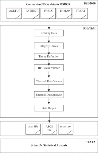
The steps of RHyThM are illustrated in the flowchart as presented in and are explained hereafter. RHyThM has been created in and operates under the MATLAB environment. Prior to the use of RHyThM, all data recorded by the BSD2000/3D system software has to be transferred from the PDOS environment to the MSDOS environment using a PDOS command.
Figure 1. Steps of the Rotterdam Hyperthermia Thermal Modulator (RHyThM).
First and second steps: Reading data and integrity check
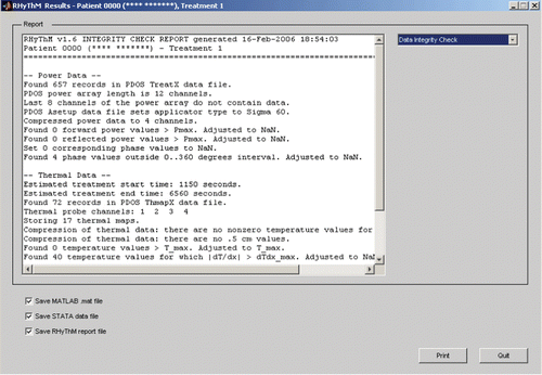
RHyThM reads all MSDOS formatted data files and checks the integrity of both the RF-power and thermal data. The integrity check values are defined in a settings-file. The results are shown in the Report-window (). For the RF-power integrity check RHyThM counts the number of records and verifies which amplifier system is used by checking the length of the power array. If the power array contains data for only four channels it is concluded that a 4-channel amplifier system (i.e. BSD2000) was used. Alternatively, if all 12 channels of the power array contain valid data it is concluded that the Dodek amplifier system (i.e. BSD2000-3D) is used. Secondly, it reads from the ‘ASETUP’ file which applicator type (Sigma 60 or Sigma Eye) is used. If the Sigma 60 is used with the 12-channel Dodek amplifier it automatically compresses the RF-power data to four channels. To rule out unrealistically high RF-powers all readings are verified against limited maximum forward and reflected power values. These maximum values are user-defined in the settings-file. Similarly, RHyThM checks whether all recorded phase values are within a 0–360° interval. From the power data history, RHyThM estimates the treatment start and treatment end time. Next, RHyThM allocates from the ‘ThmapX’-file which temperature measurement probes are used, how many thermal maps are performed and assesses the thermal map step size, i.e. 0.5 or 1.0 cm. As the temperature measurement with the Bowman probes is sensitive to sudden movements of the patients, RHyThM also checks (and counts) all temperature values on a gradient criterion in space and time, i.e. |dT/dx| should be smaller than dT/dxmax as default defined in the settings-file. If |dT/dx| values larger than dT/dxmax are found then these values are adjusted to not-a-number (NaN). Moreover, RHyThM Integrity Check logs which thermal analysis mode is used. If the user manually specifies a time horizon for the thermal analysis, the analysis horizon and the number of thermal maps within the selected time horizon are given. This selection is performed in the RF-power viewer.
Figure 2. Example of a BSD2000/3D treatment data import and integrity check by RHyThM.
Third step: Tissue definition
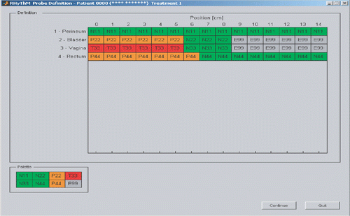
In this stage the temperature probe position for each thermometry catheter is translated into a tissue type such as normal tissue, tumour indicative or tumour contact based on the CT-scan information obtained in RT-position. If the temperature probe position is located outside the tissue it is labelled as an error (E99). In the settings-file default names are available to identify the location of the thermometry catheter, for instance bladder, vagina and rectum lumen. Based on the probe definition file, RHyThM comes up with the default tissue coding. The probe definition window (see ) allows the user to assign interactively a tissue definition to each position, using the identification code.
Figure 3. Probe definition window. Based on the CT-scan information all temperature probe positions are assigned the related tissue type. N11, N22, N33 and N44 are codes for normal tissue in perineum, bladder, vagina and rectum, respectively. P22 and P44 are codes for tumour indicative in bladder and rectum, respectively. T33 is a code for tumour contact in vagina. E99 shows the measurement is outside the tissue.
Fourth step: RF-power viewer
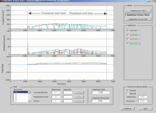
The BSD2000/3D system records forward and reflected power output and phase at 10 s intervals for all 12 RF-channels. In the RF-power viewer window () the operator can inspect the details of the total power or for individual applicator. In addition, the edit mode allows correction of erroneous power data. Three separate graphs show profiles of total forward power, total reflected power as well as phase vs. time. The effect of an on/off switch during the treatment is visible in the forward and reflected power graphs. The applicator panel indicates the number of each channel that is 1–4 for the Sigma 60 and 1–12 for the Sigma Eye. If the user identifies an irregularity in the graph such as spikes in forward and reflected power or phase measurements that is out of bound the user is able to zoom and edit the erroneous data point. Two more parameters, i.e. treatment start and end time as indicated on the profiles can be adjusted to represent the real treatment start and end time to provide a representative analysis. The user may manually specify a time horizon for the thermal analysis, which is of course also used for RF-power analyses.
Figure 4. RF-power viewer: total power view, applicator power view and applicator power edit panels of RHyThM. Forward power, reflected power and phase are shown in the upper, middle and lower graphs, respectively. The left lower panel enables the user to adjust erroneous RF-power data. The right lower panel enables the user to select a time horizon for thermal analysis.
Fifth and sixth steps: Thermal data viewer and thermal data analysis
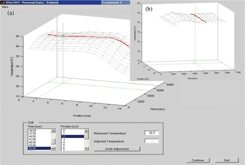
For thermal data verification RHyThM assembles a 3D interactive graph of temperature output vs. space and time (). The user can also switch to a 2D graph, which is convenient to check whether the insertion depth of the thermal mapping catheter remained constant during the whole treatment. By clicking on the 3D-graph the user can manipulate the view-angle of the graph in order to easily recognize erroneous temperature data points (). As it can be seen in the lower panel of , RHyThM provides an editor mode for individual evaluation of each data point by selecting time and position. RHyThM already automatically detects unrealistically high temperature gradients in time and space. In the edit mode the user can correct these temperature values. The thresholds for too high temporal and spatial temperature gradients are defined in the settings-file.
Figure 5. (a) Thermal data validation to check for probe movement and for unrealistically high temperature gradients in time and space. (b) Right upper legend, that is an inset, shows a sample of indefinite views of the graphical outputs (useful to recognize erroneous temperature data points). The lower panel enables the user to adjust erroneous temperature data.
Seventh step: Data output
At this time all available data is valid and is used for the thermal and power analysis. In the ‘default’ mode RHyThM calculates the following general temperature-related dose parameters:
Tmin, Tmean, T20, T50, T90 between start of the therapeutic heating period and end of the treatment.
Tmax between start of treatment and end of the treatment.
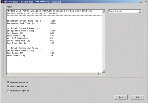
In the ‘manual’ mode the output depends on the time horizon selected by the user for the analysis. Next, RHyThM reports these temperature-related dose parameters per individual thermal map location including the tissue type. Additionally, a more integrated evaluation is obtained from the temperature data analyses per tissue type, i.e. normal, tumour indicative, tumour contact and all. For each main thermometry location, e.g. for HT in the pelvic region these are bladder, vagina and rectum, a separate report is printed and saved as a text-file. As an example, shows the results for a thermometry catheter in the vagina. Additionally, the thermal analysis includes overall tumour statistics, which are based on the measurements from all tumour points from all probes. They are listed on a separate page in the report. Furthermore, RHyThM performs an analysis on the recorded RF-power data of the whole treatment and provides several RF-power-related treatment evaluation parameters. For instance, maximum and average of forward and reflected power, as well as integrated power (kJ). Further, it records number of RF-power off-switches and calculates the total and maximum of switches off time (). In order to make the complete verified set of data available for future scientific thermal dose analysis, RHyThM stores the data as an original MATLAB data file and in ASCII format accessible for standard statistical packages. The ASCII format provides patient number, treatment number, time, location and verified temperature for each measured temperature point.
Figure 6. RHyThM Results: temperature data analysed reported for vagina. *T33 and N33 are two codes for tumour contact and normal tissue in vagina, respectively. E99 shows the measurement is outside the tissue and NaN means not-a-number.
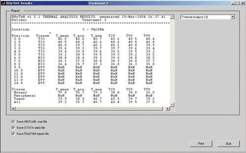
Figure 7. RF-Power analysed quantities reported by RHyThM.
Experiences when entering and analysing the treatment data
To evaluate the performance of RHyThM regarding the data integrity check a test run of RHyThM on a selected group of 22 patients with advanced cervical cancer was performed. The patients were treated by radiotherapy + HT + chemotherapy. Details respecting temperature data are presented in a submitted paper Citation[11]. Data transfer from PDOS to MSDOS was successful for 21 patients. For one patient the data of all four treatments was lost due to a reading failure of the floppy under PDOS. Additionally, it was not possible to transfer a single treatment for three other patients. Hence, of the 104 treatments performed 97 treatment records could be made available for analysis and seven treatment data records are inaccessible. Hereafter, the MSDOS data was imported in RHyThM to perform the data analysis.
For each single treatment, tissue type assignment was done according to the tissue map trajectory per thermometry catheter as provided by the HT-physician and for the insertion length as measured by the HT-technician. A steep change in the slope of the profile of the first temperature map was used to verify the precise location of the transition between in- and outside the body, as reported by the HT-technician. If necessary the location of the transition of the body was corrected to much better with the steep temperature gradient.
Overall RHyThM performed satisfactorily, was able to check integrity and validate the temperature data and performed the tissue type assignment and thermal analysis for all remaining 97 treatments. Two minor problems were experienced:
Defining tissue type and insertion length. Overall it was necessary to adapt the insertion length for 53 out of 291 thermal map trajectories (18%) or in 39 out of the 97 treatments (40%). Initially, a dislocation of the thermal probe could only be recognized in the fifth step of RHyThM, i.e. thermal data viewer. For more convenience an additional tool: ‘First Thermal Map’ was added. This part of the programme shows the first temperature map of a treatment next to the probe definition window and helps to identify the correct insertion length. Without the additional tool correction is only possible when the user quits RHyThM and starting all over again.
Detection of temperature gradients in space and time. The temperature profile is checked for too large gradients in space and time. In the current implementation a single jump or fall in the temperature causes two temperature points to be identified as NaN: the temperature point itself and the point following it. At first a sudden movement causes a steep gradient, which is correctly identified as an erroneous temperature value. However, as the movement is transient, the next measurement will be at a correct temperature, hence causing an inverse temperature gradient compared to the previous one. RHyThM, incorrectly, identifies this value as an error.
Discussion
In the authors’ opinion RHyThM is a valuable tool greatly enhancing the possibilities for analysis of temperature data obtained during HT-treatments with the BSD2000/3D system. The RHyThM programme enables easy access to the PDOS data files, performs an important integrity check of the data, connects the tissue assignment with the thermal data and includes the correction of unrealistic temperature values due to mechanical stress on the Bowman temperature probes. In comparison to earlier programmes (mostly based on eavesdropping the ASCII codes on the RS232 cable to the printer) it provides besides the thermal mapping temperature data also all other data stored by the BSD control programme. A striking feature is the availability of the forward and reflected RF-power values and the phase for each channel. Further, the current version of RHyThM provides a minimum of data analysis required to make a fast assessment of the quality of the HT-treatment delivered. In this situation this information is used as input for our weekly multi-disciplinary discussion reviewing the quality of the HT-treatments delivered as a preparation for the next treatment of each patient.
In an exploratory study Citation[11] with the intention to evaluate the performance of RHyThM, it was found that for seven out of 104 treatments, i.e. ∼7%, it was not possible to recover the data, due to a reading failure or a failure to transfer the data from PDOS to MSDOS. During the application of the HT-treatments (currently ≥3500) using the BSD2000/3D, one has regularly noticed that the data recording with the PDOS floppy disk unit is sensitive to RF-interference. Such an accident is indicated by the BSD system with the warning ‘disk error, data might be lost’. It is anticipated that, when one started analysing the data of treatments performed longer (years) ago the percentage of lost data files will grow as the reliability of the current BSD2000-3D software system has been improved significantly since 1990.
During the period of the development of RHyThM, the BSD Corporation has upgraded the PDOS control of the BSD2000-3D to a newly designed MS-Windows based platform. Major advantages of this upgrade are a better exchangeability of the hardware and, in the authors’ view even more important, a highly improved access to the treatment data. According to Turner Citation[12], the current BSD2000-3D PC-software solves most of the difficulties experienced with the older PDOS software. The files are now in comma separated value file format (.csv) allowing processing of the data for tissue assignment and mapping length validation. Integrity and quality data check are, as far as is known, not yet incorporated. Although this new PCDOS based platform provides an important improvement, the need for RHyThM remains as the majority of the deep HT centres are still using the BSD2000/3D systems, which are running under PDOS.
Independent of the software used to analyse the data (RHyThM or the new MS-Windows platform of the BSD2000 system) the correction of mechanical-based errors in the thermal mapping data, i.e. curling, sticking and slip of thermometry catheter, remains to be manual and specific for each occurrence. In the patient data analysis in the exploratory study Citation[11], one had to correct the insertion depth of the thermal catheters for 53 of 291 thermal map trajectories. Hence, in 82% the tissue assignment used in the data analysis was valid for the entire treatment duration and for the remaining 18% the tissue assignment used resembles only the tissue distribution valid at the end of the treatment.
As explained in the experiences the way that RHyThM checks on short and too steep temperature gradient in time or space has a high potential of assigning two instead of one temperature measurement as an invalid value. These limitations of RHyThM will be addressed in a future version.
The design of RHyThM does not provide functionality to calculate thermal dose expressed as equivalent minutes at 43°C. The reason for this choice is simple, RHyThM is considered only as a tool to prepare the thermal data for further analysis with more advanced statistical packages.
The availability of the full details of the RF-power applied to the patient provides the opportunity for new interesting analysis between RF-power, temperatures achieved and (indirect) pain complaints. Accurate information about the applied RF-power during the entire deep HT-treatment as provided by RHyThM might add relevant and welcome extra input in finding the proper answer to the posed problem.
Conclusion
RHyThM is a valuable tool enabling easy access to the temperature and power data recorded during deep HT-treatments performed with the BSD2000 system. RHyThM has been created in and operates under the MATLAB environment. A critical demand is the ability to transfer the PDOS formatted disks to MSDOS formatted disks. RHyThM performs an important integrity and validation check of the data, it connects the tissue assignment with the thermal data and it includes the correction of unrealistic temperature values. A unique feature is the availability of the forward and reflected RF-power values and the phase for each channel. RHyThM provides a minimum of data analysis required to make a fast assessment of the quality of the HT-treatment. Although the newest BSD2000 operating systems run under MS-Windows software, the availability of RHyThM is of interest for many HT-groups as the majority of the currently installed BSD2000 systems are still running under PDOS.
Acknowledgement
This work was supported by the Dutch Cancer Society, grant DDHK 2003-2884. The first author was financially supported by the Shahrekord University of Medical Sciences (related to the Iranian Ministry of Health, Treatment and Medical Education).
Related Research Data
References
- Shrivastava P, Luk K, Oleson J, Dewheirst M, Pajak T, Paliwal B, Perez C, Sapareto S, Saylor T, Steeves R. Hyperthermia quality assurance guidelines. Int J Radiat Oncol Biol Phys 1989; 16: 571–577
- Dewhirst M, Phillips TL, Samulski TV, Stauffer P, Shrivastava P, Paliwal B, Pajak T, Gillim M, Saponzink M, Myerson R, Waterman FM, Sapareto SA, Corry P, Cetas TC, Leeper DB, Fessenden P, Kaap D, Oleson JR, Emami B. RTOG quality assurance guidelines for clinical trials using hyperthermia. Int J Radiat Oncol Biol Phys 1990; 18: 1249–1259
- Oleson JR, Dewhirst MW, Harrelson JM, Leopold KA, Samulski TV, Tso CY. Tumor temperature distributions predict hyperthermia effect. Int J Radiat Oncol Biol Phys 1989; 16: 559–570
- Myerson RJ, Perez CA, Emami B, Straube W, Kuske RR, Leybovich L, Von Gerichten D. Tumor control in long-term survivors following superficial hyperthermia. Int J Radiat Oncol Biol Phys 1990; 18: 1123–1129
- Leopold KA, Dewheirst MW, Samulski TV, Harrelson JA, George S, Dodge R, Grant W, Clegg S, Prosnitz L, Oleson JR. Relationships among tumor temperature, treatment time and histopathological outcome using hyperthemia with radiation in soft tissue sarcomas. Int J Radiat Oncol Biol Phys 1992; 22: 989–998
- Kaap DS, Cox RS. Thermal treatments parameters are most predictive of outcome in patients with single tumor nodules per treatment field in recurrent adenocarcinoma of the breast. Int J Radiat Oncol Biol Phys 1995; 33: 887–899
- Sapareto SA, Corry PM. A proposed standard data file format for hyperthermia treatments. Int J Radiat Oncol Biol Phys 1989; 16: 613–627
- Straube WL, Myerson RJ, Moros EG. A multi-user networked database for analysis of clinical and temperature data from patients treated with simultaneous radiation and ultrasound hyperthermia. Int J Hyperthertmia 1999; 5: 419–426
- Turner PF, Tumeh A, Schaefermeyer T. BSD-2000 approach for deep local and regional hyperthermia: Physics and technology. Strahlenther Onkol 1989; 165: 738–741
- Dewhirst MW, Sneed PK. Those in gene therapy should pay closer attention to lessons from hyperthermia. Int J Radiat Oncol Biol Phys 2003; 57: 597–599
- Fatehi D, van der Zee J, van der Wal E, van Wieringen WN, van Rhoon GC. Temperature data analysis for 22 patients with advanced cervical carcinoma treated in Rotterdam using radiotherapy, hyperthermia and chemotherapy: A reference point is needed. Int J Hyperthermia 2006, Submitted
- Turner PF. 2004, Personal communication