
Abstract
Background: Isolated limb perfusion (ILP) is an established and effective treatment for advanced melanoma and soft tissue sarcomas of the extremities with a high overall response rate. The aim of this study was to describe our experience of ILP for more rare types of tumours.
Methods: Patients with Merkel cell carcinoma (MCC) (n = 4), squamous cell carcinoma (SCC) (n = 2), B-cell lymphoma (n = 1), desmoid tumours (n = 3), pigmented villonodular synovitis (PVNS) (n = 1) and giant cell tumour (n = 1) were treated with ILP and analysed retrospectively.
Results: The four patients with in-transit MCC had three complete responses (CR) and one partial response (PR); the two patients with SCC had one CR and one stable disease (SD); the patients with desmoid tumours had two PR and one SD. A CR was also observed for the patient with a giant cell tumour, but the patient with PVNS had a SD. The patient with cutaneous metastases of B-cell lymphoma showed a CR, however with rapid systemic progression. Local toxicity according to Wieberdink was grade II in 10 patients (83%) and grade III in two patients (17%).
Conclusions: These results show that ILP can be used as a treatment option also for more rare disease entities when other treatments have failed.
Introduction
Isolated limb perfusion (ILP) is an established and effective technique, introduced in 1958 by Creech and Krementz [Citation1], used for the treatment of advanced extremity tumours. The main indications for the procedure have been in-transit metastases of malignant melanoma or advanced soft tissue sarcomas (STS) [Citation2]. During ILP the extremity is surgically isolated and then perfused with chemotherapy under mild to moderate hyperthermia. The technique is based on the synergistic antineoplastic effect of high temperature and chemotherapeutic drugs. The most used agent is melphalan, which can also be combined with tumour necrosis factor-alpha (TNF-alpha), mainly for patients with bulky melanoma tumours or STS [Citation2].
For patients with melanoma in-transit metastases, ILP gives an overall response rate of approximately 90%, including a high proportion of patients with complete response (CR) [Citation3,Citation4]. For patients with STS the response rate is approximately 70% and results in the possibility of limb-sparing surgery in two thirds of the patients [Citation5–7].
ILP can also be used for other more rare locally advanced tumours confined to the extremities, such as squamous cell carcinomas (SCC) [Citation8], lymphomas [Citation9–11], desmoid tumours [Citation12] and Merkel cell tumours (MCC) [Citation13]. The aim of this study is to describe our experience of ILP for these more rare types of tumours, and also include results from the, to our knowledge, first two patients treated with pigmented villonodular synovitis (PVNS) and giant cell tumours.
Patients and methods
Patients
Between 1992 and 2015 a total of 12 patients with various locally advanced tumours were treated with ILP. The indications were Merkel cell carcinoma (n = 4), SCC (n = 2), B-cell lymphoma (n = 1), desmoid tumours (n = 3), PVNS (n = 1) and giant cell tumour (n = 1). The study was approved by the Regional Ethical Review Board at the University of Gothenburg and was in compliance with the 1975 Helsinki Declaration.
Evaluation
Clinical response was recorded prospectively at 3 months according to the World Health Organization criteria, and was evaluated clinically for superficial tumours and by MRI for deep-seated tumours [Citation14]. A complete response (CR) is defined as the disappearance of all lesions, partial response (PR) as a reduction of more than 50% of the total tumour burden, a progressive disease (PD) as an increase of more than 25% of the tumour burden or the appearance of new tumours, stable disease (SD) as a situation in which there is neither sufficient shrinkage to qualify for PR nor sufficient increase to qualify for PD. Local progression was defined as the appearance of new lesions or a progression of existing lesions within the treated limb, not including lymph node metastases. Data were collected retrospectively from medical records.
Treatment
The ILP technique has previously been described in detail [Citation15,Citation16]. ILP was carried out through either axillary, brachial, external iliac or femoral approach, with dissection and cannulation of the respective artery and vein. An inflatable tourniquet (Zimmer disposable tourniquet, Warsaw, Poland) was used in femoral perfusions, and an Esmarch bandage positioned around a Steinmann pin was used in iliac and upper limb perfusions.
Toxicity
Local toxicity was measured prospectively, and graded as the worst reaction using the Wieberdink grading system that categorises tissue reaction as grade I (no reaction), grade II (erythema or swelling), grade III (major erythema, swelling or blistering), grade IV (extended epidermolysis and/or damage to deep tissues, causing final functional disorders, risk or manifest compartment syndrome) and grade V (reaction that may necessitate amputation) [Citation17].
Results
The response rates varied according to tumour type. All MCC (n = 4) responded, with three patients having a CR (75%) and one patient a PR (25%). No patient with MCC progressed locally during the follow-up, but one patient had a recurrence outside of the perfused area after 12 months. Two patients died, the first patient was fully recuperated after the procedure, but was admitted to hospital with acute respiratory failure after 2 months. Computed tomography (CT) showed pulmonary metastases together with a pulmonary embolism. The second patient died after 70 months due to a cerebrovascular insult without any signs of disease recurrence.
The two patients with SCC responded with a SD and a CR. The patient with SD had a local recurrence after 7 months and was finally amputated 1 year later. There was one systemic recurrence 6 months later and the patient died due to metastatic disease after a total of 27 months. The patient with a CR relapsed after 4 months and continued to slowly progress locally and died after 18 months due to advanced age, but without evidence of systemic metastases.
For the three patients with desmoid tumours, one patient still has a long-standing SD after 20 months and no signs of progression. The second patient had a PR, underwent resection of the remaining tumour after 3 months, and is still alive at 63 months without any signs of disease. The third patient had a PR and developed locally progressive disease after 14 months and has since then been treated with multiple resections, and is currently alive after 145 months without evidence of local recurrence.
The treatment of a patient with an advanced cutaneous B-cell lymphoma resulted in a CR within 3 weeks. However, the patient rapidly developed a systemic progressive disease and died without evidence of local recurrence after 3 months.
A patient with PVNS in the right knee had an initial response, however not enough to be considered a PR. He subsequently developed progressive symptoms and after 4 months underwent surgery with a modular universal tumour and revision system (MUTARS®) prosthesis. The patient with a giant cell tumour had an initial CR at 3 months, but relapsed locally after 7 months leading to resection surgery with the placement of a MUTARS prosthesis. Both patients are currently alive with no evidence of disease.
In total, 82% of the patients had a mild local toxicity, grade I–II according to Wieberdink, and two patients (18%) had a grade III reaction. Both patients with the grade III reaction developed a transient peroneus paresis; however, without long-lasting deficits. There was one post-operative wound infection and two patients also developed seromas. No other complications were reported.
Discussion
ILP is an established treatment for in-transit metastases of malignant melanoma as well as for the treatment of advanced STS. However, other more rare diseases might also benefit from treatment with ILP. MCC is a rare form of a neuroendocrine tumour with a high loco-regional recurrence rate. The treatment mainly consists of a wide local excision, sentinel lymph node biopsy and adjuvant radiotherapy [Citation18]. However, when numerous local metastases are present, there are few therapeutic options. Previously a summary on the outcome of 12 patients treated with ILP or isolated limb infusion (ILI) have been published. Eleven patients had a CR, and one patient had a PR. Four patients had a regional relapse after a median time of 22 months [Citation13]. We can now confirm these good results with all of our four treated patients responding to ILP.
Cutaneous squamous cell carcinoma (SCC) is an epithelial skin tumour with a local recurrence rate of up to 7%. The therapeutic options are limited in cases where surgery and radiotherapy have failed, with systemic therapies having a limited success [Citation19]. There is one previous report of ILP for SSC (n = 12), the overall histopathological response rate was 92%, and 25% developed local recurrence after a median time of 5 months [Citation8]. In our two patients with SCC, one patient had a SD and the other a CR; however, both experienced local progressive disease within 4–7 months.
Desmoid tumours are a rare benign soft tissue disease without metastatic behaviour. However, they are often characterised by local aggressiveness and it usually infiltrates the surrounding tissues causing severe disability [Citation12]. A previous report summarising the results of 25 patients from three different centres showed an overall response rate of 72%; however, only two patients with a CR. The functional outcome was good, with 16 patients not having any physical limitations, and amputation was prevented in all but three patients [Citation20]. One of our three patients had a long-lasting SD without any other treatment. The other two patients were treated with ILP as a neoadjuvant approach to enable surgery, they both responded with a PR and underwent resection 3 months later. Unfortunately, one patient relapsed after a little more than 1 year, but the other patient is still alive after 63 months without any signs of relapse.
Figure 1. These images show a Merkel cell carcinoma before and 6 weeks after isolated limb perfusion. Almost all tumours have disappeared and the last necrotic tumour fell off; at 3 months there was no evidence of disease left. Image reproduced with permission from the patient.
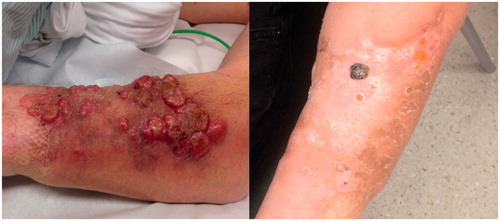
Table 1. A summary of 12 patients treated with isolated limb perfusion.
PVNS is a rare proliferative disease that includes the synovial joints, tendon sheaths and bursa membranes. It is usually mono articular and mostly linked to the knee joint. Giant cell tumour is also a rare proliferative disease, often considered the soft tissue counterpart of PVNS, with lesions most commonly found around the knee. These tumours mainly occur in younger patients and the tumour is often locally aggressive causing progressive damage in bone and joint structures. The treatment is based on the removal of all abnormal synovial tissue to stop discomfort, reduce risks of joint destruction, and prevent local relapse [Citation21]. In the two cases reported, both patients had undergone several treatment regimens before, and ILP was considered a last resort before more extensive surgery. The patient with a giant cell tumour responded with a long-lasting CR, and still no progression after more than 9 years. In the patient with PVNS, only a moderate response was seen, and the patient then underwent resection of the joint with a MUTARS prosthesis. To our knowledge, this is the first report presenting outcomes of ILP for these rare diseases.
Lymphomas can also present with cutaneous manifestations, and the diffuse large B-cell lymphoma is an aggressive cutaneous B-cell lymphoma accounting for approximately 6% of all cutaneous lymphomas. It tends to spread to lymph nodes and extracutaneous sites and is associated with a relatively poor prognosis with a 5-year survival of approximately 50% [Citation22]. ILP or ILI has demonstrated an overall response rate of 100% for both T- and B-cell lymphomas in four different previous case reports [Citation9–11,Citation23]. Together with our case, these five patients with therapy refractory disease experienced an impressive clinical response after regional treatment. This warrants further research into the treatment of cutaneous lymphomas, where ILP might be a treatment that should be considered in earlier stages of the disease.
The toxicity profile after ILP has been well described previously [Citation3,Citation24], and the local toxicity and complications after ILP in this series do not seem to be different.
Taken together, ILP is an established treatment for melanoma and STS. However, ILP can also be effective for other disease entities, especially SCC, desmoid tumours, cutaneous lymphomas and Merkel cell tumours. We here also report, to our knowledge, the first cases of ILP treatment for PVNS and giant cell tumours. Further structured and joint research efforts into the use of ILP for these more rare tumours are warranted in the future.
Disclosure statement
The authors declare no conflict of interest. The authors alone are responsible for the content and writing of the paper.
Related Research Data
References
- Creech O, Jr, Krementz ET, Ryan RF, Winblad JN. Chemotherapy of cancer: regional perfusion utilizing an extracorporeal circuit. Ann Surg 1958;148:616–32.
- Bhangu A, Broom L, Nepogodiev D, Gourevitch D, Desai A. Outcomes of isolated limb perfusion in the treatment of extremity soft tissue sarcoma: a systematic review. Eur J Surg Oncol 2013;39:311–19.
- Moreno-Ramirez D, de la Cruz-Merino L, Ferrandiz L, Villegas-Portero R, Nieto-Garcia A. Isolated limb perfusion for malignant melanoma: systematic review on effectiveness and safety. Oncologist 2010;15:416–27.
- Olofsson R, Mattsson J, Lindner P. Long-term follow-up of 163 consecutive patients treated with isolated limb perfusion for in-transit metastases of malignant melanoma. Int J Hyperthermia 2013;29:551–7.
- Grunhagen DJ, de Wilt JH, van Geel AN, Verhoef C, Eggermont AM. Isolated limb perfusion with TNF-alpha and melphalan in locally advanced soft tissue sarcomas of the extremities. Recent Results Cancer Res 2009;179:257–70.
- Olofsson R, Bergh P, Berlin O, Engstrom K, Gunterberg B, Hansson M, et al. Long-term outcome of isolated limb perfusion in advanced soft tissue sarcoma of the extremity. Ann Surg Oncol 2012;19:1800–7.
- Deroose JP, Grunhagen DJ, de Wilt JH, Eggermont AM, Verhoef C. Treatment modifications in tumour necrosis factor-alpha (TNF)-based isolated limb perfusion in patients with advanced extremity soft tissue sarcomas. Eur J Cancer 2015;51:367–73.
- Olieman AF, Lienard D, Eggermont AM, Kroon BB, Lejeune FJ, Hoekstra HJ, et al. Hyperthermic isolated limb perfusion with tumor necrosis factor alpha, interferon gamma, and melphalan for locally advanced nonmelanoma skin tumors of the extremities: a multicenter study. Arch Surg 1999;134:303–7.
- Paramo JC, Benavides C, Tang LW, Martinez A, Cabello-Inchausti B, Davila E, et al. Complete remission of previously intractable peripheral cutaneous T-cell lymphoma of the lower extremity using isolated hyperthermic limb perfusion with melphalan (1-phenylalanine mustard). Cancer Invest 2004;22:545–9.
- Kobold S, Killic N, Lutkens T, Bokemeyer C, Fiedler W. Isolated limb perfusion with melphalan for the treatment of intractable primary cutaneous diffuse large B-cell lymphoma leg type. Acta Haematol 2010;123:179–81.
- Jansen RF, van Geel BN, van der Zee J, Hagenbeek A, Levendag PC. Intractible cutaneous non-Hodgkin's lymphoma of the lower limb. Complete remission after sequential regional isolated hyperthermic perfusion and perfusion with 1-phenylalanine-mustard (melphalan, L-Pam). Cancer 1989;64:392–5.
- Grunhagen DJ, de Wilt JH, Verhoef C, van Geel AN, Eggermont AM. TNF-based isolated limb perfusion in unresectable extremity desmoid tumours. Eur J Surg Oncol 2005;31:912–16.
- Zeitouni NC, Giordano CN, Kane JM III. In-transit Merkel cell carcinoma treated with isolated limb perfusion or isolated limb infusion: a case series of 12 patients. Dermatol Surg 2011;37:357–64.
- World Health Organization. WHO handbook for reporting results of cancer treatment. Geneva: World Health Organization, 1979.
- Olieman AF, Pras E, van Ginkel RJ, Molenaar WM, Schraffordt Koops H, Hoekstra HJ. Feasibility and efficacy of external beam radiotherapy after hyperthermic isolated limb perfusion with TNF-alpha and melphalan for limb-saving treatment in locally advanced extremity soft-tissue sarcoma. Int J Radiat Oncol Biol Phys 1998;40:807–14.
- Schraffordt Koops H, Oldhoff J, Oosterhuis JW, Beekhuis H. Isolated regional perfusion in malignant melanoma of the extremities. World J Surg 1987;11:527–33.
- Wieberdink J, Benckhuysen C, Braat RP, van Slooten EA, Olthuis GA. Dosimetry in isolation perfusion of the limbs by assessment of perfused tissue volume and grading of toxic tissue reactions. Eur J Cancer Clin Oncol 1982;18:905–10.
- Hughes MP, Hardee ME, Cornelius LA, Hutchins LF, Becker JC, Gao L. Merkel cell carcinoma: epidemiology, target, and therapy. Curr Dermatol Rep 2014;3:46–53.
- Lansbury L, Bath-Hextall F, Perkins W, Stanton W, Leonardi-Bee J. Interventions for non-metastatic squamous cell carcinoma of the skin: systematic review and pooled analysis of observational studies. BMJ 2013;347:f6153.
- van Broekhoven DL, Deroose JP, Bonvalot S, Gronchi A, Grunhagen DJ, Eggermont AM, et al. Isolated limb perfusion using tumour necrosis factor alpha and melphalan in patients with advanced aggressive fibromatosis. Br J Surg 2014;101:1674–80.
- Di Grazia S, Succi G, Fragetta F, Perrotta RE. Giant cell tumor of tendon sheath: study of 64 cases and review of literature. G Chir 2013;34:149–52.
- Vermeer MH, Willemze R. Recent advances in primary cutaneous B-cell lymphomas. Curr Opin Oncol 2014;26:230–6.
- Elhassadi E, Egan E, O'Sullivan G, Mohamed R. Isolated limb infusion with cytotoxic agent for treatment of localized refractory cutaneous T-cell lymphoma. Clin Lab Haematol 2006;28:279–81.
- Vrouenraets BC, Eggermont AM, Hart AA, Klaase JM, van Geel AN, Nieweg OE, et al. Regional toxicity after isolated limb perfusion with melphalan and tumour necrosis factor-alpha versus toxicity after melphalan alone. Eur J Surg Oncol 2001;27:390–5.