Abstract
Objective
Non-communicating extradural spinal arachnoid cysts (NEACs) are extremely rare aetiology of symptomatic spinal cord compression. The aim of this study was to address their pathogenesis, optimum management strategy and outcome through systematic review of existing published studies.
Materials and method
We have found 13 eligible publications by searching through PubMed, ScienceDirect, and Google Scholar databases, published from inception to December 2020. We have analysed the data of 21 patients extracted from those 13 publications by IBM SPSS version 23.
Results
According to our analysis congenital predisposition, trauma, and previous surgery history are the aetiology of NEAC. Clinical presentation of cyst depends upon the location and extent of compression or involvement of the neurovascular structures. Paraparesis with variable degree of sensory disturbance was seen among patients. Based on neuroimaging findings, NEACs are most commonly found at dorsal and dorsolumbar region. Magnetic resonance imaging (MRI) is the diagnostic modalities of choice and CT myelography can demonstrate the communication with the subarachnoid space. Recurrence rate of cyst after surgery is very low as only one out of twenty patients showed recurrence. If dural defect is not accurately addressed, the recurrence rate increased significantly.
Conclusions
Our study has highlighted aetiology, treatment strategies, and neurological outcome of NEAC. These findings may help neurosurgeons to manage this rare surgical entity for favourable outcome.
Disclosure statement
No potential conflict of interest was reported by the author(s).
Figure 5. Per operative photograph demonstrates an extradural, large lobulated cystic lesion after laminectomy of D11-L2 (A) and complete excision of the cyst (B).
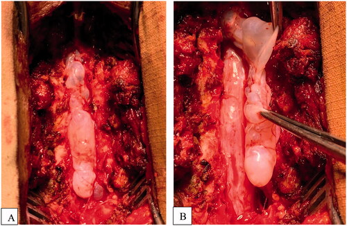
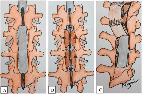
Figure 6. Schematic picture demonstrates surgical treatment modalities for non-communicating extradural arachnoid cyst; laminectomy (A), Recapping T saw laminoplasty (B), vascularized pedicled laminoplasty (C).