
Abstract
ABSTRACT—The fossil record of odontocetes (toothed cetaceans) is relatively scarce during the Oligocene and early Miocene compared with later in the Miocene and Pliocene; most of the odontocete families from these epochs are known by a limited number of species and specimens. Among those, Squalodelphinidae is a family of small- to medium-sized platanistoids with single-rooted teeth, which until now has included only four genera based on diagnostic material, from the early Miocene of Europe, Argentina, and North America. Recent field work in the Pisco-Ica desert, southern coast of Peru, has resulted in the discovery of several marine vertebrate-rich localities in various levels of the late Oligocene–early Miocene Chilcatay Formation. Based on three specimens from Ullujaya and Zamaca, including two well-preserved skulls with periotics, we describe a new squalodelphinid genus and species, Huaridelphis raimondii. This new species increases the early Miocene diversity of the family and is also its smallest known member. It further differs from other squalodelphinids by its thin antorbital process of the frontal, abruptly tapering rostrum, and higher tooth count. A more fragmentary skull, from Zamaca, is referred to Squalodelphinidae aff. H. raimondii. This skull provides information on the morphology of the tympanic, malleus, and incus, currently unknown in H. raimondii. Focusing on platanistoids with single-rooted teeth, our phylogenetic analysis suggests that Squalodelphinidae are monophyletic and confirms the sister-group relationship between the latter and Platanistidae. The relationships within Squalodelphinidae are not fully resolved, but H. raimondii might be one of the earliest diverging taxa.
INTRODUCTION
This paper represents the first of a series of contributions on the fossil odontocetes discovered by us during the last few years in the upper Oligocene–lower Miocene Chilcatay Formation (Pisco-Ica Basin, Peru; DeVries, Citation1998, 2001) (). Preliminary observations, both in the field and on the collected material under preparation, indicate a high diversity documented by well-preserved fossils. Some levels of the Chilcatay Formation display an exceptional wealth of fossil marine vertebrates, similar to that in the overlying younger Pisco Formation (e.g., Muizon, Citation1984, 1988a, 1993; Lambert et al., Citation2008a, 2009, 2013; Bianucci et al., Citation2010; Lambert et al., Citation2010a, 2010b; Lambert and Muizon, 2013). The time interval covered by the Chilcatay Formation spans an important phase of the odontocete radiation, with the appearance of several major extinct and extant clades, but this phase is poorly documented in the fossil record (Fordyce and Muizon, 2001; Uhen and Pyenson, Citation2007; Uhen, Citation2008). Consequently, the description of this new material could clarify some crucial aspects of odontocete evolutionary history. In this paper, we describe a new genus and species of Squalodelphinidae, based on well-preserved specimens (including two almost complete skulls) from the early Miocene levels of the Chilcatay Formation outcropping in the new localities of Ullujaya and Zamaca.
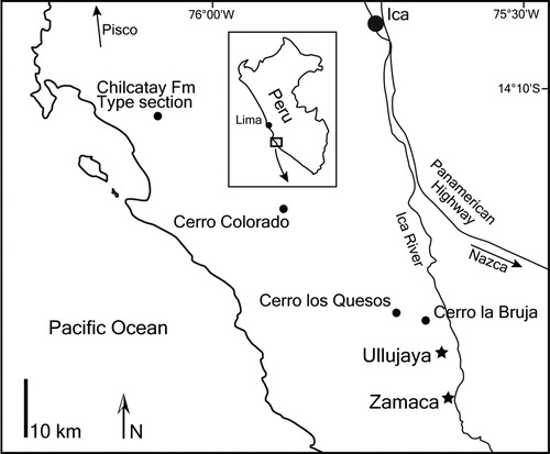
FIGURE 1. Map of the Pisco-Ica desert, southern coast of Peru, showing the two localities of the Chilcatay Formation where squalodelphinids, including Huaridelphis raimondii, n. gen. et sp., were found: Ullujaya and Zamaca. Marine vertebrate-rich localities of the Mio-Pliocene Pisco Formation are also indicated: Cerro Colorado, Cerro los Quesos, and Cerro la Bruja.
Squalodelphinidae are an extinct Miocene family of marine odontocetes characterized by a moderately elongated and tapered rostrum, posterior cheek teeth being single-rooted but retaining accessory denticles, and marked skull asymmetry. Based on some peculiar features, mostly of the ear bones and scapula, squalodelphinids are considered closely related to the family Platanistidae, inside the superfamily Platanistoidea sensu Muizon, Citation1987, although the relationships among the squalodelphinids and between the squalodelphinids and the other platanistoids are still debated (see below for discussion; Muizon, Citation1987; Fordyce, Citation1994; Barnes, Citation2006; Barnes and Reynolds, Citation2009; Geisler et al., Citation2011, 2012). In any case, the closest extant relative of the squalodelphinids is the south Asian river dolphin Platanista gangetica, representing the only surviving platanistoid (Bianucci et al., Citation2013). The fossil record of squalodelphinids is rather limited. Besides a skull associated with a mandible and other more fragmentary specimens referred to the type genus and species Squalodelphis fabianii Dal Piaz, Citation1917 (early Miocene, northern Italy) (Dal Piaz, Citation1917; Pilleri, Citation1985), the most significant fossils assigned to this family are (1) three specimens from the early Miocene of southern Argentina referred to Notocetus vanbenedeni Moreno, Citation1892: one skull with mandibles, another skull with a tympanic bulla, mandibles, and a few postcranial bones, and fragments of skull with ear bones, teeth, scapula, and a few other postcranial elements (Moreno, Citation1892; True, Citation1910; Muizon, Citation1987); and (2) one fragmentary skull referred to Medocinia tetragorhina Delfortrie, Citation1875, from the early Miocene of southern France (Muizon, Citation1988b). More fragmentary remains referred in the past to this family and other significant specimens tentatively assigned to this family are discussed in detail below.
Besides the N. vanbenedeni specimens from Argentina, to our knowledge the only other South American fossil odontocete remains tentatively referred to squalodelphinids are a scapula (Sánchez-Villagra et al., Citation2001) and poorly diagnostic skulls fragments, with a few vertebrae and ribs (Cozzuol and Aguilera, Citation2008) from the early Miocene of Venezuela. Hence, the new squalodelphinid from Peru reported here represents an important addition to our knowledge of this extinct family. The cladistic analysis investigating the phylogenetic affinities of this new genus and species gives us the opportunity to propose a new phylogeny for the squalodelphinids and related platanistoids with single-rooted teeth.
MATERIALS
Institutional Abbreviations—AMNH, American Museum of Natural History, New York, U.S.A.; BDNLTM, Bünde Doberg und Tabak Museum, Bünde, Germany; IRSNB, Institut royal des Sciences naturelles de Belgique, Brussels, Belgium; MGP, Museo di Geologia e Paleontologia, Padua, Italy; MNHL, Muséum d’Histoire naturelle de Lyon, Lyon, France; MNHN, Muséum national d’Histoire naturelle, Paris, France; MUSM, Museo de Historia Natural, Universidad Nacional Mayor de San Marco, Lima, Peru; MLP, Museo de La Plata, La Plata, Argentina; RMNH, Naturalis, Leiden, The Netherlands; USNM, United States National Museum of Natural History, Smithsonian Institution, Washington, D.C., U.S.A.
Anatomical Measurement Abbreviations—BZW, Bizygomatic width of skull; CBL, condylobasal length of skull.
Anatomical Terminology—We mostly follow Muizon (Citation1987), Fordyce (Citation1994), and Mead and Fordyce (Citation2009) for the terminology of skull elements and Rommel (Citation1990) for vertebrae.
List of Specimens Directly Examined for This Work—Albertocetus meffordorum USNM 525001; Eosqualodon langewieschei BDNLTM 326; Eurhinodelphis cocheteuxi IRSNB M.294, 295, 296, 297, 299, 1856, 1857; E. longirostris IRSNB M.342, 1858; Huaridelphis raimondii, n. gen. et sp., MUSM 1396, 1403, 599; Medocinia tetragorhina (cast MNHN); Notocetus vanbenedeni MLP 5-5; Phocageneus venustus USNM 21039; Platanista gangetica IRSNB 1507; MNHN A7943; RMNH 31169, USNM 172409, 23456, Pomatodelphininae USNM 206006; Pomatodelphininae cf. Zarhachis USNM 214759; Pomatodelphis bobengi USNM 299695; Pomatodelphis sp. USNM 187414; Prosqualodon australis MLP 5-8; Prosqualodon davidis (cast USNM 467596); Simocetus rayi USNM 256517; Squalodelphinidae USNM 475596; Squalodelphinidae aff. Huaridelphis raimondii MUSM 603; Squalodelphis fabianii MGP 26134, 26141, 26378; Squalodon bariensis MGP 26081, MNHL Dr15; Squalodon bellunensis MGP 17715, 26091, 26131, 26322; Squalodon calvertensis USNM 10484, 328343; Squalodon whitmorei USNM 183023; Waipatia maerewhenua cast USNM; Xiphiacetus bossi IRSNB M.367, USNM 8842, 10464, 10711, 10714, 16581; X. cristatus IRSNB M.361, 1893, 1894, 1895, 1896, USNM 13436, 21303, 21360, 21363; Zarhinocetus errabundus cast USNM; Zarhachis flagellator USNM 10911, 10485; Ziphiodelphis abeli MGP 26187; Z. sigmoideus MGP 26395; Zygorhiza kochii USNM 4679, 11962.
SYSTEMATIC PALEONTOLOGY
Order CETACEA Brisson, Citation1762
Suborder ODONTOCETI Flower, Citation1867
Superfamily PLATANISTOIDEA Gray, Citation1863
Remarks on the Definition of the Platanistoidea— Since the proposal of Muizon (Citation1987) to include the families Platanistidae, Squalodelphinidae, and Squalodontidae in the superfamily Platanistoidea, the content of the latter has been controversial. In a subsequent work, based respectively on the morphology of the scapula (loss of coracoid process and acromion on the anterior edge of the bone) and the morphology of the periotic, Muizon (Citation1991) included Prosqualodon and Dalpiazina in Platanistoidea. Waipatia, for which no scapula is known, entered the superfamily in the first software-assisted phylogenetic analysis of fossil and extant odontocetes (Fordyce, Citation1994). With a larger morphological matrix, Geisler and Sanders (Citation2003) excluded Notocetus, Prosqualodon, and Squalodon from the superfamily, and included other river dolphin lineages (iniids, lipotids, and pontoporiids) as well as Eurhinodelphis. It should be noted that the diagnostic platanistoid characters of the scapula have not yet been observed in any member of a river dolphin lineage or in a eurhinodelphinid (Muizon, Citation1991, 1994; pers. observ.). In two steps, Barnes (Citation2006) and Barnes and Reynolds (Citation2009) placed the allodelphinids Allodelphis and Zarhinocetus in a superfamily incorporating Platanistidae, Squalodelphinidae, Squalodontidae, and Waipatiidae. Recently, with a supermatrix containing both molecular and morphological data, Geisler et al. (Citation2011) did not find support for a monophyletic superfamily Platanistoidea. The definition of Platanistoidea and the relationships between included families are beyond the scope of this work; they will be investigated in detail in a future study. For that reason, we choose to provisionally maintain in the superfamily the less debated families, namely, Allodelphinidae, Platanistidae, and Squalodelphinidae, with a question mark for Squalodontidae and Waipatiidae. And because our group of interest is squalodelphinids, we will mostly comment on presumed members of the superfamily having lost double-rooted teeth.
Among platanistoids, platanistids, squalodelphinids, and at least some allodelphinids (Zarhinocetus) share a cranium distinctly shorter than wide, with a ratio between cranium length (longitudinal, from occipital condyles to level of antorbital notches) and postorbital width <0.90. These taxa also share deep fossa in orbit roof (possibly present in Zarhinocetus, unknown in Allodelphis), which seemingly connects the pterygoid sinus fossa to a crest on the supraorbital process of platanistids; elevated antorbital region, distinctly higher than the dorsal margin of rostrum base in lateral view (obscured in Zarhinocetus by the thick lateral margin of the rostrum at the antorbital notch; unknown in Allodelphis); vertex distinctly shifted to the left compared with the longitudinal midline of the skull (present in Zarhinocetus; difficult to detect in Allodelphis); and loss of double-rooted posterior teeth (present in Zarhinocetus; unknown in Allodelphis).
Furthermore, platanistids and squalodelphinids share marked asymmetry of premaxillae on the rostrum, at some distance anterior to premaxillary foramina, with the right premaxilla narrower than the left in dorsal view (the opposite occurs in some squalodontids, e.g., Squalodon bariensis and S. bellunensis); posterior infraorbital foramen(ina) along the vertex more medial than the lateral-most margin of the premaxilla in the cranium; thick zygomatic process of the squamosal, with the ratio between maximum distance from the anteroventral margin of the zygomatic process to the posterodorsal margin, in lateral view, and vertical distance from the lower margin of the occipital condyles to the cranial vertex greater than 0.38; development of an articular rim on the lateral surface of the periotic (hook-like in platanistids); and elongated anterior spine on the tympanic bulla, which is associated with a marked anterolateral convexity. Finally, most platanistids and squalodelphinids share a deep longitudinal groove on the posterior apex of the premaxilla (possibly homologous to the premaxillary cleft in Waipatia).
Family SQUALODELPHINIDAE Dal Piaz, Citation1917
Emended Diagnosis—Squalodelphinidae differ from Allodelphinidae and Platanistidae except Platanista by having a proportionally shorter rostrum (rostrum length/condylobasal length <0.70); Allodelphinidae and Platanistidae by lacking a deep lateral groove on the rostrum and a deeper, ‘V’-shaped, left antorbital notch, related to anteriorly pointed antorbital process (right notch usually shallower). Squalodelphinidae differ from Platanistidae by having an antorbital-supraorbital region lacking a distinct crest; a square-shaped pars cochlearis; a large, dorsally facing, aperture for the cochlear aqueduct of the periotic; median furrow of the tympanic affecting the whole length of bone, including the anterior spine (except in Squalodelphinidae aff. Huaridelphis raimondii MUSM 603); apical extension of the manubrium of the malleus (except in Squalodelphinidae aff. Huaridelphis raimondii MUSM 603); retention of ornamentation on posterior teeth; and strong development of the dorsal transverse process of the atlas and extreme reduction of its ventral process.
Remarks on Previous Attributions to Squalodelphinidae— The holotype of the only species of the squalodelphinid genus Phocageneus, P. venustus, is an isolated single-rooted tooth from Virginia (Leidy, Citation1869). Later, a partial skeleton from the Calvert Formation, Maryland, USNM 21039, including a part of the mandible, the ear bones, teeth, vertebrae, and ribs, was referred to the species, based on the similar tooth morphology (Kellogg, Citation1957). In addition, a fragmentary skull from the Miocene of the Lee Creek Mine, USNM 475596, with associated tooth and fragment of periotic was tentatively attributed to the genus Phocageneus together with other more fragmentary specimens, including five isolated periotics (Whitmore and Kaltenbach, Citation2008). The ear bones of USNM 21039 and the skull of USNM 475596 clearly bear resemblances with other squalodelphinid taxa, and we agree with their family attribution. Even if the correlation with the holotype of P. venustus is only based on the poorly diagnostic tooth morphology, the specimen USNM 21039 was long considered as belonging to the species, being more often used in comparisons and discussions than the holotype, especially for the ear bones. Therefore, we follow a conservative approach, considering the attribution of the specimen USNM 21039 to the species P. venustus as valid. The other specimen, USNM 475596, will be mentioned below as squalodelphinid USNM 475596, pending a detailed review of its relationships with other squalodelphinids and/or the discovery of more complete specimens. It should be noted that among squalodelphinids, USNM 475596 was only compared with Notocetus and Squalodelphis, not with Medocinia (Whitmore and Kaltenbach, Citation2008). The latter shares some interesting similarities with the Maryland skull: the frontal much anteroventrally projected on the antorbital process, with a consequently limited overlap of the frontal by the maxilla in this area; and the general outline of the swollen zygomatic process of the squamosal.
The holotype of ‘Prosqualodon’ marplesi, an incomplete skull from the early Miocene of New Zealand with associated ear bones, teeth, parts of the mandible, vertebrae, ribs, one scapula, and one ulna (Dickson, Citation1964), was later referred to the genus Notocetus, based on the morphology of the vertex, the nuchal crest, the supraoccipital, the atlas, and the periotic (Fordyce, Citation1994). Very schematically illustrated in both papers, the skull displays some differences with Notocetus vanbenedeni. The posterior apex of the premaxilla is wider in ‘P.’ marplesi; the medial sutures on the vertex are not shifted to the left side; the frontal is considerably thinner in the proportionally longer and lower orbital region; and the zygomatic process of the squamosal is much less dorsoventrally thickened, with a more distinctly concave anteroventral margin. In addition, the ventral transverse process of the atlas of ‘P.’ marplesi is more developed than in Notocetus vanbenedeni AMNH 9485. The cervical vertebra figured by Dickson (Citation1964:) shares similarities with the sixth cervical of the Phocageneus venustus USNM 21039 (True, Citation1910:pl. 7, ), but with a larger transverse foramen. The scapula, bearing platanistoid characters, does not display major differences with the scapula of AMNH 29060, referred to Notocetus vanbenedeni by Muizon (Citation1987:fig. 13c). The periotic (Fordyce, Citation1994:fig. 10) displays similarities with squalodelphinids. Most of the differences at the level of the skull can also be applied to other squalodelphinids. Pending direct observation of the specimen or the publication of more detailed illustrations of the skull, we question its attribution to the genus Notocetus.
FIGURE 2. Map of the Chilcatay Formation locality Ullujaya, where the holotype of Huaridelphis raimondii, n. gen. et sp., was found together with other cetacean remains, marine turtles, and fishes. Cerro Buque, from where a section of the Chilcatay Formation was published (Alván De la Cruz, 2008), is a short distance northwest to Ullujaya. Curved lines represent limits of more consolidated beds visible from aerial views.
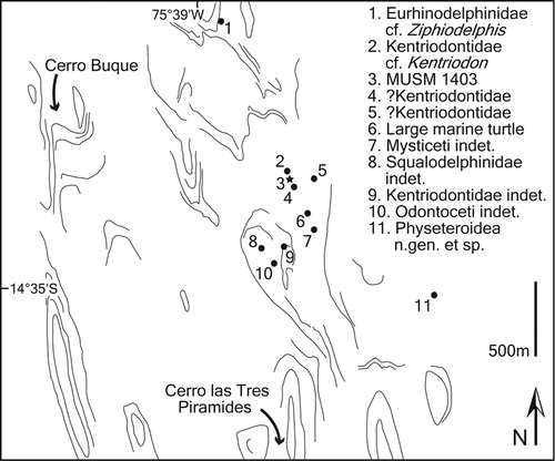
FIGURE 3. Huaridelphis raimondii, n. gen. et sp., MUSM 1396 (holotype), skull in dorsal (A, B, C; detail of the vertex) and left lateral (D) views. Diagonal solid lines indicate major breaks. Scale bar for A, B, D equals 10 cm. Scale bar for C equals 2 cm.
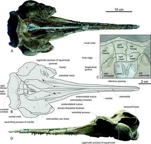
FIGURE 4. Huaridelphis raimondii, n. gen. et sp., MUSM 1396 (holotype), skull in ventral (A, B), posterior (C), anterior (D), and right lateral (E, F) views. Diagonal solid lines indicate major breaks.
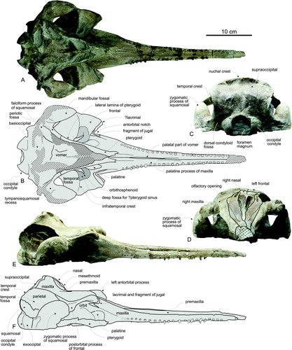
FIGURE 5. Huaridelphis raimondii, n. gen. et sp., MUSM 1403, skull in dorsal (A, B; detail of the vertex) and left lateral (C) views; symphyseal portion of mandible in left lateral (D) and dorsal (E) views; mandibular tooth in lateral view (F); incomplete atlas in anterior (G) and ventral (H) views; axis in anterior (I), ventral (J), posterior (K), and right lateral (L) views; four cervical vertebrae in anterior views (M–P); four thoracic vertebrae in posterior (Q, W), anterior (S, T, V), and right lateral (R, U, X) views; almost complete right rib in anterior (ZC) and posterior (ZD) views; proximal fragment of left rib in anterior view (ZE). Huaridelphis raimondii n. gen. et sp., MUSM 599, caudal vertebra in anterior (Y) and left lateral (Z) views; posterior tooth in lateral (ZA) and lingual (ZB) views. Diagonal solid lines indicate major breaks. Scale bar for A, C, D, and E equals 10 cm. Scale bars for F, ZA, and ZB equal 1 cm. Scale bar for G–Z and ZC–ZE equals 5 cm.
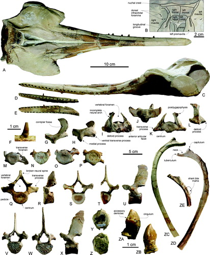
FIGURE 6. Huaridelphis raimondii, n. gen. et sp., MUSM 1396 (holotype), incomplete right periotic in dorsal (A, B), ventral (C, D), and lateral (E, F) views. Huaridelphis raimondii, n. gen. et sp., MUSM 599, left periotic in dorsal (G, H), dorsolateral (I, J), ventral (K, L), medial (M, N), and lateral (O, P) views. Diagonal solid lines indicate major breaks.
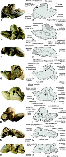
FIGURE 7. Squalodelphinidae aff. Huaridelphis raimondii, MUSM 603, incomplete skull in dorsal (A) and left lateral (B) views; left periotic articulated to the skull in ventral view (C); left malleus in posterior (D, E), and medial (F, G) views; left incus in medial (H, K), lateral (I, L), and dorsal (J, M) views; left tympanic bulla in medial (N, R), ventral (O, S), dorsal (P, T), and lateral (Q, U) views. Diagonal solid lines indicate major breaks. Scale bar for A and B equals 10 cm. Scale bars for C and N–U equal 2 cm. Scale bar for D–M equals 1 cm.
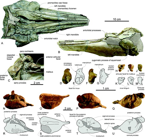
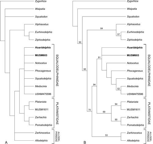
FIGURE 8. Consensus tree (A) and 50% majority consensus tree (B) of 60 equally parsimonious cladograms showing the relationships of Huaridelphis raimondii, n. gen. et sp., with the other Platanistoidea having lost double-rooted teeth. Tree length = 55, consistency index = 0.76, and retention index = 0.90. Numbers associated with the nodes in B are bootstrap values. See text for discussion and Appendices 1 and 2 for description of characters and data matrix.
HUARIDELPHIS RAIMONDII, n. gen. et sp.
()
Holotype—MUSM 1396, well-preserved skull with the ventral-most portion of the basicranium worn along an horizontal plane (basioccipital crests, ventral part of exoccipitals, and postglenoid processes of squamosals missing), with associated fragmented right periotic.
Type Locality—Ullujaya (, 2), Pisco-Ica Basin, 58 km SSE from Ica, 1.5 km N from Cerro Las Tres Piramides (see Alván De la Cruz, 2008:). Approximate geographic coordinates: S14°34′30″–W75°38′40″.
Type Horizon—Chilcatay Formation, latest Oligocene to early Miocene (Dunbar et al., Citation1990; DeVries, Citation1998, 2001). A section has been published at Cerro Buque, 1.65 km NWW to the type locality (Alván De la Cruz, 2008); based on sedimentology and shark teeth (presence of Isurus desori and Carcharocles chubutensis, absence of Carcharocles megalodon), the beds of the Chilcatay Formation in this section were dated to early Miocene. In the type locality and approximately in the same horizon of the holotype, we found a rich vertebrate fossil assemblage represented by other odontocetes (e.g., possible eurhinodelphinid cf. Ziphiodelphis, kentriodontids, stem physeteroid), a mysticete, marine turtles, sharks, and teleostean fishes ().
Trophic relationships between odontocetes and sharks are documented; in addition to the close association of odontocete bones and shark teeth, bite marks are observed in two ribs of MUSM 1403, a specimen referred to Huaridelphis raimondii.
Etymology—From ‘Huari,’ ancient culture of the south-central Andes and coastal area of Peru (500–1000 AD), and from ‘delphis,’ the Latin word for dolphin. Gender masculine. The species name honors Antonio Raimondi (1826–1890), an Italian scientist who first documented fossil whales from Peru (Bianucci, Citation2010).
Diagnosis—Huaridelphis raimondii differs from all the other squalodelphinids in smaller size (as seen in BZW and CBL); thin antorbital process of the frontal, barely thicker than the antorbital process of the maxilla in lateral view. It further differs from Notocetus and Squalodelphis in more abrupt anterior tapering of rostrum in dorsal view (see quantification below); higher tooth count, more than 28 teeth per row; short and less robust postorbital process of frontal; anteroventral slope of flat dorsal surface of vertex; dorsoventral compression of periotic at level of pars cochlearis and superior process, producing a flat dorsal surface; and posterior process of periotic more ventrally bent, with posterodorsal margin forming a right angle with dorsal surface of body of bone. It further differs from Medocinia and Squalodelphis in dorsal opening of mesorostral groove narrower than premaxilla in rostrum base; and wider dorsal exposure of maxilla at rostrum base (premaxilla nearly reaches lateral margin of rostrum in Medocinia and Squalodelphis).
Referred Specimens—MUSM 1403, well-preserved skull with the ventral portion of the basicranium worn along a subhorizontal plane (right squamosal, occipital condyles, basioccipital, basisphenoid, and paroccipital processes of the exoccipitals missing), with associated anterior symphyseal portion of mandible and posterior part of left dentary, partly preserved vertebrae including a fragment of atlas, axis, four additional cervicals, and four to five partly preserved thoracics, one nearly complete rib, and several smaller rib fragments. Found in the type locality Ullujaya. Accurate geographic coordinates: S14°34′36″–W75°38′39″. MUSM 599, fragmentary skull including the rostrum base, part of the vertex, the supraoccipital shield, the occipital condyles, the squamosals, the basioccipital, and the left periotic, and one caudal vertebra. Found in the locality of Zamaca, Pisco-Ica Basin, along the Ica River, 8 km SSE from the type locality Ullujaya.
DESCRIPTION AND COMPARISON
Skull
The skull of Huaridelphis raimondii is small (); CBL is 494 mm in the holotype, with BZW of 207 mm. This is smaller than in all other known squalodelphinids (holotype of Notocetus vanbenedeni MLP 5-8: CBL = 582 mm, BZW = ca. 235; N. vanbenedeni AMNH 9485: CBL = 634, BZW = ca. 254 mm; holotype of Squalodelphis fabianii: CBL = 640 mm, BZW = 263 mm; the skull of the holotype of Medocinia tetragorhina is too fragmentary for providing estimates of these measurements, but other skull measurements, for example, the width at rostrum base, are greater than in Huaridelphis raimondii). The rostrum is moderately elongated, constituting 67% of CBL in the holotype, a value in the range (63–70%) of Notocetus and Squalodelphis. The rostrum is considerably longer in all the known fossil platanistids (e.g., 84% in Zarhachis flagellator; Kellogg, Citation1924) and allodelphinids. The rostrum is only slightly dorsoventrally flattened, contra pomatodelphinines, in which the rostrum is much wider than high, and in platanistines, in which the rostrum is higher than wide (Barnes, Citation2002, 2006). The anterior two-thirds of the rostrum are slender, with a more abrupt tapering in the proximal third of the rostrum than in Notocetus and Squalodelphis (); in other words, the anterior part of the rostrum is proportionally narrower in Huaridelphis raimondii compared with the wide rostrum base.
TABLE 1. Measurements (in mm) on the skulls of Huaridelphis raimondii, n. gen. et sp., compared with two skulls of Notocetus vanbenedeni (measurements partly taken from True, Citation1910).
The cranium is proportionally short, with a ratio between length of the cranium (taken from the level of the antorbital notches to the occipital condyles) and BZW of 79.2% in the holotype. The cranium is roughly as long as wide in Squalodon and Waipatia, whereas the proportions of the cranium are closer to Huaridelphis in other squalodelphinids, allodelphinids, platanistids, and Prosqualodon.
The antorbital notches are deep and ‘V’-shaped. They are distinctly asymmetrical: the right antorbital notch is located more posteriorly than the left, and the lateral and medial borders of the right antorbital notch form a more open angle (60° in the holotype and 68° in MUSM 1403) than the equivalent angle on the left side (50° in the holotype and 55° in MUSM 1403). The ratio between right and left angles is around 1.2. A similar morphology and asymmetry of the antorbital notches is observed in Notocetus and Squalodelphis, although in both Notocetus and Squalodelphis the antorbital notches are wider and the ratio between the right and left angles is greater in Notocetus (ca. 1.7) and slightly smaller in Squalodelphis (ca. 1.1). Only the left deep and ‘V’-shaped antorbital notch is known in Medocinia. The asymmetry of the antorbital notches observed in Huaridelphis, Notocetus, and Squalodelphis is likely related to the asymmetry of the maxillary crests and antorbital processes: the left antorbital process is more elevated dorsally and longer anteriorly than the right process. A similar asymmetry is recorded in the frontal/maxillary crests of fossil platanistids, with the left crest more developed than the right in Pomatodelphis (e.g., USNM 187414) and Zarhachis (e.g., USNM 10911), whereas a higher right maxillary crest is at least occasionally observed in the strongly modified Platanista (e.g., IRSNB 1507, MNHN A7943). Asymmetry is also observed in the premaxillary sac fossae, the vertex, and surrounding bones laterally shifted on the right side (see below). All these elements of asymmetry are also observed in Notocetus, Squalodelphis, and platanistids. The temporal fossa of Huaridelphis has a shape and extent roughly similar to other squalodelphinids, anteriorly shorter and lower than in Squalodon and Waipatia.
Premaxilla—At the dorsoventrally flattened apex of the rostrum, the premaxilla bears the three anterior-most dental alveoli, as seen on the right side of MUSM 1403. The dorsal surface of each premaxilla at the anterior apex is excavated by a narrow sulcus a few centimeters long exiting forward from a small foramen. The two premaxillae are dorsally joined above the mesorostral groove for about half the length of the rostrum, whereas the groove is open until the apex in Notocetus (see Moreno, Citation1892; True, Citation1910); the medial opening of the groove is distinctly narrower than in Medocinia and Squalodelphis, and the widening towards the bony nares is progressive, with a ‘V’-shaped anterior limit of the nares. At 100–110 mm anterior to the antorbital notch, the right premaxilla is slightly narrower than the left, a condition also observed, but more pronounced, in pomatodelphinines and Platanista (see Kellogg, Citation1924, 1957, 1959; Lambert, Citation2006; Lee et al., Citation2012; pers. observ.). In parallel to the posterior widening of the rostrum, the premaxillae widen towards the proximal part of the rostrum, with a convex lateral margin, as in Medocinia, Notocetus, and even more pronounced in Squalodelphis. A roughly similar widening is possibly present in some specimens of Pomatodelphis, but not in Platanista, Prepomatodelphis, and Zarhachis. In this area the right premaxilla is distinctly wider than the left in both the holotype and MUSM 1403. As in other squalodelphinids, allodelphinids, and pomatodelphinines, each premaxilla displays a marked medial slope at this level, making a depressed area between the antorbital notches, which resembles the prenarial basin of several extinct and extant ziphiids (Lambert, Citation2005a). A similar medial depression is also present, but less marked, in Squalodon and Eosqualodon. Each premaxilla is pierced by a single premaxillary foramen. The transverse line through right and left foramina is anterior to the level of the antorbital notches. The ratio between the longitudinal distance from the level of the premaxillary foramina to the level of the right antorbital notch and the width of the rostrum base varies between 0.10 and 0.20, close to the condition in Notocetus (ratio between 0.20 and 0.27), whereas the premaxillary foramina are more anterior in Squalodelphis (ratio ca. 0.40). The anteromedial sulcus is long, discernible for more than 100 mm anterior to the foramen. Among squalodelphinids, a similarly long sulcus is also observed in Notocetus. The posteromedial sulcus nearly reaches the anterior limit of the bony nares and the posterolateral sulcus ends at about mid-length of the bony nares. The premaxillary sac fossa is roughly flat, with a medial slope. An angle of the medial margin of the premaxillae marks the anterior limit of the bony nares; this angle is more pronounced than in Squalodon or Waipatia. The ascending process of the premaxilla narrows abruptly and ends as a pointed apex on the right side, before the level of the nasal-frontal suture. On the left side the premaxilla is slightly longer and its wider apex contacts the frontal. In the pomatodelphinines Pomatodelphis, Prepomatodelphis, and Zarhachis and in Allodelphis, this posterior part of the premaxilla is more expanded, whereas it is narrower in Platanista. It should be noted that this part of the premaxilla is also wide in some other archaic odontocetes, such as Albertocetus and Xenorophus (see Uhen, Citation2008). The posterior apices of both premaxillae bear a deep longitudinal groove laterally margined by a thick ridge in the premaxilla. A similar combination of a groove and a thickened lateral margin is observed in Notocetus, Prepomatodelphis (commented in Barnes, Citation2006, char. 41, therein), Zarhachis (see Kellogg, Citation1926), and possibly Squalodelphis and the poorly preserved Medocinia. This groove may be homologous to the premaxillary cleft described in Waipatia (Fordyce, Citation1994), as well as to the elongated and shallow depression in the posterior part of the premaxilla of the highly modified Platanista.
Maxilla—In dorsal view, the maxilla is narrower than the premaxilla for the whole posterior third of the rostrum. Nevertheless, it is proportionally wider than in Medocinia, Notocetus, and Squalodelphis. Differing from platanistids and allodelphinids (as well as some other long-snouted odontocetes such as eurhinodelphinids and eoplatanistids), the lateral maxilla-premaxilla suture on the rostrum is not located in a deep lateral groove. The lateral margin of the rostrum at its base is thick and it increases steeply towards the antobital notch, as seen in lateral view. This margin is moderately convex in dorsal view, similar to Notocetus and Squalodelphis. The posterior wall of the deep and ‘V’-shaped antorbital notch (see below for a comment on the asymmetry of the notches) is made of the maxilla dorsally and the jugal ventrally. In platanistids, the notch is shallower, less clearly individualized, and often obscured by the maxillary/frontal crest. Just posteromedial to the antorbital notch, each maxilla is pierced by one or two small dorsal infraorbital foramina, followed anteriorly by a sulcus towards the antorbital notch. The antorbital region is elevated compared with the rostrum base, as in other squalodelphinids and platanistids, but more than in Allodelphis, Squalodon, and Waipatia. This thickening does not correspond to an individualized crest, differing from the high crest made of the maxilla and/or frontal in platanistids, even if a slight ridge marks the dorsolateral margin of each maxilla above the orbit, likely corresponding to the outer limit of a facial muscle. A series of deep oblique grooves marks the dorsal surface of the left antorbital process of the holotype (A). First thought to have been produced by a predator or scavenger, similar grooves were observed on the left process of USNM 526604, an undescribed platanistoid from the Calvert Formation, and might correspond to a genuine anatomical feature. The thin posterolateral part of the supraorbital process, roughly complete on the right side of MUSM 1403 (A), narrows posteriorly, leaving uncovered the posterior part of the temporal fossa. Linked to the conspicuous lateral shift of the vertex to the left side, the posterior part of the right maxilla is wider and less pointed backwards than on the left side, a feature also observed in Notocetus and Squalodelphis. One or two posterior dorsal infraorbital foramina are located at a short distance lateral to the posterior tip of the premaxilla, along the vertex (B). As in Notocetus and the platanistids, these foramina are more medial than the lateral-most margin of the premaxilla; this is not the case in Allodelphis, Squalodon, and Zarhinocetus. Due to the even wider premaxilla in this area, the posterior dorsal infraorbital foramen is just posterior to the maxilla-premaxilla suture in the platanistids Platanista, Pomatodelphis, Prepomatodelphis, and Zarhachis.
Including the three anterior alveoli that pierce the premaxilla, 28 and 29–30 alveoli for single-rooted teeth were counted on each upper tooth row for the holotype and MUSM 1403, respectively. This tooth count is higher than in Notocetus (22–23; Moreno, Citation1892; True, Citation1910) and Squalodelphis (15), but much smaller than in pomatodelphinines. The alveoli are small, with a diameter ranging from 3.7 mm for the smallest posterior alveoli to 5.5 mm for the largest anterior alveoli. This is smaller than in Notocetus and Squalodelphis. The spacing of the alveoli is wider posteriorly (6–7 mm), decreasing irregularly in more anterior alveoli (3–6 mm).
Mesethmoid—On the posterior wall of each bony naris, the mesethmoid is pierced by an elongated crescent-like fenestra starting at the dorsolateral corner of the wall, directed ventromedially then ventrolaterally (D). Such a pair of openings is commonly observed in platanistoids, for example, Notocetus, Pomatodelphis, Squalodon, and Zarhachis (see Kellogg, Citation1926; Dickson, Citation1964; Hoch, Citation2000), in Waipatia (Fordyce, Citation1994), and in one eurhinodelphinid, together with a possible ethmoturbinal (Schizodelphis morckhoviensis IRSNB M.343; Lambert, Citation2004). Hoch (Citation2000) proposed an olfactory interpretation for this pair of cavities, which probably represent reduced olfactory openings. No olfactory fontanelles have been observed in any extant adult odontocete, including Platanista, and the reduction and subsequent loss of the olfactory system is thought to have occurred in parallel in different odontocete lineages (Geisler and Sanders, Citation2003; McGowen et al., Citation2008; Godfrey et al., Citation2013). The dorsal margin of the mesethmoid roughly reaches the level of the anterodorsal margin of the nasal.
Nasal—Each small nasal is shorter and narrower than the corresponding frontal on the moderately elevated vertex (more elevated than in Allodelphis and Squalodon). The right nasal is longitudinally shorter and transversely narrower than the left (C, 5B). The nasal-frontal suture is somewhat irregular, but with a general transverse direction, as in Pomatodelphis or Waipatia. In Notocetus, the suture is more anteromedially pointed, whereas it is posteromedially pointed in Medocinia (region not clear in Squalodelphis). The nasals are lower than the frontals; together, their flat dorsal surfaces form an anteroventrally inclined plane (slope best seen in lateral view), as in Pomatodelphis and the highly modified Platanista. In contrast, in Notocetus, the vertex is roughly horizontal; it seems to slope posteroventrally in Squalodelphis; and it is more dorsally convex in Zarhachis. As a whole, the vertex is shorter than in Allodelphis, Squalodon, and Waipatia, as in other squalodelphinids and platanistids.
Frontal—Because the vertex is strongly shifted to the left side, the suture between frontals is far left from the midline of the skull. In relation with this shift, the left frontal exhibits a longitudinally shorter and transversely wider exposure at the vertex than the right frontal, partly due to the anterior projection of the lateral portion of the nuchal crest. The vertex is similarly shifted to the left in at least Platanista, Pomatodelphis, Notocetus, and Squalodelphis; this feature is less pronounced in Squalodon, Waipatia, and Zarhachis. Nevertheless, the shorter and wider exposure of the left frontal at the vertex is only similarly observed in Notocetus and Squalodelphis.
The slightly anteroventrally projected antorbital process of the frontal is only moderately dorsoventrally thickened, less than in Notocetus and Squalodelphis; it is less anteroventrally developed than in Medocinia and the possibly related USNM 475596. The postorbital process is also less robust, corresponding to a proportionally larger orbit than in other squalodelphinids. The ventral surface of the orbit roof is hollowed by a large fossa, with an abrupt lateral border at about 20–25 mm from the lateral margin of the frontal, and occupying the whole length from the lacrimal to the postorbital crest (A, B). Also present in Notocetus, and possibly in Zarhinocetus, this deep fossa is interpreted as the support for an extension of the pterygoid sinus in the orbit region. In Squalodelphis and Medocinia, this part of the skull is covered with sediment; therefore, this character cannot be assessed. A similar fossa, likely homologous, is even deeper in Pomatodelphis (e.g., P. bobengi USMN 299695) and Zarhachis (e.g., Z. flagellator USNM 10911), seemingly connecting the ventral surface of the orbit roof with the crest in the antorbital-supraorbital region. This interpretation is supported by the morphology of the pterygoid sinus in the extant Platanista. Indeed, in the latter, Fraser and Purves (Citation1960) propose a continuum between the pterygoid sinus cavity and the sinus in the maxillary crest, with bony channels connecting the two regions. The condition in squalodelphinids, with a moderately elevated antorbital region and a fossa in the orbit roof, might be considered as an intermediate grade towards the development of a large maxillary and/or frontal crest connected to a deeper sinus fossa in the orbit roof of platanistids. No such fossa has been detected in Squalodon, Waipatia, or other archaic odontocetes. The condition observed in squalodelphinids and platanistids, a single fossa in the frontal groove, is interpreted as not homologous to the fossae for the pre- and/or postorbital lobes of the pterygoid sinus on anterior and posterior flanks of the frontal groove of many delphinidans and some other odontocete taxa (e.g., several eurhinodelphinids; Lambert, Citation2005b), even if phocoenids display a somewhat similar extension of the preorbital lobe of the sinus into a space between maxilla and frontal (Fraser and Purves, Citation1960).
Supraoccipital—The anterodorsal margin of the supraoccipital is slightly posteriorly convex, with a short medial posterior projection. A similar projection is present in adult specimens of several extant delphinid species (external occipital crest sensu Fordyce and Mead, 2009). Just below the prominent nuchal crest, the supraoccipital shield is concave on a short height, before the two convex surfaces corresponding to the cerebral hemispheres. In dorsal view, the temporal crest is only posteriorly projected on a short distance, somewhat less than in Pomatodelphis and Zarhachis, and much less than in Platanista, the latter displaying a much narrower supraoccipital shield than other platanistoids.
Palatine—The extent of the palatine in the palate is interpreted as similar to what is observed in pomatodelphinines: limited exposure lateral to the lateral lamina of the pterygoid (see Muizon, Citation1987, for Pomatodelphis), with an anterior apex slightly anterior to the antorbital notch in the holotype of Huaridelphis raimondii (A, B). Previously thought to represent a synapomorphy of the platanistids (Muizon, Citation1987), the lateral exposure of the palatine seems to be a feature also present in Medocinia and Notocetus (Muizon, Citation1988b; pers. observ.; character not assessable in Squalodelphis). In Platanista, the palatine is completely covered by the pterygoid. The lateral lamina of the palatine borders the infraorbital foramen medially and reaches posteriorly to a level beyond two-thirds of orbit length, ventrally overhanging the deep fossa for the pterygoid sinus in the frontal groove.
Pterygoid—The pterygoid is long and narrow on the palate, with an apex 60 mm anterior to the antorbital notch; right and left apices are separated by a wedge of maxillae. As mentioned above, the pterygoid directly contacts the maxilla for most of its rostral portion. The pterygoid sinus fossa is similarly elongated, until a level more than 35 mm anterior to the antorbital notch. The fossa is also long in other squalodelphinids and platanistids; it is not anterior to the level of the antorbital notch in Squalodon, and it is even shorter in Waipatia. The lateral lamina is a continuous high plate posteriorly contacting the falciform process of the squamosal, as in all the other platanistoids, including Notocetus and Squalodelphis.
Jugal-Lacrimal—Only a thin and long oblique element along the anteromedial margin of the antorbital process is detected in the holotype, bearing a thin crest (A, B). It corresponds either to the jugal alone, or to jugal + lacrimal. The second interpretation would match the condition reported in Waipatia (see Fordyce, Citation1994:). The jugal sends a narrow anteromedial projection along the medial wall of the antorbital notch.
Squamosal—In lateral view, the zygomatic process of the squamosal is strongly swollen. Its dorsal and posterior margins are highly convex, as in Medocinia and Pomatodelphis, but more so than in Notocetus. Compared with Huaridelphis, the subhorizontal part of the dorsal margin of the zygomatic process is considerably longer and the dorsal margin is dorsoventrally lower in Platanista, Squalodon, and Waipatia, whereas a more distinct posterodorsal angulation is present in Prepomatodelphis and Zarhachis. As in other squalodelphinids, platanistids, and some specimens of Squalodon, the anteroventral margin of the process is rectilinear to slightly convex (not taking account of the postglenoid process), due to a ventral bulge of the lateral surface, whereas it is clearly concave in Allodelphis and Waipatia. To quantify the robustness of the zygomatic process, we calculated a ratio between the maximum distance from the anteroventral margin of the zygomatic process to the posterodorsal margin, in lateral view, and the vertical distance from the lower margin of the occipital condyles to the vertex of the skull. This ratio is higher than 0.38 in all the measured squalodelphinids (including Huaridelphis) and platanistids (Platanista, Pomatodelphis, and Zarhachis), whereas it is lower or equal to 0.38 in other presumed platanistoids (Allodelphis, Squalodon, and Waipatia), with the exception of Zarhinocetus errabundus. As in other squalodelphinids, platanistids, and Squalodon, the postglenoid process, only preserved in MUSM 599, is an anteroposteriorly flattened and short plate, anteroventrally pointed, and more slender in lateral view than in Allodelphis, eurhinodelphinids, Simocetus, and Waipatia. The mandibular fossa is vast, laterally closed by the ventrally bulging thin lateral wall of the zygomatic process. The tympanosquamosal recess is a shallow and narrow depression, short anteriorly along the falciform process and extending on the medial wall of the postglenoid process. The falciform process is a dorsoventrally high plate, widely contacting the lateral lamina of the pterygoid sinus.
Exoccipital—The occipital condyles are large and salient, with an individualized neck and a deep dorsal condyloid fossa. The ventral-most parts of the exoccipitals, including the paroccipital processes, are not preserved in any of the three known specimens of Huaridelphis.
Basioccipital—The ventral margins of the basioccipital crests, preserved in MUSM 599, are thick and moderately divergent posteriorly, with an angle between the crests of about 65 degrees.
Alisphenoid—The foramen ovale is widely separated by a bony bridge from the reduced posterior lacerate foramen, but the identity of the constituting bones cannot be established with certainty. On the ventral surface of the alisphenoid, the pterygoid sinus fossa is a slightly concave area anteromedial to the foramen ovale.
Periotic—The two known periotics (MUSM 1396: incomplete right periotic, total length as preserved = 33.7 mm; MUSM 599: nearly complete left periotic, total length as preserved = 35.4 mm) are similar in shape (). As for the skull, these periotics are smaller than in other squalodelphinids.
The periotic of Huaridelphis is similar to Notocetus and Squalodelphis in several characters: (1) anterior process elongated, not transversely thickened, moderately dorsally inflated, and only slightly bent anteromedially (less than in platanistids); (2) posterior process ventrally bent, bearing a narrow and smooth posterior bullar facet (incomplete in both periotics but probably originally elongated as in the other squalodelphinids) and a distinct articular rim; (3) pars cochlearis square-shaped in dorsal and ventral views; and (4) large, thin-edged dorsal opening of the cochlear aqueduct, which faces dorsally. A distinctive feature of the periotic of Huaridelphis, compared with Notocetus and Squalodelphis, is the dorsoventral compression of the pars cochlearis and of the body of the periotic; the compression of the latter is evidenced by a wide, flat dorsal surface lateral to the pars cochlearis (superior process sensu Kasuya, Citation1973). Moreover, the posterior process is more ventrally bent than in Notocetus and Squalodelphis, with its posterodorsal surface forming a straight angle with the dorsal surface of the body of the periotic. Among the other platanistoids, a similar right angle is observed in Phocageneus, Platanista, and Zarhachis.
As in the other squalodelphinids, the anterior bullar facet covers most of the length of the anterior process; it is transversely deeply concave, limited by prominent medial and lateral margins. Lateral to the anterior bullar facet, a well-developed anteroexternal sulcus is visible. On the medial surface of the anterior process is an irregular protuberance, possibly a small piece of bone fused to the periotic. A similar structure of unknown origin is observed in Xenorophus and in a squalodontid-like periotic by Luo and Marsh (1996:E, F, tuberosity of anterior process), as well as in the other squalodelphinids, the squalodontids, and some eurhinodelphinids. The ventral margin of this protuberance is marked by a narrow groove, specially visible in MUSM 1396, likely corresponding to one of the ‘anterointernal sulci’ noted in Waipatia by Fordyce (Citation1994). The fovea epitubaria is a longitudinally concave depression between the anterior bullar facet and the mallear fossa. The latter is wide, circular, distinctly concave, anteromedially oriented, and bounded laterally by a prominent lateral tuberosity, followed posteriorly by a deep hiatus epitympanicus. On the lateral margin of the posterior process is a prominent articular rim more developed than in Notocetus, less pointed than in Squalodelphis, and probably more reduced than the easily broken prominent hook-like articular process of platanistids (see Muizon, Citation1987).
As in Notocetus and Squalodelphis, the internal acoustic meatus is almost circular, deep, and tubular. An anterolateral notch in the outline of the meatus delimits the dorsal opening of the facial canal, which is located slightly anterolateral to the spiral cribriform tract. A similar location of the dorsal opening of the facial canal is observed in Phocageneus and Squalodelphis, whereas in Notocetus this foramen is less anterolaterally shifted and the internal acoustic meatus does not display a distinct notch. A narrower and longer notch is seen in Squalodon and Waipatia, both having a dorsal opening of the facial canal clearly more anteriorly located, whereas in Platanista and a Miocene platanistine from the Amazonian Basin (Bianucci et al., Citation2013) the dorsal opening of the facial canal is lateral to the spiral cribriform tract, with an oval internal acoustic meatus lacking an anterior notch. The foramen singulare is close to the opening of the facial canal, and both are separated from the spiral cribriform tract by a distinct transverse crest. As in the other squalodelphinids, the aperture for the cochlear aqueduct is wide and circular and it opens on the dorsal surface of the pars cochlearis, whereas the aperture for the endolymphatic duct is a small fissure. The fenestra rotunda is large and semicircular. The stapedial muscle fossa is deep, and its posterior opening is transversely wider than high.
Mandible
The symphyseal portion of the mandible is more than 168 mm long in MUSM 1403 (D, E), with the two dentaries nearly completely fused. The symphysis is similarly ankylosed in Notocetus, Phocageneus, and Squalodelphis. As for the rostrum, this part is dorsoventrally flattened, with a section wider than high for the whole preserved anterior 80 mm. Eighteen circular alveoli are counted on 161 mm of the right alveolar groove; their diameter ranges from 4 to 5 mm, with an irregular spacing (3–7 mm). The first right and left alveoli are anterolaterally directed and separated by 4 mm. Several mental foramina are present on each side, within a very shallow lateral groove.
Teeth
As in other squalodelphinids, allodelphinids, and platanistids, the teeth are conical and single-rooted, whereas the posterior teeth of Squalodon, Waipatia, and other archaic odontocetes remain double-rooted.
One detached tooth of MUSM 599 (ZA, ZB), presumably a right upper posterior, has a low crown (crown height 6.4 mm) roughly equal to its maximum mesiodistal diameter (6.35 mm). Its transverse section is buccolingually flattened with sharp mesial and distal keels. Both keels are slightly crenulated. The apex of the crown is pointed. An ‘S’-shaped cingulum on the lingual side bears several accessory denticles. Additional denticles are observed above the line of the cingulum on the lingual surface. The surface of the enamel is rough, with an ornamentation made of partly anastomosed grooves. This crown is similar to crowns of posterior teeth of Phocageneus venustus USNM 21039 (Kellogg, Citation1957:pl. 3, , 6, fourth and sixth lower teeth counting forward), an isolated tooth of aff. P. venustus (Whitmore and Kaltenbach, Citation2008:), and posterior teeth of Squalodelphis fabianii (Dal Piaz, Citation1917:pl. 5, –6). The cingulum is more developed than in the holotype of P. venustus (Kellogg, Citation1957:pl. 3, –3). The crown is slightly lower than in one posterior tooth of Notocetus vanbenedeni (Muizon, Citation1987:b, c). In Notocetus, one to three accessory denticles are observed on the distal keel, whereas these denticles seem to be lost in Phocageneus, Squalodelphis (Muizon, Citation1987), and Huaridelphis. Posterior teeth of platanistids, including Platanista, also have a low crown (Lambert et al., Citation2008b; pers. observ.), but the teeth are simpler and lack cingula, accessory denticles, and papillae.
A more anterior lower tooth of MUSM 1403 (F), the right ninth counting backward, displays a simple, elongated, moderately posteromedially curved, and conical crown with a less marked ornamentation made of more regular thin and low longitudinal grooves. The mesiodistal and buccolingual diameters of the crown base are respectively 3.7 and 3.35 mm. The morphology of this tooth is also similar to more anterior teeth of N. vanbenedeni and Squalodelphis fabianii (Dal Piaz, Citation1917:pl. 5, –11; Muizon, Citation1987:a; pers. observ.), corroborating the retention of some degree of heterodonty, both in terms of proportions and development of ornamentation (grooves, accessory denticles, cingulum), in squalodelphinids.
Vertebrae
As in other platanistoids, atlas and axis are not fused and both are anteroposteriorly long (medial length of the ventral surface of the atlas and axis respectively 31 and 37 mm). The degree of preservation of the ventral and dorsal transverse processes of the partly preserved atlas (G, H) does not allow an estimation of their length, even if the ventral process was probably shorter than in Zarhachis, possibly more similar to Notocetus (see True, Citation1910). The dorsal transverse process is thicker dorsoventrally than in Zarhachis, and dorsal and ventral processes are less divergent than in Zarhachis, closer to Phocageneus. The medial process projecting posteriorly from the posteroventral margin of the atlas is broad, as in Notocetus, Phocageneus, and Zarhachis.
In addition to the measurements presented in , the distance between the lateral margins of the anterior articular facets of the axis is 69.5 mm. The shape of the axis of MUSM 1403 (I–L) generally matches the axis of USNM 206006, an undescribed platanistid from the Calvert Formation showing affinities with Zarhachis. The transverse process is distinctly less ventrally projected in MUSM 1403, more similar, even if shorter, to the possible platanistid Araeodelphis (see Kellogg, Citation1957) and Platanista. The centrum is less ventromedially pointed, more regularly oval in posterior view than in the platanistid USNM 206006. The neural spine is missing.
TABLE 2. Measurements (in mm) of vertebrae of Huaridelphis raimondii, n. gen. et sp., MUSM 1403 (cervicals and thoracics) and MUSM 599 (caudal).
One of the other preserved cervicals (C; M) displays similarities with the third cervical of the P. venustus USNM 21039 (Kellogg, Citation1957:pl. 7, ). In MUSM 1403, with a maximum diameter of 8.5 mm, the transverse foramen for the vertebrarterial canal is located at a lower level relative to the centrum than in Phocageneus, with a dorsoventrally thinner parapophysis. In Platanista, the transverse foramen is much smaller in the third cervical and laterally open in the next vertebrae. The centrum is more transversely flattened here than in Phocageneus. The three other cervicals preserved (D–F; N–P) are too fragmentary to allow a comparison. Only some measurements are provided in .
A series of four well-preserved thoracic vertebrae (A–D; Q–X) is close to the first thoracics of P. venustus USNM 21039. The anterior-most vertebra A (Q-R) is shorter than T1 in P. venustus. The centrum becomes more ventrally pointed in anterior/posterior view backwards, more than in T3 or T4 of P. venustus. The preserved neural spines of the two last vertebrae C and D (U–X) of this series tend to be slightly tilted posteriorly, more than in T3–T5 of P. venustus and closer to the thoracics of Platanista. The anterior thoracic vertebrae of Notocetus illustrated by True (Citation1910) bear longer transverse processes.
With a centrum slightly higher than wide, the caudal vertebra of MUSM 599 (Y, Z) probably originates from the tail stock, just before the fluke (sensu Buchholtz and Schur, Citation2004). Pairs of ventral protuberances indicate the articulation of chevron bones.
More complete vertebral columns, both for Huaridelphis and other squalodelphinids, are needed for a detailed comparison.
Ribs
The preserved ribs are less transversely flattened than in Platanista. One nearly complete, double-headed, right rib (ZC, ZD), with a preserved maximum length of 260 mm, has a prominent tuberculum and a long neck; it does not differ significantly from two ribs of Notocetus (True, Citation1910:pl. 4, , 4), also close, even if more slender, to the fourth to seventh ribs of Zarhachis (Kellogg, Citation1924:pl. 15, 16). Another proximal rib fragment (ZE), more robust with a nearly square section of the body, probably occupied a more posterior position in the rib cage.
SQUALODELPHINIDAE aff. HUARIDELPHIS RAIMONDII
()
Referred Specimen—MUSM 603, skull lacking the anterior portion of the rostrum, associated to a partial mandible. Both periotics and the right tympanic are still attached to the skull, whereas the left tympanic bulla, malleus, and incus are detached. Found in the locality of Zamaca, Pisco-Ica Basin, along the Ica River, 8 km SSE from Ullujaya. Because a specimen of Huaridelphis raimondii (MUSM 599) was also found in this locality, we suspect that the locality of Zamaca corresponds to levels roughly temporally equivalent to the levels of Ullujaya, in the early Miocene of the Chilcatay Formation.
DESCRIPTION AND COMPARISON
With a cranium more heavily damaged than MUSM 1396 and MUSM 1403, this specimen (A, B) shares several similarities with Huaridelphis raimondii: the size and proportions of the cranium are roughly similar; the antorbital process is similarly elevated and weakly thickened; and the zygomatic process of the squamosal has the same outline in lateral view. However, the width of the rostrum, as well as the width of the premaxillae on the rostrum, decrease less abruptly anteriorly than in Huaridelphis raimondii (see ). Furthermore, the lateral margin of the premaxilla is less distinctly convex in the proximal region of the rostrum, differing from all other squalodelphinids. The antorbital notch is also shallower, more widely open as in Notocetus.
TABLE 3. Comparison of rostral measurements (width of rostrum and premaxillae at different levels, in mm) in the squalodelphinids Huaridelphis raimondii, n. gen. et sp., MUSM 1396 and aff. Huaridelphis raimondii MUSM 603.
The periotics are similar in size and shape to those of Huaridelphis raimondii MUSM 1396 and MUSM 1403 (C). The posterior process, complete in both periotics, exhibits a narrow and elongated posterior bullar facet, as in the other squalodelphinids. The left tympanic bulla, malleus, and incus deserve particular attention, because they are unknown in the specimens referred to Huaridelphis raimondii.
The tympanic bulla (N–U) is peculiar in its thin and extremely elongated anterior spine, representing 27% of the tympanic length (ca. 20% in Squalodelphis fabianii MGP 26134 and in the squalodelphinid USNM 21036; ca. 14% in Notocetus vanbenedeni AMNH 29026). Differing from the other squalodelphinids (especially Phocageneus and Squalodelphis), the median furrow does not extend anteriorly on the anterior spine, and, as in Notocetus and unlike Phocageneus and Squalodelphis, the lateral furrow is not deep. The outer posterior prominence is posteriorly shorter than the inner posterior prominence, whereas the outer and inner posterior prominences have approximately the same posterior extent in Notocetus, Phocageneus, and Squalodelphis). By contrast, in the platanistids Platanista, Pomatodelphis, and Zarhachis, the outer posterior prominence extends farther posteriorly than the inner posterior prominence. The dorsal margin of the involucrum is convex in medial view, but this margin is not as elevated as in Pomatodelphis.
The malleus (D–G) does not display the apical extension of the manubrium observed by Muizon (Citation1987) in Notocetus, Phocageneus, and Squalodelphis. On the whole, the tuberculum is not pointed and elevated as in the other squalodelphinids, but hook-shaped as in Pomatodelphis, with an outline even more similar to Squalodon (see Muizon, Citation1991:fig. 14).
The incus (H–M) is similar to Squalodelphis in having a short and tapered crus longum and a relatively wide articular facet for the malleus. The broken base of the crus breve is located at about half the length of the crus longum. This is probably the plesiomorphic condition among odontocetes, differing from the condition observed, for example, in the physeteroids, with a crus breve located close to the lateral margin of the articular facet for the malleus.
Based on these limited observations, with both similarities and differences with Huaridelphis raimondii, and the fragmented state of the specimen, we propose to identify it as Squalodelphinidae indet., aff. Huaridelphis raimondii, pending the discovery of more complete specimens.
PHYLOGENY
The aim of the cladistic analysis was to investigate the phylogenetic relationships of Huaridelphis raimondii, n. gen., n. sp., and MUSM 603 (a specimen referred here to Squalodelphinidae aff. Huaridelphis raimondii) with the other Squalodelphinidae, as well as the relationships of the latter with Allodelphinidae and Platanistidae, the other Platanistoidea (sensu Fordyce, Citation1994; Muizon, Citation1987; Barnes, Citation2006) lacking double-rooted teeth. We think that a much broader taxon sample would be needed to elucidate relationships with other presumable platanistoids retaining double-rooted teeth. The considered Squalodelphinidae are, in addition to Huaridelphis and MUSM 603, four genera (Medocinia, Notocetus, Phocageneus, and Squalodelphis) and one indeterminate specimen (USNM 475596; see above for the discussion). Allodelphinidae includes Allodelphis and Zarhinocetus, following Barnes and Reynolds (Citation2009). Platanistidae includes Platanista, Pomatodelphis, Zarhachis, and MUSM 1611 (an isolated periotic from the Amazonian Basin; see Bianucci et al., Citation2013). The genus Prepomatodelphis, also assigned to Platanistidae by Barnes (Citation2002), was not included in the cladistic analysis because of the difficulty to code this more fragmentarily known taxon based on published data alone. Because previous studies proposed relationships between the eurhinodelphinid Xiphiacetus and part of the platanistoids (Geisler and Sanders, Citation2003; Geisler et al., Citation2011), we include in the analysis the eurhinodelphinids Eurhinodelphis, Xiphiacetus, and Ziphiodelphis. Presumed platanistoids with double-rooted teeth Squalodon and Waipatia are also coded, whereas the basilosaurid Zygorhiza is used as outgroup. Thirty-seven morphological characters (Appendix 1), partly taken and modified from previous analyses (e.g., Muizon, Citation1987, 1988b, 1994; Fordyce, Citation1994; Lambert, Citation2005c; Bianucci et al., Citation2013), were coded for the 19 taxa (data matrix in Appendix 2). All characters are binary with the exception of five multistate characters (5, 16, 20, 22, 29). All characters are parsimony-informative in this analysis. The analysis was executed, considering all characters as non-additive (unordered) and unweighted, with the software PAUP (version 4.0b10; Swofford, Citation2001), using the heuristic search option, optimized by ACCTRAN using the tree bisection and reconnection (TBR) algorithm.
The cladistic analysis produced 60 equally parsimonious trees, with tree length = 55, consistency index (CI) = 0.76, and retention index (RI) = 0.90. The consensus tree, the 50% majority consensus tree, and the bootstrap values are presented in and discussed below.
The cladistic analysis supports the monophyly of the clade formed by Allodelphinidae + Platanistidae + Squalodelphinidae, representing members of the superfamily Platanistoidea without double-rooted teeth. The monophyly of this large clade is supported by a bootstrap value of 75 and by the following 11 synapomorphies: (1) rostral suture between premaxilla and maxilla deeply grooved (char. 3, state 1; reversal to state 0 in the Squalodelphinidae); (2) elevation of antorbital region, distinctly higher than the dorsal margin of the rostrum base in lateral view (char. 8, state 1; absent in Allodelphis); (3) widening of the cranium (char. 11, state 1); (4) presence of a deep fossa in the orbit roof (char. 13, state 1); (5) vertex distinctly shifted to the left compared with the sagittal plane of the skull (char. 14, state 1); (6) reduction of the ventral exposure of the palatine (char. 16, states 1 and 2; absent in Zarhinocetus); (7) hamular fossa of the pterygoid sinus extended anteriorly on the palatal surface of the rostrum (char. 17, state 1); (8) presence of an articular rim on the lateral surface of the periotic (char. 20, states 1 and 2); (9) elongation of the anterior spine on the tympanic bulla, associated to a marked anterolateral convexity (char. 27, state 1); (10) loss of double-rooted posterior teeth (char. 32, state 1); and (11) tooth count greater than 25 (char. 34, state 1; reversal to state 0 in Notocetus and Squalodelphis).
This clade is sister group to the Eurhinodelphinidae, a result that partly confirms several previous analyses (Geisler and Sanders, Citation2003; Geisler et al., Citation2011, 2012). However, as mentioned above, a larger sample, including among others Eoplatanista, physeteroids, ziphiids, Prosqualodon, and other odontocetes with double-rooted teeth, is needed to test this relationship.
The Squalodelphinidae appear to be sister group to the Platanistidae, as suggested in several previous works (e.g., Muizon, Citation1987; Fordyce, Citation1994), with Allodelphinidae in a more stemward position. The clade Platanistidae + Squalodelphinidae has a bootstrap value of 94 and is diagnosed by the following three synapomorphies: (1) widening of the premaxillae at the rostrum base (char. 5, states 1 and 2); (2) posterior infraorbital foramen (ina) along the vertex more medial than the lateral-most margin of the premaxilla in the cranium (char. 12, state 1); and (3) thickening of the zygomatic process of the squamosal (char. 18, state 1).
The monophyly of Squalodelphinidae is supported by a bootstrap value of 91 and by the following five synapomorphies: (1) deeper, ‘V’-shaped, left antorbital notch, related to an anteriorly pointed antorbital process (char. 7, state 1); (2) circle-shaped dorsal margin of the zygomatic process of the squamosal in lateral view (char. 19, state 1; reversal to state 0 in Notocetus and Squalodelphis); (3) square-shaped pars cochlearis of the periotic (char. 21, state 1); (4) large, thin-edged dorsal opening of the cochlear aqueduct of the periotic, which faces dorsally (char. 22, state 1); and (5) strong development of the dorsal transverse process of the atlas and extreme reduction of its ventral process (char. 35, state 1). Moreover, squalodelphinids seemingly lost the deep lateral groove along the rostral suture between premaxilla and maxilla (char. 2, state 1), observed in all other platanistoids lacking double-rooted teeth.
Relationships within squalodelphinids are unresolved, as evidenced by the consensus tree of all 60 cladograms (A) and by the bootstrap analysis. Nevertheless, the 50% majority consensus of the 60 trees (B) provides a tentative reconstruction of the relationships within the squalodelphinids, with Huaridelphis and MUSM 603 being the first to diverge, followed by Notocetus and by the clade formed by Medocinia, Phocageneus, Squalodelphis, and USNM 475996. Although Huaridelphis and MUSM 603 are the first diverging squalodelphinids, they are not conclusively sister groups. Consequently, their generic affinities as hypothesized in the systematic section are not strongly supported in the phylogenetic analysis. Analyzing the 60 equally parsimonious trees, the early divergence of Huaridelphis and MUSM 603 is supported in 52 trees (87%). Among these 52 trees, 20 (33% of the total) identify MUSM 603 as the earliest diverging taxon, 18 (30%) place Huaridelphis as sister group to MUSM 603, and 14 (23%) identify Huaridelphis as the earliest diverging taxon. In the remaining eight trees (13%), MUSM 603 is the earliest diverging taxon, with two possibilities for Huaridelphis: (1) a polytomy with Phocageneus and the clade containing all other squalodelphinids (8%), or (2) an intermediate position between the earlier divergence of Phocageneus and the clade containing all other squalodelphinids (5%). These last two topologies contradict the 50% majority consensus of the 60 trees, evidencing that further data (e.g., additional characters from more complete specimens of Phocageneus) should be added to improve the resolution of the phylogenetic analysis. A sister-group relationship between Medocinia and USNM 475996 is proposed in the 50% majority consensus of the 60 trees, as already suggested in the systematic section. However, it is weakly supported, with a bootstrap value of 52. With a bootstrap value of 94, the close relationships between Platanista and MUSM 1611, an isolated periotic from the Miocene of Peruvian Amazonia, confirms the preliminary phylogeny of Bianucci et al. (Citation2013).
CONCLUSIONS
Based on three specimens, including two well-preserved skulls, from early Miocene localities of the Chilcatay Formation (Ullujaya and Zamaca), Pisco-Ica desert, southern coast of Peru, we describe a new genus and species of Squalodelphinidae, Huaridelphis raimondii. In addition to periotic characters, H. raimondii differs from other known squalodelphinids in, among others, its smaller size, the thin antorbital process of the frontal, the more abrupt tapering of the rostrum, and the higher tooth count. Another fragmentary skull from the Chilcatay Formation in Zamaca is referred to Squalodelphinidae aff. Huaridelphis raimondii. It brings additional information on the morphology of the tympanic, malleus, and incus, not yet known in H. raimondii. Our phylogenetic analysis of platanistoids with single-rooted teeth suggests that the family Squalodelphinidae is monophyletic; the analysis also confirms the sister-group relationship between the latter and Platanistidae. The relationships within Squalodelphinidae are not fully resolved, but H. raimondii might be one of the first diverging taxa of the family.
ACKNOWLEDGMENTS
We wish to thank C. Bens, D. J. Bohaska, L. Del Favero, M. Fornasiero, G. Lenglet, C. de Muizon, N. D. Pyenson, H. van Grouw, and R. Salas-Gismondi for providing us access to specimens under their care. Many thanks to W. Aguirre, A. J. Altamirano, E. Díaz, R. Salas-Gismondi, J. Tejada, N. Valencia, and R. Varas for their help during field work, for the preparation and curation of the Peruvian specimens described here, and for making us feel welcome during each of our visits at the Museo. Several of our field campaigns in Peru would not have been possible without the support of C. de Muizon, K. Post, J. Reumer, the MNHN Paris, and the NMR Rotterdam. We also thank J. H. Geisler, C. de Muizon (the two reviewers), and E. M. G. Fitzgerald (the editor) for their constructive comments and suggestions. The work of O.L. at the IRSNB was funded by a Return Grant of the Belgian Federal Science Policy Office from 2012 to April 2013.
Handling editor: Erich Fitzgerald.
Related Research Data
LITERATURE CITED
- Alván De la Cruz, A. 2008. Geología de Ocucaje: aportes en la sedimentología y paleontología de Lomas de Ullujaya (Ica, Perú). Revista del Instituto de Investigaciones FIGMMG 11(21):51–59.
- Barnes, L. G. 2002. An Early Miocene long-snouted marine platanistid dolphin (Mammalia, Cetacea, Odontoceti) from the Korneuburg Basin (Austria). Beiträge zur Paläontologie 27:407–418.
- Barnes, L. G. 2006. A phylogenetic analysis of the superfamily Platanistoidea (Mammalia, Cetacea, Odontoceti). Beiträge zur Paläontologie 30:25–42.
- Barnes, L. G., and R. E. Reynolds. 2009. A new species of early Miocene allodelphinid dolphin (Cetacea, Odontoceti, Platanistoidea) from Cajon Pass, Southern California, U.S.A. Museum of Northern Arizona Bulletin 65:483–507.
- Bianucci, G. 2010. Esplorazioni e nuove scoperte nel deserto del Perù: i cetacei fossili di Cerro Colorado e Cerro los Quesos. Quaderni del Museo di Storia Naturale di Livorno 23:3–12.
- Bianucci, G., O. Lambert, and K. Post. 2010. High concentration of long-snouted beaked whales (genus Messapicetus) from the Miocene of Peru. Palaeontology 53:1077–1098.
- Bianucci, G., O. Lambert, R. Salas-Gismondi, J. Tejada, F. Pujos, M. Urbina, and P.-O. Antoine. 2013. A Miocene relative of the Ganges River dolphin (Odontoceti, Platanistidae) from the Amazonian Basin. Journal of Vertebrate Paleontology 33:741–745.
- Brisson, M.-J. 1762. Regnum Animale in classes IX distributum, sine synopsis methodica. Theodorum Haak, Paris, 296 pp.
- Buchholtz, E. A., and S. A. Schur. 2004. Vertebral osteology in Delphinidae (Cetacea). Zoological Journal of the Linnean Society 140:383–401.
- Cozzuol, M. A., and O. A. Aguilera. 2008. Cetacean remains from the Neogene of northwestern Venezuela. Paläontologische Zeitschrift 82:196–203.
- Dal Piaz, G. 1917. Gli Odontoceti del Miocene Bellunese. Parte terza Squalodelphis. Memorie dell’Istituto Geologico della Reale Università di Padova 5:1–34.
- Delfortrie, E. 1875. Un dauphin d’espèce nouvelle dans les faluns du Sud-Ouest. Actes de la Société Linnéenne de Bordeaux 30(2):3–7.
- DeVries, T. J. 1998. Oligocene deposition and Cenozoic sequence boundaries in the Pisco Basin (Peru). Journal of South American Earth Sciences 11:217–231.
- DeVries, T. J. 2001. Molluscan evidence for an Oligocene-Miocene age of ‘Paracas’ beds in Southern Peru. Boletín de la Sociedad Geológica del Perú 92:57–65.
- Dickson, M. R. 1964. The skull and other remains of Prosqualodon marplesi, a new species of fossil whale. New Zealand Journal of Geology and Geophysics 7:626–635.
- Dunbar, R. B., R. C. Marty, and P. A. Baker. 1990. Cenozoic marine sedimentation in the Sechura and Pisco basins, Peru. Palaeogeography, Palaeoclimatology, Palaeoecology 77:235–261.
- Flower, W. H. 1867. Description of the skeleton of Inia geoffrensis and the skull of Pontoporia blainvillii, with remarks on the systematic position of these animals in the Order Cetacea. Transactions of the Zoological Society of London 6:87–116.
- Fordyce, R. E. 1994. Waipatia maerewhenua, new genus and new species (Waipatiidae, new family), an archaic late Oligocene dolphin from New Zealand; in A. Berta and T. A. Deméré (eds.), Contributions in marine mammal paleontology honoring Frank C. Whitmore, Jr. Proceedings of the San Diego Society of Natural History 29:147–178.
- Fordyce, R. E., and C. de Muizon. 2001. Evolutionary history of cetaceans: a review; pp. 169–233 in J.-M. Mazin and V. de Buffrénil (eds.), Secondary Adaptation of Tetrapods to Life in Water. Verlag Dr. Friedrich Pfeil, Munich.
- Fraser, F. C., and P. E. Purves. 1960. Hearing in cetaceans: evolution of the accessory air sacs and the structure of the outer and middle ear in recent cetaceans. Bulletin of the British Museum (Natural History), Zoology 7:1–140.
- Geisler, J. H., and A. E. Sanders. 2003. Morphological evidence for the phylogeny of Cetacea. Journal of Mammalian Evolution 10:23–129.
- Geisler, J. H., S. J. Godfrey, and O.Lambert. 2012. A new genus and species of Late Miocene inioid (Cetacea: Odontoceti) from the Meherrin River, North Carolina, U.S.A. Journal of Vertebrate Paleontology 32:198–211.
- Geisler, J. H., M. R. MacGowen, G. Yang, and J. Gatesy. 2011. A supermatrix analysis of genomic, morphological, and paleontological data for crown Cetacea. BMC Evolutionary Biology 11:112.
- Godfrey, S. J., J. Geisler, and E. M. G. Fitzgerald. 2013. On the olfactory anatomy in an archaic whale (Protocetidae, Cetacea) and the minke whale Balaenoptera acutorostrata (Balaenopteridae, Cetacea). The Anatomical Record 296:257–272.
- Gray, J. E. 1863. On the arrangement of the cetaceans. Proceedings of the Zoological Society of London 1863:197–202.
- Hoch, E. 2000. Olfaction in whales: evidence from a young odontocete of the Late Oligocene North Sea. Historical Biology 14:67–89.
- Kasuya, T. 1973. Systematic consideration of recent toothed whales based on the morphology of tympano-periotic bone. The Scientific Reports of the Whales Research Institute 25:1–103.
- Kellogg, R. 1924. A fossil porpoise from the Calvert Formation of Maryland. Proceedings of the United States National Museum 63(14):1–39.
- Kellogg, R. 1926. Supplementary observations on the skull of the fossil porpoise Zarhachis flagellator Cope. Proceedings of the United States National Museum 67(28):1–18.
- Kellogg, R., 1957. Two additional Miocene porpoises from the Calvert Cliffs, Maryland. Proceedings of the United States National Museum 107:279–337.
- Kellogg, R., 1959. Description of the skull of Pomatodelphis inaequalis Allen. Bulletin of the Museum of Comparative Zoology, Cambridge 121:3–26.
- Lambert, O. 2004. Systematic revision of the Miocene long-snouted dolphin Eurhinodelphis longirostris du Bus, 1872 (Cetacea, Odontoceti, Eurhinodelphinidae). Bulletin de l’Institut Royal des Sciences Naturelles de Belgique, Sciences de la Terre 74:147–174.
- Lambert, O. 2005a. Systematics and phylogeny of the fossil beaked whales Ziphirostrum du Bus, 1868 and Choneziphius Duvernoy, 1851 (Cetacea, Odontoceti), from the Neogene of Antwerp (North of Belgium). Geodiversitas 27:443–497.
- Lambert, O. 2005b. Review of the Miocene long-snouted dolphin Priscodelphinus cristatus du Bus, 1872 (Cetacea, Odontoceti) and phylogeny among eurhinodelphinids. Bulletin de l’Institut Royal des Sciences Naturelles de Belgique, Sciences de la Terre 75:211–235.
- Lambert, O. 2005c. Phylogenetic affinities of the long-snouted dolphin Eurhinodelphis (Cetacea, Odontoceti) from the Miocene of Antwerp. Palaeontology 48:653–679.
- Lambert, O. 2006. First record of a platanistid (Cetacea, Odontoceti) in the North Sea Basin: a review of Cyrtodelphis Abel, 1899 from the Miocene of Belgium. Oryctos 6:69–79.
- Lambert, O., and C. de Muizon. 2013. A new long-snouted species of the Miocene pontoporiid dolphin Brachydelphis and a review of the Mio-Pliocene marine mammal levels in the Sacaco Basin, Peru. Journal of Vertebrate Paleontology 33:709–721.
- Lambert, O., G. Bianucci, and C. de Muizon. 2008a. A new stem-sperm whale (Cetacea, Odontoceti, Physeteroidea) from the Latest Miocene of Peru. Comptes Rendus Palevol, 7:361–369.
- Lambert, O., G. Bianucci, and K. Post, 2009. A new beaked whale (Odontoceti, Ziphiidae) from the middle Miocene of Peru. Journal of Vertebrate Paleontology 29:910–922.
- Lambert, O., G. Bianucci, and K. Post. 2010a. Tusk-bearing beaked whales from the Miocene of Peru: sexual dimorphism in fossil ziphiids? Journal of Mammalogy 91:19–26.
- Lambert, O., C. de Muizon, and G. Bianucci. 2013. The most basal beaked whale Ninoziphius platyrostris Muizon, 1983: clues on the evolutionary history of the family Ziphiidae (Cetacea: Odontoceti). Zoological Journal of the Linnean Society 167:569–598.
- Lambert, O., J. Schlögl, and M. Kováč. 2008b. Middle Miocene toothed whale with Platanista-like teeth from the Vienna Basin (Western Carpathians, Slovakia). Neues Jahrbuch für Geologie und Paläontologie 250:157–166.
- Lambert, O., G. Bianucci, K. Post, C. de Muizon, R. Salas-Gismondi, M. Urbina, and J. Reumer. 2010b. The giant bite of a new raptorial sperm whale from the Miocene epoch of Peru. Nature 466:105–108.
- Lee, Y.-N., H. Ichishima, and D. K. Choi. 2012. First record of a platanistoid cetacean from the middle Miocene of South Korea. Journal of Vertebrate Paleontology 32:231–234.
- Leidy, J. 1869. The extinct mammalian fauna of Dakota and Nebraska, including an account of some allied forms from other localities, together with a synopsis of the mammalian remains of North America. Journal of the Academy of Natural Sciences of Philadelphia, new series 7:1–472.
- Luo, Z., and K. Marsh. 1996. The petrosal and inner ear structure of a fossil kogiine whale (Odontoceti, Mammalia). Journal of Vertebrate Paleontology 16:328–348.
- McGowen, M. R., C. Clark, and J. Gatesy. 2008. The vestigial olfactory receptor subgenome of odontocete whales: phylogenetic congruence between gene-tree reconciliation and supermatrix methods. Systematic Biology 57:574–590.
- Mead, J. G., and R. E. Fordyce. 2009. The therian skull: a lexicon with emphasis on the odontocetes. Smithsonian Contributions to Zoology 627:1–248.
- Moreno, F. P. 1892. Noticias sobre algunos cetáceos fósiles y actuales de la República Argentina. Revista del Museo de La Plata 3:383–400.
- Muizon, C. de. 1984. Les Vertébrés de la Formation Pisco (Pérou). Deuxième partie: les Odontocètes (Cetacea, Mammalia) du Pliocène inférieur du Sud-Sacaco. Travaux de l’Institut Français d’Etudes Andines 27:1–188.
- Muizon, C. de. 1987. The affinities of Notocetus vanbenedeni, an Early Miocene platanistoid (Cetacea Mammalia) from Patagonia, southern Argentina. American Museum Novitates 2904:1–27.
- Muizon, C. de. 1988a. Les vertébrés de la Formation Pisco (Pérou). Troisième partie: les odontocètes (Cetacea, Mammalia) du Miocène. Travaux de l’Institut Français d’Etudes Andines 42:1–244.
- Muizon, C. de. 1988b. Le polyphylétisme des Acrodelphidae, odontocètes longirostres du Miocène européen. Bulletin du Muséum National d’Histoire Naturelle, Paris 10:31–88.
- Muizon, C. de. 1991. A new Ziphiidae (Cetacea) from the Early Miocene of Washington State (USA) and phylogenetic analysis of the major groups of odontocetes. Bulletin du Muséum National d’Histoire Naturelle, Paris 12:279–326.
- Muizon, C. de. 1993. Walrus-like feeding adaptation in a new cetacean from the Pliocene of Peru. Nature 365:745–748.
- Muizon, C. de. 1994. Are the squalodonts related to the platanistoids?; in A. Berta and T. A. Deméré (eds.), Contributions in marine mammal paleontology honoring Frank C. Whitmore, Jr. Proceedings of the San Diego Society of Natural History 29:135–146.
- Pilleri, G. 1985. The Miocene Cetacea of the Belluno sandstones (eastern southern Alps). Memorie di Scienze Geologiche 36:1–87.
- Rommel, S. A. 1990. Osteology of the bottlenose dolphin; pp. 29–49 in S. Leatherwood and R. R. Reeves (eds.), The Bottlenose Dolphin. Academic Press, San Diego, California.
- Sánchez-Villagra, M. R., Z. Gasparini, R. Lozsán, J. M. Moody, and M. D. Uhen. 2001. New discoveries of vertebrates from a near-shore marine fauna from the Early Miocene of northwestern Venezuela. Paläontologische Zeitschrift 75:227–232.
- Swofford, D. L. 2001. PAUP*. Phylogenetic Analysis Using Parsimony (*And Other Methods), version 4b10. Sinauer Associates, Sunderland, Massachusetts.
- True, F. W. 1910. Description of a skull and some vertebrae of the fossil cetacean Diochotichus vanbenedeni from Santa Cruz, Patagonia. Bulletin of the American Museum of Natural History 28:19–32.
- Uhen, M. D. 2008. A new Xenorophus-like odontocete cetacean from the Oligocene of North Carolina and a discussion of the basal odontocete radiation. Journal of Systematic Palaeontology 6:433–452.
- Uhen, M. D., and N. D. Pyenson. 2007. Diversity estimates, biases, and historiographic effects: resolving cetacean diversity in the Tertiary. Palaeontologia Electronica 10(2):1–22. Available at http://palaeo-electronica.org/2007_2/00123/index.html
- Whitmore, F. C., Jr., and J. A. Kaltenbach. 2008. Neogene Cetacea of the Lee Creek Phosphate Mine, North Carolina. Virginia Museum of Natural History Special Publication 14:181–269.
APPENDIX 1. List of the characters used in phylogenetic analysis. Characters are polarized with respect to Zygorhiza as the outgroup.
Rostrum elongation (Bianucci et al., Citation2010, modified): short, ratio between rostrum length and condylobasal length <0.70 (0); elongated, ratio >0.70 (1).
Apex of the rostrum constituted by the only premaxillae on more than 10% of its total length and lacking alveoli (Lambert, Citation2005c): absent (0); present (1).
Lateral rostral suture between premaxilla and maxilla deeply grooved (Fordyce, Citation1994): no (0); yes (1).
Marked asymmetry of the premaxillae on the rostrum, at some distance anterior to the premaxillary foramina, with the right premaxilla distinctly narrower than the left in dorsal view: absent (0); present (1).
Widening of the premaxillae at the rostrum base: narrow premaxillae, ratio between the width of the rostrum and the transverse width of the premaxillae at the antorbital notch <0.60 (0); wide premaxillae, ratio between 0.60 and 0.75 (1); extremely wide premaxillae nearly reaching the lateral margin of the rostrum, ratio >0.75 (2).
Dorsal opening of the mesorostral groove anterior to the rostrum base (Geisler and Sanders, Citation2003, modified): narrower than the premaxilla (0); wider than the premaxilla (1).
Deep, ‘V’-shaped, left antorbital notch, related to an anteriorly pointed antorbital process: no (0); yes (1).
Elevated antorbital region, distinctly higher than the dorsal margin of the rostrum base in lateral view: no (0); yes (1).
Distinct dorsal crest in the antorbital-supraorbital region: no (0); yes (1).
Thickening of the antorbital process of the frontal: absent, ratio between the height of the antorbital process of the frontal, measured in lateral view perpendicular to the maxillary-frontal suture above the orbit, and the vertical distance from the lower margin of the occipital condyles to the vertex of the skull <0.25 (0); present, ratio >0.30 (1).
Widening of the cranium: cranium roughly as long as wide or longer than wide with ratio between cranium length (longitudinal, from occipital condyles to level of antorbital notches) and postorbital width >0.90 (0); cranium distinctly shorter than wide with ratio <0.90 (1).
Posterior infraorbital foramen(ina) along the vertex more medial than the lateral-most margin of the premaxilla in the cranium: no (0); yes (1).
Deep fossa in the frontal on orbit roof, at the level of the frontal groove: no (0); yes (1).
Vertex distinctly shifted to the left compared with the sagittal plane of the skull: no (0); yes (1).
Transverse premaxillary crest on the vertex (Lambert, Citation2005c): absent (0); present (1).
Ventral exposure of the palatine (Muizon, Citation1987, modified): palatine widely exposed anterior to the pterygoid (0); palatine only exposed laterally to the lateral lamina of the pterygoid (1); palatine completely covered by the pterygoid (2).
Hamular fossa of the pterygoid sinus (Lambert et al., Citation2013): small, not reaching anteriorly the level of the antorbital notch (0); wide, extending anteriorly on the palatal surface of the rostrum (1).
Thickening of the zygomatic process of the squamosal: absent, ratio between the maximum distance from the anteroventral margin of the zygomatic process to the posterodorsal margin, in lateral view, and the vertical distance from the lower margin of the occipital condyles to the vertex of the skull <0.35 (0); present, ratio >0.35 (1).
Circle-shaped dorsal outline of the zygomatic process of the squamosal in lateral view: no (0); yes (1).
Articular rim on the lateral surface of the periotic (Muizon, Citation1987, modified): absent (0); present (1); present and hook-like (2).
Pars cochlearis of the periotic square-shaped in ventral view (Muizon, Citation1987): no (0); yes (1).
Aperture of the cochlear aqueduct of the periotic (Muizon, Citation1987, modified; Bianucci et al., Citation2013): small (0); very small (1); large and thin-edged (2).
Aperture of the cochlear aqueduct of the periotic (Muizon, Citation1987, modified): faces mediodorsally (0); faces dorsally (1).
Transverse thickening of the anterior process of the periotic (Muizon, Citation1987): no (0); yes (1).
Internal auditory meatus of the periotic oval, with the dorsal opening for the facial canal lateral to the spiral cribriform tract (Bianucci et al., Citation2013): no (0); yes (1).
Separate ossicle at the apex of the anterior process of the periotic (Bianucci et al., Citation2013): no (0); yes (1).
Elongated anterior spine on the tympanic bulla, associated to a marked anterolateral convexity (Muizon, Citation1987): no (0); yes (1).
Ventral groove of the tympanic affecting the whole length of the bone, including the anterior spine (Muizon, Citation1987): no (0); yes (1).
Extent of the inner and outer posterior prominences of the tympanic: both prominences with approximately the same posterior extent (0); outer posterior prominence posteriorly longer than the inner posterior prominence (1); outer posterior prominence posteriorly shorter than the inner posterior prominence (2).
Dorsal margin of the involucrum of the tympanic cut by a median indentation, in medial view (Lambert, Citation2005c): absent (0); present (1).
Apical extension of the manubrium of the malleus (Muizon, Citation1987): no (0); yes (1).
Loss of double-rooted posterior teeth: (Muizon, Citation1987): no (0); yes (1).
Retention of accessory denticles on posterior teeth (Muizon, Citation1987, modified): yes (0); no (1).
Tooth count per upper or lower row: <25 (0); >25 (1).
Strong development of the dorsal transverse process of the atlas and extreme reduction of its ventral process (Muizon, Citation1987): no (0); yes (1).
Great reduction of coracoid process of the scapula (Muizon, Citation1987, 1991, 1994): no (0); yes (1).
Great reduction or loss of supraspinatus fossa, with acromion located on anterior edge of scapula (Muizon, Citation1987, 1991, 1994): no (0); yes (1).