Exposure to particles that have deposited on surfaces is common in occupational and residential environments. Lack of an accurate tool for assessing particle size distribution and loading (mass per unit area) on carpet fibers available for exposure contributes to the uncertainty associated with current risk assessment models. This research presents a new, direct image analysis method (IAM) for measuring particle size distribution and loading on carpet fibers. New and old carpet fibers loaded with Arizona Test Dust were used to test the method. Carpet fibers were removed from the bulk carpet, mounted on substrates, and scanning electron microscopy (SEM) images were collected. Particle size distribution and total mass were calculated from the processed images. The Arizona Test Dust (ATD) size distribution on fibers from two different carpets had mass median diameters of 3.6 ( ± 1.2) and 4.1 (±0.7) μm, similar to that for bulk ATD, 4.0 (± 0.5) μm. Total ATD mass available on new carpet fibers calculated by IAM were statistically correlated with the mass collected on micro-vacuum samples (R 2 = 0.95). Direct comparison of the aerodynamic diameters measured by IAM with those measured automatically by the SEM showed a slight negative bias due to image resolution problems for the smallest particles.
INTRODUCTION
Adults and children are exposed to particulate matter from flooring and other surfaces in their homes and offices everyday. The source of this particulate matter stems from indoor generation, shoe tracked-in dusts, and penetration of ambient particles that deposit on the flooring and other horizontal surfaces. All or a fraction of these particles may be available for inhalation exposure through resuspension or dermal exposure through contact with the surface.
Several researchers have calculated particle resuspension emission rates (CitationMontoya and Hildemann 2001; CitationButtner et al. 2002; CitationFerro et al. 2004) and dermal transfer rates (CitationLewis et al. 1994; CitationNishioka et al. 2002) from a variety of surfaces. However, comparison of the rates between studies is difficult because the quantity of dust available on the surface was not quantified or was measured by different techniques. A simple, inexpensive method to measure the mass available on a surface would provide a means to normalize emission rate data to allow comparison across studies.
Accurate assessment of the particle mass available for exposure is needed to reduce the uncertainty associated with the source terms in exposure models (CitationFenske et al. 1991). These models currently quantify the exposure mass inhaled or contacted as a fixed value instead of fraction of the mass available, contributing to the uncertainty in the risk assessment (CitationU.S. EPA 1997). Emission or risk factors, represented as the ratio of quantity released from a source to the quantity available for exposure, are calculated from these models. Common instrumentation or established methods determine the quantity released for inhalation or dermal exposure (CitationRodes and Thornburg 2005; CitationEdwards and Lioy 1999). However, determining the quantity of particulate matter available is made difficult by differences in surface types and environmental conditions. There is evidence that exposure concentrations are linearly correlated with the mass available, but the linear correlation may not hold for all scenarios (unpublished data; CitationThornburg et al. 2004).
Determining the particle mass available for exposure from hard indoor surfaces, such as linoleum, is fairly simple. Wipe, vacuum, or dermal press methods collect a representative sample from these surfaces because of their two-dimensional structure and weak adhesion forces (CitationQue Hee et al. 1985; CitationLu and Fenske 1999; CitationBrouwer et al. 1999). Collecting a representative sample from carpets to determine mass available for exposure is more difficult. Carpet provides depth that may allow migration of the particles, especially those > 10 μm, downward towards the backing such that the particles are not an exposure risk. Therefore, only particles on the upper portion of the carpet fibers may be available for exposure. Sample collection mechanisms also affect the mass available estimate. Particles adhering to carpet fibers that may not be collected with a vacuum sample may be collected easily with a wipe sample. The vacuum beater bar and suction may not be sufficient to overcome adhesion forces that may be neutralized by the damp material used for wipe samples. The conundrum is determining which method, vacuum or wipe, collects a representative sample of the mass available.
The challenge in determining the particle mass on carpet fibers available for exposure is finding an accurate method. Wipe and vacuum methods collect in situ samples that may not be representative of the mass available because of variations in particle-fiber adhesion forces and generation mechanisms. Electrostatic, Van der Waals, and surface tension adhesion forces hold particles onto carpet fibers. The strength and importance of these forces depends on carpet age, fiber surface coatings, percent moisture, particle diameter, particle structure, and particle composition. Old carpets coated with oils and new carpets with an electret charge have the strongest adhesion forces (unpublished data; CitationThornburg et al. 2004). The generation mechanism is directly related to the exposure method. Dermal exposure may result from a hand-press onto the fibers, whereas inhalation exposure may result from resuspension of particles via walking. The force per square inch applied and the contact area will be different for each mechanism and have differing effect on the magnitude of the exposure concentration. For example, wipe and vacuum methods have independent application forces and contact areas that may not be correlated to the exposure method.
Therefore, a need exists for a method to assess the number, mass, and size distribution of particles on carpet fibers available for exposure. Sample collection must not dislodge particles from the fibers so the quantity of particles available for exposure can be measured accurately. This research presents an image analysis method (IAM) for accurately measuring quantity and size distribution of particles on carpet fibers available for exposure using scanning electron microscopy (SEM). Automatic, computer controlled SEM (CCSEM) also provides particle size distribution and loading data by measuring individual particles within a defined area. CCSEM is the standard method for obtaining this data from surfaces. Although automated, CCSEM requires two to four hours of SEM time to obtain the data from a single fiber as individual particles are sized and counted. Manually obtaining low magnification images requires about 30 minutes per fiber and the measurement of the particle size distribution with an image processing program requires another 30 minutes. The result is a reduction in required SEM time and a cost savings.
SAMPLE COLLECTION AND ANALYSIS
Fiber Removal and Mounting
Fibers were removed from the carpets and mounted on SEM stubs. Multiple one-square-inch sections from two carpet types were removed and transported to the laboratory where individual fibers were selected. When removal of a small section is not practical, such as inside occupied residences, individual fibers could be removed directly from the bulk carpet. Stainless steel tweezers with very fine points designed for electronics work handled the fibers. The selected fiber tip, approximately the top 1 mm, was grabbed with the tweezers and pulled taut. Using the other hand, the surrounding fibers were pushed downward to expose the base of the selected fiber. The selected fiber base was cut with a common fine tip, 45ˆ micro-scalpel. A second pair of tweezers then positioned the fiber base onto a SEM stub with double-sided, conductive tape. Handling the fiber base was not a concern because of the low probability that particles that deep in the carpet were available for exposure. The fiber was laid straight on the SEM stub. If necessary to fit the entire fiber onto the SEM stub, the top of the fiber was curved into a U shape. Performing these steps under a magnifying glass simplifies the procedure and is recommended.
Image Analysis Method (IAM)
The IAM was based on the method developed by CitationWagner and Leith (2001). A Personal SEM (Aspex LLC, Delmont, PA) operating at working distance of 16 mm, 20 kV accelerating voltage, and 30% spot size captured images of the fibers. Backscatter images at 880× magnification were ideal for identifying and sizing particles between 0.4 and 12 μ m. Image field dimensions were 101 μm by 101 μm, with 5.059 pixels per μ m. Carpet fiber diameters were between 35 and 50 μm; occupying up to 25% of the image perimeter. Normally, particles were attached to the carpet fiber; the occasional particle not on the fiber always was within the image field. Thirty images were collected along the top 4 to 6 mm of the fiber, excluding the portion contacted by the tweezers. Images were collected for every other field.
Images were processed using Scion Image version Beta 4.0.2 (Scion Corporation, Frederick, MD). Particles were separated from the background via thresholding, interior holes in the particles were filled, and phantom particles were removed during image processing. Phantom particles were single pixels generated while highlighting particles from the background via thresholding. Phantom particles were removed by removing pixels surrounded by 5 or more “white,” non-particle pixels. This process removed a maximum of 2 pixels from real particles containing less than 5 pixels; larger particles were not affected by the removal process. This pixel reduction in the smallest particles was offset by adding one pixel to every particle. The software could not add a pixel only to the smallest particles.
Individual particles were automatically counted and their projected area measured by the software. The projected area measured was the number of whole pixels within the identified particle. Particles partially within the image field were ignored. Any resulting bias on the size distribution should be minimal due to the small fraction of the image perimeter contacted by the fiber. Agglomerates were treated as a single particle. Projected area diameters from all images of a fiber were combined into a single size distribution. Variability in the size distribution was estimated form analysis of multiple fibers from the same carpet section.
Projected area diameters (dpa) were converted to equivalent diameters (de) then to aerodynamic diameters (da) using the following equations (CitationDavies 1979; CitationNoll et al. 1988)
Particle count data were divided into 13 size bins. The number of bins chosen was balanced against providing adequate number of particles per bin for good counting statistics and having sufficient number of bins for calculating a smooth size distribution. IAM size bin limits were selected to match those of a Model 3321 Aerodynamic Particle Sizer (TSI Inc., Minneapolis, MN). IAM particle count data were converted to mass loadings using the SiO2 density and the equation for sphere volume.
From the size distribution calculated in each image, the total mass, MIAM, of particles on a known area of carpet was calculated using:
VALIDATION METHODS
The IAM was validated two ways. The dust size distribution and concentration deposited onto different types of carpet fibers was compared against bulk dust. Additionally, the resuspended size distribution measured on filters via IAM was compared against the size distribution measured with a time-of-flight instrument.
The IAM for measuring particle mass available was validated by measurement of particle mass loaded onto carpet. Two test carpets loaded with 0 to 10 μ m Arizona Test Dust (CMD = 4.036 μ m, Variance = 0.23 μ m) were used. New carpet loaded with ATD in the laboratory was evaluated. Older carpet previously seeded with 0 to 10 μ m ATD from the U.S. EPA Test House in Cary, NC also was used. Elemental analysis by XRF confirmed ATD was the dominant particle type found on this carpet. The EPA Test House carpet represented an older, soiled carpet. Carpet fibers were collected and analyzed using the IAM procedure. Carpet fibers were collected from five different 6.5 cm2 sections of the 13 m2 piece of new carpet. Ten fibers were collected from each section, for a total of 50 fibers. Ten fibers from each of five sections (50 fibers total) also were collected from a 14 m2 piece of EPA Test House carpet. Additional fibers were needed from new carpet to obtain satisfactory particle counting statistics following a stratified counting procedure (CitationHinds 1982). The IAM calculated size distributions were compared against the known size distribution of the bulk ATD, provided and certified by the manufacturer.
The mass available estimates (MIAM) for new carpet was compared against the ATD mass collected using the ASTM D5755-03 micro-vacuum procedure (CitationASTM 1995). Two micro-vacuum samples were collected for each 1 in2 section of new carpet only. MIAM for new carpet was hypothesized to be related to the mass collected via micro-vacuum because the electrostatic forces holding the particles to the carpet fibers were weak. The old, soiled EPA test house carpet had a heavy coating of oil(s) that increased the variability in surface tension adhesion force between the particles and fibers such that the error in micro-vacuum mass within a one square inch section prevented a statistically valid comparison against MIAM.
Measurement of particle size distribution resuspended from carpet provided a second, independent method for validation of the SEM calculated particle size distribution. A volunteer walking across a 1 m2 section of carpet for 5 minutes resuspended particles. PM10 gravimetric samples were collected on 47 mm Teflo filters at 20 Lpm with two URG Model 2000 filter packs (University Research Glassware, Chapel Hill, NC) during the particle resuspension. The particle size distribution measured on the filters by IAM was compared against that measured by a Model 3321 Aerodynamic Particle Sizer. The URG units and APS were collocated at the edge of the 1 m2 section. All samples were collected at a height of 0.5 m. Only data from resuspension tests on the EPA Test House carpet were analyzed. Insufficient counts of particles larger than 7 μ m were resuspended from new carpet to satisfy the stratified counting criteria for calculation of an accurate size distribution by IAM.
The SEM also measured particle projected areas within an image field and automatically calculated aerodynamic diameters, da, using the flowing formula:
RESULTS
Concerns that particles approaching 10 μ m would be dislodged from the fiber during removal and mounting were unfounded. While there was evidence particles were removed where the tweezers contacted the fiber, the IAM did not include this area in the analysis.
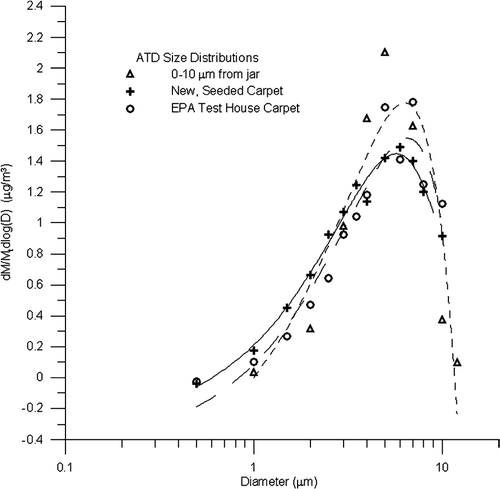
IAM measured size distributions of ATD on individual fibers from new and EPA Test House carpet are shown in . For comparison, the certified size distribution of bulk ATD is provided. Average mass median diameters were 4.0 (± 0.5), 3.6 (± 1.2), and 4.1 (± 0.7) μ m for bulk ATD, new carpet, and EPA carpet. Differences between the three size distributions measured were statistically insignificant (p > 0.05).
FIG. 1 IAM size distributions of 0–10 μ m Arizona Test Dust measured from bulk dust direct from jar, carpet fibers removed from new, seeded carpet, and carpet fibers removed from U.S. EPA Test House carpet. Bulk dust distribution is the certified size distribution provided by the manufacturer. Each distribution normalized by their respective total mass.
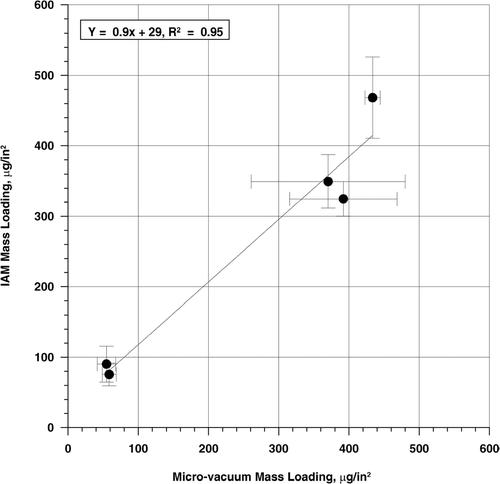
The strong linear relationship between mass loading (mass per unit area sampled) measured on the fibers via IAM and gravimetric analysis of filters collected by the ASTM micro-vacuum method provides evidence that the two methods are comparable (). The IAM and micro-vacuum mass loadings had essentially the same amount of variation in the estimates, as indicated by the similar standard deviations shown in . The comparability between the two methods suggests the calculated mass loadings are representative of the true loading available for resuspension.
FIG. 2 Comparison of ATD mass loading on new carpet measured gravimetrically and via IAM. Two fibers per 1 in2 carpet section were analyzed via IAM. Two filter samples per 1 in2 carpet section were collected via the ASTM D5755 micro-vacuum technique for gravimetric analysis. Carpet seeded with 0–10 μ m Arizona Test Dust.
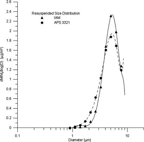
Size distributions of resuspended ATD measured by IAM of the 47 mm filters and the APS are shown in . Size distributions matched almost identically, with mass median diameters of 4.1 (± 1.0) and 4.3 (± 0.8) measured by IAM and APS, respectively.
FIG. 3 Size distribution of resuspended Arizona Test Dust measured by IAM and APS 3321. Heavily loaded carpet from EPA test house was the carpet fiber source. Each distribution normalized by their respective total mass.
Aerodynamic diameters for approximately 2000 particles were measured automatically by the computer controlled SEM (CCSEM) for comparison against the corresponding IAM diameter (). The regression slope of 1.15 was statistically different from unity (p < 0.0001) and the intercept of −0.82 was statistically different from zero (p < 0.0001). Although the count median diameters for IAM (1.28 μ m) and CCSEM (1.38 μ m) were similar, there were differences in measured diameters at the largest and smallest particle sizes. IAM aerodynamic diameters were greater than the CCSEM aerodynamic diameters for the upper end of the size distribution (> 6 μ m), and smaller for the lower end of the size distribution (< 3 μ m). These differences possibly resulted from the differences in conversion factors and image processing techniques.
FIG. 4 Comparison of aerodynamic diameters measured by IAM and CCSEM.
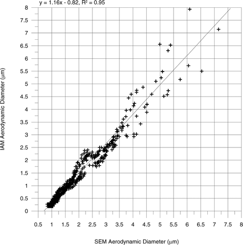
One source is the difference in scaling factors used to convert the particle size to aerodynamic diameters. The CCSEM scaling factor was 0.81, whereas multiplication of the constants in Equations (Equation1) and (Equation2) yields 0.69. The IAM scaling factor used for submicron particles will be slightly higher than 0.69 after inclusion of the Cunningham correction factor.
Another cause for the difference was an artifact caused by the type and size of particles being measured from the SEM images collected for the IAM. In backscattered electron images, feature contrast increases with average atomic number. Particles having low average atomic number, including silicates, are more difficult to distinguish from the carbonaceous carpet fiber background than particles of higher average atomic number. Hence, the IAM threshold level in the image processing software must be set such that phantom particles were generated to adequately distinguish silicate particles from the background. As described previously, removing the phantom particles also decreased the area of real, submicron particles. The step that added pixel(s) to the submicron particles also unnecessarily added pixels to larger particles. The IAM procedure was optimized to balance the number of pixels added against the accuracy of the particle diameters at the ends of the distribution. As a result, the negative offset in the smallest particles was not completely corrected and a positive offset was introduced in the largest particles.
CONCLUSIONS
A new method for measuring particle size distribution and mass available on carpet fibers was developed. The method prepares a carpet fiber for scanning electron microscopy and processes collected SEM images to accurately determine the particle mass and size available for exposure. The current image processing procedure generated a small offset in measured aerodynamic diameter for particles less than 3 μ m or greater than 6 μ m. The difference did not affect the overall size distribution or mass measurements collected in this research because the mass distribution was log-normally distributed with less than 10% greater than 6 μ m. However, application of the IAM to particle size distributions with a greater percentage of particle mass greater than 6 μ m or counts less than 3 μ m would generate significant errors unless the image processing parameters were customized for that specific size distribution. Higher resolution images or better image processing software will be investigated as means to eliminate the bias with a standard set of image processing parameters.
The method is applicable to particles of different composition and structure. This method originally was developed to identify lead particles in carpets from remediated homes. Any metallic particle will be clearly visible under backscatter imaging and easy to size. Ongoing research on particles with high aspect ratios indicates these particles also are easily identified, counted, and sized using this procedure. Future applications to explore include bacterial spores, fungal spores, and pollens. SEM image collection and processing parameters can be customized for dusts with different size distributions and compositions.
The utility of the method will yield improved models by for use in risk assessment and fundamental research. Uncertainty in exposure models will be reduced because the exposure concentration will be directly related to the mass available. Also, the method provides a new tool for conducting fundamental research to explore the mechanisms affecting particle adhesion to surfaces that determine mass availability.
Acknowledgments
Method development was funded by U.S. Department of Housing and Urban Development under Cooperative Agreement # NCLHR0022-97. SEM images were funded by U.S. Environmental Protection Agency under Contract 68-D-00-206. Validation of the method was funded by U.S. EPA under Contract 3C-R185-NALX. Although reviewed by U.S. EPA, this paper may not necessarily reflect official Agency policy.
Related Research Data
REFERENCES
- ASTM . 1995 . Standard Test Method for Microvacuum Sampling and Indirect Analysis of Dust by Transmission Electron Microscopy for Asbestos Structure Number Concentrations , West Conshohocken, PA : ASTM .
- Brouwer , D. H. , Kroese , R. and Van Hemmen , J. J. 1999 . Transfer of Contaminants from Surfaces to Hands: Experimental Assessment of Linearity of the Exposure Process, Adherence to the Skin, and Area Exposed During Fixed Pressure and Repeated Contact with Surfaces Contaminated with a Powder . App. Occup. And Environ. Hyg , 14 : 231 – 239 . [CSA]
- Buttner , M. P. , Cruz-Perez , P. , Stetzenbach , L. D. , Garrett , P. J. and Luedtke , A. E. 2002 . Measurement of Airborne Fungal Spore Dispersal from Three Types of Flooring Materials . Aerobiologia , 18 : 1 – 11 . [CROSSREF] [CSA]
- Davies , C. N. 1979 . Particle-Fluid Interaction . J. Aerosol Sci , 10 : 477 – 513 . [CROSSREF] [CSA]
- Edwards , R. D. and Lioy , P. J. 1999 . The EL Sampler: A Press Sampler for the Quantitative Estimation of Dermal Exposure to Pesticides . J. Exposure Anal. Environ. Epid. , 9 : 521 – 529 . [CROSSREF] [CSA]
- Fenske , R. A. , Curry , P. B. , Wandelmaier , F. and Ritter , L. 1991 . Development of Dermal and Respiratory Sampling Procedures for Human Exposure to Pesticides in Indoor Environments . J. Exposure Anal. Environ. Epid. , 1 : 11 – 30 . [CSA]
- Ferro , A. R. , Kopperund , R. J. and Hildemann , L. M. 2004 . Source Strengths for Indoor Human Activities that Resuspend Particulate Matter . Environ. Sci. Technol , 38 : 1769 – 1764 . [CROSSREF] [CSA]
- Hinds , W. C. 1982 . Aerosol Technology: Properties, Behavior, and Measurement of Airborne Particles. , p. 359 : John Wiley & Sons, New York .
- Lewis , R. G. , Fortmann , R. C. and Camann , D. E. 1994 . Evaluation of Methods for Monitoring the Potential Exposure of Small Children to Pesticides in the Residential Environment . Arch. Environ. Contam. Toxicol. , 26 : 37 – 46 . [PUBMED] [INFOTRIEVE] [CROSSREF] [CSA]
- Lu , C. and Fenske , R. A. 1999 . Dermal Transfer of Chlorpyrifos Residues from Residential Surfaces: Comparison of Hand Press, Hand Drag, Wipe, and Polyurethane Foam Roller Measurements after Broadcast and Aerosol Pesticide Applications . Environ. Health Perspect , 107 : 463 – 468 . [PUBMED] [INFOTRIEVE] [CSA]
- Montoya , L. D. and Hildemann , L. M. 2001 . Evolution of the Mass Distribution of Resuspended Cat Allergen (Fel d 1) Indoors Following a Disturbance . Atm. Environ , 35 : 859 – 866 . [CSA]
- Nishioka , M. G. , Lewis , R. G. , Brinkman , M. C. and Burkholder , H. M. 2002 . Transfer of Lawn Applied Pesticides from Carpet: Comparison of Semivolatile Chlorpyrifos with Nonvolatile Chlorothalonil . Bull. Environ. Contam. Toxicol. , 68 : 64 – 71 . [PUBMED] [INFOTRIEVE] [CSA]
- Noll , K. E. , Fang , K. Y. P. and Watkins , L. A. 1988 . Characterization of the Deposition of Particles from the Atmosphere to a Flat Plate . Atm. Environ. , 22 : 1461 – 1468 . [CROSSREF] [CSA]
- Que Hee , S. S. , Peace , B. , Clark , C. S. , Boyle , J. R. , Bornschein , R. L. and Hammond , P. B. 1985 . Evolution of Efficient Methods to Sample Lead Sources, Such as House Dust and Hand Dust, in Homes of Children . Environ. Res , 38 : 77 – 95 . [PUBMED] [INFOTRIEVE] [CROSSREF] [CSA]
- Rodes , C. E. and Thornburg , J. 2005 . “ Breathing Zone Exposure Assessment ” . In Aerosols Handbook: Measurement, Dosimetry, and Health Effects , Edited by: Ruzer , L. S. and Harley , N. H. 61 – 74 . Boca Raton, FL : CRC Press .
- Thornburg , J. and Rodes , C. E. 2004 . “ Resuspension of Particulate Matter from Carpeted Flooring Surfaces ” . In RTI Report #0886 Research Triangle Park, NC
- U.S. EPA . 1997 . Exposure Factors Handbook , Washington, DC : U.S. EPA Office Research and Development . EPA/600/P-95/002Fa
- Wagner , J. and Leith , D. 2001 . Passive Aerosol Sampler. Part II: Wind Tunnel Experiments . Aerosol. Sci. and Technol , 34 : 193 – 201 . [CROSSREF] [CSA]