Abstract
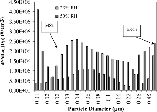
This study investigated the removal efficiency of viable signals of aerosolized bacteria and viruses, stabilized with respect to relative humidity, by simple glass microfiber filters. When examined over an extended time period, relative humidity affected both the size distribution of the stabilized aerosols and the removal efficiency of aerosolized microorganisms by simple glass microfiber filters. The size distribution of the generated humidity stabilized aerosol particles differed, with 50% relative humidity having a greater number of small diameter particles ( < .02 microns) than aerosols generated at 23% relative humidity, and 23% relative humidity having more particles in the range between .02 and .3 microns than 50%. The removal efficiency of aerosols containing viable bacteria (E. coli) and bacterial viruses (MS2) initially showed greater than 5 logs of removal (99.999%) at both 23% and 50% RHs for both aerosolized microorganisms. Increased RH was related to improved removal of viable aerosolized bacteria and viruses at all timepoints measured over a 60-minute test period. RH had more impact upon removal efficiencies for MS2 bacteriophages than E. coli bacteria, with removal efficiencies for MS2 at 50% RH declining after 30 minutes to levels seen at 23% RH. Some interesting findings of this study were that the two microorganisms that were mixed into a single cocktail at similar concentrations to generate the aerosol apparently did not associate, to a large extent, in the same aerosol particles, as inferred by significant differences in their removal behavior at higher RH of 50%. This study shows that the relative humidity of an aerosol-containing stream should be considered as an important experimental control variable, and that the removal of aerosolized viruses cannot be predicted from bacteria.
INTRODUCTION
It has been recognized that indoor bioaerosols can cause sick building syndrome (SBS) and building-related illness (BRI) (CitationAnderson 1992; CitationBurge 1995). BRIs are generally related to allergic reactions and infections (CitationDennis 1990). In testimony to the United States House of Representatives about the aircraft cabin environment, it was stated that bacterial and viral aerosols were common cabin air contaminants, and that there was evidence of transmission on aircraft of infectious disease agents, even the bacterium that causes tuberculosis (CitationHoward 2003). Further, occupationally acquired TB has become a serious concern since 1990. TB strains are acquiring multiple antibiotic resistance and the disease is considered re-emerging (CDC 1990). However, bacteria are not the only aerosolized disease agents of concern in closed spaces. Another emerging disease, severe acute respiratory syndrome (SARS), caused by a newly detected coronavirus, has become prevalent in Asia and is expected to spread. Studies have shown a potential for SARS to be transmitted by airborne routes, although close contact is required and it is thought that the low humidity of cabin air will minimize the potential for transmission. These re-emerging and newly emerging diseases have been challenging researchers to devise better ways to control indoor bioaerosol contamination, while meeting energy and cost-effective design constraints. There are three recommended strategies for reducing pollutants in the indoor air: source control, ventilation, and air cleaning. Source control is limited because not all pollutant sources can be identified and practically eliminated, or reduced. Even though air cleaning devices using ultraviolet radiation have reduced airborne bacteria in residences, schools, and hospitals (CitationLai 1999; CitationMorris 1972), it is believed that the use of the air cleaning devices alone cannot assure adequate air quality, particularly when the significant sources are present and the ventilation is inadequate, such as the typical indoor environment. Ventilation can be effectively implemented using higher air exchange rate. However, air exchange rate can only be increased so far due to the costs of heating or cooling large volumes of incoming air. Therefore, treatment of circulated indoor air using filtration in addition to mechanical ventilation and other air cleaning devices is common, especially for commercial aircraft.
There are two basic types of filters for removing particulate matter from the atmosphere, electrostatic and mechanical. Electrostatic filters with high removal efficiency are generally used exclusively at industrial sites, while mechanical filters can be widely used in residential and commercial buildings and aircraft. In general, a HEPA filter is defined as having a minimum efficiency of 99.97% removal of small particles (approximately equal to 0.3 μ m) from air. These filters may be constructed from a variety of materials. Researchers have focused on synthesizing filters from novel materials to increase the removal efficiency of various pollutants (CitationJagtoyen et al. 1996; CitationPowell et al. 2000; CitationSun et al. 2001). However, earlier studies on glass fiber, one of the cheapest synthesized materials, found that simple glass microfiber filters had high removal efficiencies and removal of bioaerosols from the indoor air at least as great as the novel and expensive materials developed later (CitationDecker et al. 1951; CitationDecker et al. 1954). Therefore, the use of glass microfiber filters in addition to increased ventilation rates may provide an additional, inexpensive measure of protection.
Decker et al. (Citation1951 and Citation1954) showed the potential of inexpensive glass microfiber filters for removing bioaerosols from air. The glass microfiber filter is a depth filter that relies upon removal by a tortuous path through the media for nominal sizes. This type of binder-free glass microfiber filter was specified in official methods for the testing of suspended solids in water pollution monitoring, and has demonstrated very high loading capacity and retention efficiency at high fluid flowrates. Glass fiber filters have been used in the gravimetric determination of airborne particulate matter, stack sampling, and absorption methods of air pollution monitoring (CitationDecker et al. 1954).
The one-pass microbial challenge experiments of Decker et al. (Citation1951 and Citation1954) were not optimally designed, or controlled, for evaluating biological test aerosols removal from continuously operating systems. T3 bacteriophages were used to represent the removal of other viruses that could be aerosolized. The removal efficiency using binder-free glass fiber paper reached 99.99%. However, this phage group can be easily inactivated by damage to its fragile tail fibers and is not considered an appropriate surrogate for more hardy viruses. The choice of a non-hardy surrogate can result in the over-estimation of filter removal efficiencies and underdesign of air-treatment systems, so it is worthwhile to re-investigate the glass microfiber filter removals with a more suitable bacteriophage group. The one-pass filter experiments did not allow for filter breakthrough to be investigated. Lastly, relative humidity was not considered as a control variable. In an effort to add to this preliminary work, and expand the body of knowledge on removal of aerosolized microorganisms, this study used a hardier bacteriophage (MS2) combined with a system that allowed for the continuous control of relative humidity in the generated aerosol stream to investigate breakthrough of viable microorganisms applied to inexpensive, glass microfiber filters.
The selection of benign microorganisms to model the behavior of pathogens in engineered systems is a common practice, and the surrogate microorganisms are chosen to closely model or mimic the physical attributes of the pathogens of concern. E. coli has been successfully employed as a bacterial surrogate in many bioaerosol studies (CitationPhillips et al. 1964; CitationDavis and Morishita 2005; CitationTanner et al. 2005). However, it is well known that bacteria are not appropriate surrogates for investigating virus behavior, so another surrogate is required. The male specific, bacteriophage group designated as MS2 has been used in other aerosol studies (CitationFoarde et al. 1999). This group has been used as surrogates for studying the behavior of human enteric viruses in aqueous systems with good success (CitationKott et al. 1974; CitationHavelaar et al. 1995; CitationGerba and Goyal 1981). Because this phage group is less likely to adsorb than poliovirus, its adsorptive behavior is thought to provide a conservative estimate of sorption to activated carbon and other charged surfaces (CitationPowell et al. 2000).
For all the bioaerosol studies referenced prior that sought a culturable signal, the surrogate bacteria or bacterial virus group was generated individually. However, this research wanted to capture a more realistic scenario with both types of microorganisms aerosolized simultaneously from a single source. Utilizing a bacterial strain that does not possess the capacity to produce a pili, with a bacteriophage that requires the pili to attach to the bacteria, allows the mixture of both bacteria and bacteriophages into a single cocktail for aerosol generation.
The objective of this study was to design, construct, and evaluate a system for investigating the removal efficiency of simple glass microfiber filter materials for the viable signal from aerosolized bacteria and bacteriophages under different relative humidities and add to the current body of knowledge. The filter challenge system and the protocols developed for its use were evaluated for their potential to serve in future investigations of new chemically treated, biocidal filter materials by this initial study with simpler glass microfiber filters.
METHODS
Filters
Filters used for the aerosol challenge were simple Whatman GF/A glass microfiber filters (Fisher cat#:1827047, Whatman, Clifton, NJ). These glass microfiber filters are made of borosilicate glass without binder, have a liquid particle retention size of 1.6 μ m, a diameter of 47 mm, a thickness of 0.26 mm, high loading capacity, a wet burst strength of 0.29 psi, a Hertzberg filtration speed of 62, and demonstrated low resistance to airflow. Teflon filters (Model FGLP04700, Millipore Corporation, Bedford, MA; charge-neutral filter media, nominal pore size of 0.2 μ m, diameter of 47 mm) were used after the impinger as part of the signal collection system to ensure maximum capture of aerosolized microorganisms before exhausting the air flow into a fume hood.
Test Microorganisms
Bacteria
A non-pathogenic strain of Escherichia coli (E. coli) was used as the surrogate for pathogenic Gram-negative bacteria. E. coli is a rod-shaped, 1∼ 3 μ m, facultative anaerobe that colonizes the lower gut of animals, but also survives when released into the environment (CitationBlattner et al. 1997). A pure culture of Escherichia coli CN13 (E. coli CN13) was acquired from the American Type Culture Collection (ATCC) (ATCC#: 11303, Manassas, VA) and cultured in Tryptic Soy Broth (TSB) overnight at 37°C until log-phase growth was observed. Standard pour plate, double-layer agar assay was used for viable counting. Briefly, 0.1 mL of sample, or a dilution thereof, was added to a culture tube containing 5-mL of liquefied 0.7% top agar that had been autoclaved then cooled to 48°C ± 1.0°C. The mixture was swirled then poured evenly over a solidified 1.5% bottom agar layer held in a Petri dish. After cooling to harden the top agar layer, the Petri dish was incubated 18–24 hours at 37°C. Bacterial colonies were counted with the aid of a magnified backlit counter. All samples were assayed in duplicate, or triplicate, for each dilution examined. The average number of colonies present on the Petri dish showing the most countable dilution was used to calculate the concentration of bacterial colonies per mL of original sample (CFU/mL). The concentration of E. coli in the overnight stock was approximately 1 × 10⁹ cfu/mL and this solution was put into the nebulizer without dilution for aerosol generation and filter testing.
Bacteriophages and Host
MS2 are non-enveloped, RNA containing, icosahedral bacteriophages with a molecular weight of 3.6 × 10⁶,and diameter of about 27 nm, and its structure is close to that of naked human enteric viruses (CitationVan Duin et al. 1988). Stocks of the F-specific RNA coliphage MS2 (ATCC 15597-B1) group were prepared by scraping soft, top agar that showed confluent lysis into small volumes of phosphate buffered saline (PBS) solution. Equal volumes of the crude mixture and chloroform were blended into an emulsion, then centrifuged at 5000 × g for 20 minutes until a clear, straw-colored layer of high titer of approximately 1010 plaque forming units (PFU)/ml virus-containing supernatant could be recovered. This virus stock was refrigerated at 4°C for periods of no longer than 2 months until used in the experiments (CitationBrion and Silverstein 1999). Immediately before each set of experiments, aliquots of virus stock were diluted into PBS and retitered as a positive control. All samples and stocks were assayed by the double agar layer technique as described by CitationAdams (1959), with host E. coli strain F-amp cultured to log-phase in TSB with streptomycin and ampicillin added to amounts specified in EPA method 1602. Nutrient agar (Ref#:214010, Difco) was used for assay agars and clearly defined plaques that formed in the bacterial lawn of the top layer of 0.7% agar resting on a support layer of 1.5% agar were counted and recorded as plaque forming units (PFU) after 18–24 h incubation at 37°C with the aid of a magnified backlit counter. All samples were assayed in triplicate for each dilution examined with the average number of plaques present in the most countable dilution used to calculate PFU per mL in the original sample.
Test Systems
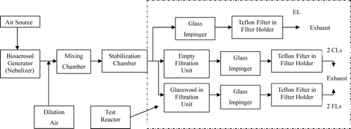
The experimental set-up overview is shown in . This system provided the means to generate aerosols containing airborne bacteria and viruses, a stabilization chamber to control humidity and stabilize the aerosols produced by the nebulizer, and a manifold system that allowed for steady-state operation with multiple filter lines receiving equalized flows at a stable flow rate. The Air Source was industrial-grade compressed air delivered to the system by a dual-stage regulator through a high-efficiency-particulate-air (HEPA) filter (Model Hepa-cap 75, Fisher Scientific, Pittsburgh, PA), and a calibrated flow meter. In order to lower the humidity in the stabilization chamber, dried dilution air was added right after the nebulizer. The source of the dried dilution air was from laboratory compressed air line passed through a drying tube filled with DriRite, a HEPA filter, and a calibrated flow meter. Aerosols were generated by an air-jet nebulizer (Collision six-jet, BGI Inc., Waltham, MA) operated at 10 psi (CitationIp and Niven 1994). The nebulizer was filled with 50 mL of a mixture of stock cultures of E. coli(48.5 mL 1 × 10⁹ cfu/mL), and MS2 (0.5 mL approximately 2.0 × 10¹¹ pfu/mL), so that the final concentration of bacteria and bacteriophages was approximately 1 × 10⁹ cfu/mL and 2 × 10⁹pfu/mL, respectively.
The aerosols with airborne microorganisms were then delivered from the nebulizer discharge port into the mixing chamber through a length of 2-cm flexible polypropylene tubing. The mixing tube was a grounded copper tube, 83 cm long and 2.67 cm in diameter that allowed for turbulent flow. The stabilization chamber received the mixed aerosol stream and provided a region of slower flow where larger aerosol particles could sediment, smaller particles mature, and humidity control be applied, and this chamber was a cylinder made of plexi-glass with dimensions of 8.9 cm diameter and 35.6 cm length. The residence times for the mixing chamber and stabilization chambers were 5 and 16 seconds, respectively, at 6 L/minute used for this study. The temperature and relative humidity inside the stabilization chamber were measured by a thermohygrometer (Model DHTD, Fisher Scientific, Pittsburgh, PA).
After time in the stabilization chamber, air stream flowed into the test reactor section of the system. The first step in the test reactor was a flow manifold attached to 3 lines: an Equilibrium Line (EL), Control Line (CL), and Filtration Line (FL). Each of the CL and FL had another manifold in place after the filtration unit (where the glass microfiber filter could be held) that created two, replicate flow paths for the collection of aerosols by an impinger followed by a Teflon filter before exhausting into the hood. These multiple paths were used to accomplish collection of control and filtered samples without interrupting the established flow regime through the CL and FL established for each experiment. One pathway would be maintained in active flow mode while the other was valved off allowing for new impingers and Teflon filters to be put into place while the experiment continued. The EL was used to allow the system to come to steady-state before testing by running at set conditions for at least 5 minutes and discharging the total airflow out of the EL to the interior of an operating fume hood for exhaust after removal of airborne microorganisms by a collection system consisting of a liquid filled (10 mL PBS) impinger (cat#:225-36-2, SKC) followed by a Teflon filter. The time to establish steady state conditions was established by initial CO2 gas experiments (CitationWang 2005). The CL had the same collection system as the EL to collect the airborne microorganisms into a liquid filled impinger, after passing through an empty (no glass microfiber filter in place) filter holder. As mentioned for the EL, all air flow through the CL passed through a Teflon filter before exhausting into the hood. The FL was where the airborne microorganisms and accompanying aerosols were filtered through glass microfiber filters held in autoclavable 47-mm filter holders. Viable airborne microorganisms that passed through the glass microfiber filters held in the FL filtration unit were captured in a collection unit consisting of a liquid filled (10 mL PBS) impinger (cat#:22-536-2, SKC), followed by a Teflon filter as described above. The flowrates passing through EL, CL, and FL were 6, 3, and 3 L/min, respectively.
Filter Material Testing at Different Relative Humidities
The filter challenge system was first evaluated for flow equality and leakage using CO2. The test reactor's stability within the CL or FL lines, and reproducibility between CL and FL lines were evaluated using a mixture of nebulizer aerosolized microorganisms. After these initial evaluations that showed the system could be operated with equal flow and loading through all lines and pathways, the effect of humidity on glass microfiber filter removal of aerosolized microorganisms was then evaluated. This was done by collecting the nebulizer generated aerosols from the air flow that passed simultaneously through the CL and FL with impinger based collection systems placed on the various lines out of the filtration units and comparing this signal. The FL filtration unit was loaded with glass microfiber filters and challenged under two different relative humidities, one dry (23%) and one humid (50%), with aerosols that contained viable surrogate microorganisms. After the system was stabilized for five minutes with all flow going out the EL, the manifold valves to the CL and FL were opened, and the valve to the EL was closed. Then continuous aerosols collection over discrete periods (10 minutes initially increasing by 10-minute intervals to 60 minutes total) was achieved by swapping impingers and Teflon filter holders into and out of the two identical CL and FL paths. After aerosol collection, the Teflon filters were placed in a 50 mL conical tube containing 10 mL PBS, secured on a vortex machine (Cat. No.: 12-812, Fisher Scientific) and shaken for five minutes to remove any additional bacteria and viruses that may have passed through the impinger. The recovered PBS was combined with the 10 mL of PBS from the glass impinger, making the total final volume of sample 20 mL. The combined solution was then analyzed for viable E. coli and MS2. The removal efficiency of tested glass microfiber filter (%RE) was obtained by Equation (Equation1).
-
NCL: Total viable microbes collected in CL by bioaerosol collection unit (#cfu or #pfu)
-
NFL: Total viable microbes collected in FL by bioaerosol collection unit after passing through tested glass microfiber filter (#cfu or #pfu)
This calculation allowed for the removal occurring from filtration through the glass microfiber filters to be separated from the losses of aerosolized microorganisms to the system.
Glass microfiber filter material was the only filter material tested, and the temperature in stability chamber was maintained at room temperature (20–22°C) for each test. The flow rate was constant at 3 L/min. There were two variables: Relative Humidity and microorganism type. For each relative humidity and microorganism, filtration efficiency over time was evaluated. Each experiment was repeated three times and the average results calculated. The experimental conditions are summarized in . All the media, buffer solution, tested filters, the filter holders, the connection tubes, and impingers were autoclaved before each experimental run.
TABLE 1 Experimental conditions
Under the methodology and testing conditions described as above, the method of the detection limit (MDL) was 1.1 microorganisms per Liter air.
Weight Gain Testing of Glass Microfiber Filter at Different Relative Humidities
The system was set up with glass microfiber filters that had been dried and brought to a steady weight in a desiccator, and placed into the filtration units on the FL to measure the weight gain of the filters over time. The system was run at 23% and 50% relative humidity with two filters evaluated for each humidity simultaneously, one held in each of the two flow paths of the FL. After a 10-min filtration time, the filters were taken from filtration units and weighed. Then the filters were immediately placed back into the same filtration units for another 10-min filtration interval. This process was repeated six times over a period of 60 minutes. The percentage of weight gain of the filter was calculated by Equation (Equation2).
-
W0: The weight of the tested filter before 10-minute filtration (g)
-
W10: The weight of the tested filter after 10-minute filtration (g)
Statistical Analysis
All data (#microorganism/mL) for each 10-minute interval were log-transformed before statistical analysis per microbial convention to normalize data distributions. A statistical software package (SigmaStat) was used for all statistical analyses. Removal efficiencies under different relative humidities were compared to see if there was any significant difference by t-test and ANOVA. The hypothesis testing, using the Student t distribution, was set for a null hypothesis of equal values, or equal removal efficiency, under the two different relative humidities, two-sided, at α = 0.05. The setting for the ANOVA was α = 0.05.
RESULTS
System Integrity
A mass balance system evaluation using CO2 gas as a conservative tracer showed that it took approximately 1.6 × system detention time for the system to reach steady-state. The system maintained integrity (no leakage) at 10 psi for 2 hours. Evaluation of the reproducibility and stability of multiple test reactor lines and pathways for bioaerosols containing E. coli and MS2 was done, and showed that high concentration of microorganisms referred as bioaerosol signal was consistently generated and passed evenly throughout the reactor. Both the control line (CL) and filtration line (FL) showed consistency of signal over long time intervals. The system mass balance did show approximately 90% of the calculated viable microorganism signal (based upon the amount of fluid that left the nebulizer multiplied by the viable titer in the nebulizer fluid) was lost, presumably to condensation of large aerosol droplets in the mixing and stabilization chambers of the system, as well as system tubing. However, the measured viable concentrations captured by the collection systems (impinger + Teflon filter) on each line and pathway within a line showed no significant difference. For E. coli and MS2, calculated viable microorganism signal losses for E. coli and MS2 were similar, at 92% and 90%, respectively. Detailed results and discussion regarding the complete system evaluation can be read elsewhere (CitationWang 2005).
Microorganism Capture Efficiency of Impinger
The collection system consisted of an impinger filled with 10-mL of PBS solution followed by a Teflon filter in an autoclavable filter holder. The microorganism capture efficiency of the impinger was investigated under the steady conditions set in this study. For both microorganisms captured by the entire collection system in this study, 99.4% of E. coli and 96.4% of MS2 were captured by the impinger with 10-mL PBS solution under 10 psi at 3 L/min actual flow rate. The remaining microorganisms that passed through the impinger were caught on the Teflon filter.
Aerosol Size Distribution
As shown in , the size distribution was dispersed over a large range and the size distribution of bioaerosols, before entering the filter measured at 23% and 50% relative humidity using a wide-range particle spectrometer (WPS, Model#: 1000XP WPS Configuration A, Shoreview, MN, 55126), was quite different. At 50% relative humidity, there were a greater number of small diameter particles (< .02 microns) than aerosols generated at 23% relative humidity. At 23% relative humidity, there were more particles in the range between 0.02 and 0.3 microns than 50%. Since the solution in the nebulizer was comprised of nutrient broth, it had a lot of salt that would aerosolize. Therefore, the size of E. coli and MS2 could not be related to the peaks in the size distribution specifically and the size distribution alone cannot tell if E. coli and MS2 were associated in the same droplets or separated from each other before they were captured by the filters tested in this study. What is quite clear is that under the different relative humidities, the distribution shifts with a great number of particles smaller than the MS2 bacteriophages present at higher humidity. The increase in very small size particles at 50% RH shown in might be due to hygroscopic growth of nuclei particles that were not detectable at 23% RH, since test organisms were suspended in PBS containing sodium chloride and other salts that were hygroscopic, which would cause particle size changing with RH.
Bioaerosol Removal Efficiency of Glass Microfiber Filter at Different Relative Humidities
Input Signals
The concentration of viable microorganisms as an input signal to the tested filters was continuously monitored by being assayed directly from the fluid obtained from CL every 10 minutes. ANOVA results showed a consistent signal input for filter testing per 10-minute period (). The mean signal of E. coli and MS2 per 10-minutes during filter testing was 108.75 cfu and 107.88 pfu caught per 10-minute loading period, respectively.
TABLE 2 Loading signal strength for E. coli, and MS2 during filtration
General Observations of Bioaerosol Removal Efficiency by Glass Microfiber Filters at 23% and 50% Relative Humidity
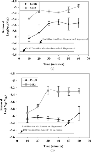
The removal efficiency (Log (NFL/NCL)) of glass microfiber filters for capturing viable, aerosolized E. coli and MS2 at 23% and 50% relative humidity are shown in and , respectively. Both figures clearly show that the bioaerosol removal efficiency of simple glass microfiber filters with relatively large nominal pore sizes (1.6 μ m) for both microorganisms was initially greater than 5 logs of removal (99.999%) at both 23% and 50% relative humidities. E. coli nearly achieved its theoretical maximum removal at the beginning of the filtration at both relative humidities, while MS2 was farther from the theoretical maximum removal. Removal efficiencies for E. coli were always greater than for MS2. MS2 initial removal efficiency was clearly impacted by relative humidity, with less removal at the first time interval measured at 23% relative humidity (5.4 logs) as compared to 50% (5.9 logs).
E. coli Removal Efficiency Over Time at Different Relative Humidities
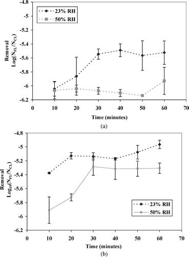
Replotting the removals displayed in allows for a visual comparison of the effect of relative humidity on each type of aerosolized microorganism. As shown in , for E. coli, the initial removal efficiency at the two different humidities is similar, but changes as time passes with removals at 23% relative humidity decreasing with increased filter time. The E. coli removal at 50% relative humidity is relatively constant for the entire time period while the removal at 23% relative humidity quickly decreases then stabilizes after 30 minutes. In general, greater removal was noted at 50% relative humidity than 23%. A paired t-test showed that relative humidity was a significant factor affecting the removal efficiency of E. coli removal for all points after the initial 10-minute sampling (p = 0.116).
MS2 Removal Efficiency Over Time at Different Relative Humidities
The effects of relative humidity on virus removal as modeled by MS2 are illustrated in and is the inverse of the pattern noted for E. coli bacteria. The initial removal efficiency for MS2 was quite different with respects to humidity. 50% relative humidity initially had the greatest removal (5.9 logs), but this decreased after 20 minutes, approaching the removal at 23% relative humidity (5.2 logs). The overall removal efficiency at 50% relative humidity was significantly greater than at 23% at the first two 10-minute intervals (paired-t test, p = 0.0085). However, the significance of this difference did not persist for the timepoints after that, even though the sixth 10-minute sampling was close to significant. It seems that relative humidity effects viral removal rate of using glass microfiber filtration initially, but as the filter saturates, removal becomes more constant. Whereas for E. coli bacteria, filter rate was not initially different, but became different with filter saturation. Clearly, filter saturation impacts virus removal by glass microfiber filtration differently than bacterial in these studies.
Weight Gain of Filter and Bioaerosol Removal Efficiency at Different Relative Humidities
Weight Gain of Filters
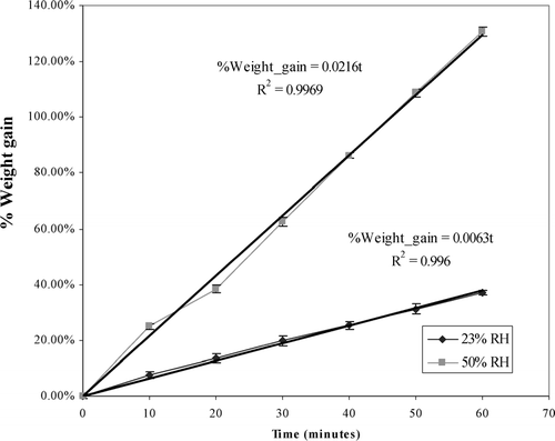
From the prior results, it was inferred that conditions within the glass microfiber filter were changing as time progressed. Some type of filter saturation was taking place and weight gain by the filter, presumably from fluids deposited from the aerosol stream, was a rational choice for investigation. The fluid content adsorbed to the glass microfiber filter during the time intervals used at two different relative humidity was measured. The change of weight content for the glass microfiber filters in percentage is shown in . During the 60-min filtration, the fluid content absorbed by glass microfiber filters kept increasing over time for both humidities, but the total fluid content gained by glass microfiber filters was quite different. At the end of the 60 minute filtration period, the filter weight gain at 23% relative humidity was only 37% of the initial filter weight, while it reached 130% at 50% relative humidity. It was hypothesized that this difference in filter weight gain was related to the differences seen in the removal of bacteria and viruses over time.
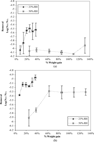
Relationship between Weight Gain and Removal Efficiency for E. coli
As shown on , the removal efficiency for E. coli at 23% relative humidity decreased with the increase in percentage of weight gain of the glass microfiber filter until the percentage of weight gain of the filter reached approximately 20%. Then, the removal efficiency was no longer a function of the percentage of weight gain anymore. At 50% relative humidity, the percentage of weight gain of glass microfiber filter was not a function of the removal efficiency since the percentage of water gain of filter had already reached over 20% at the first filtration interval. It appears from this observation that a weight gain of 20% is required for consistent removal by glasswool filtration, and that filtration efficiency is greater at higher humidity for E. coli bacteria.
Relationship between Weight Gain and Bioaerosol Removal Efficiency for MS2
As shown in , the removal efficiency for MS2 at both relative humidities examined decreased with weight gain in the filter. The removal efficiency for MS2 at 23% relative humidity decreased slightly with increased weight gain during the whole one-hour filtration period till the maximum 40% weight gain had been obtained. Looking at the removal results for 50% relative humidity, removal decreased as fluid retained in the filter increased until a level of 60% gain had been reached. Then the viral removal efficiency for MS2 became stabilized. It appears that filter saturation as measured by weight gain of the filter for MS2 removal requires a higher fluid content than for E. coli bacteria.
DISCUSSIONS
The Effect of Humidity on Bioaerosol Removal by Glass Microfiber Filter
As expected, the removal efficiency for the bacteria E. coli was the greatest at both humidities when compared to the virus MS2. This is thought to be due to the larger size of the E. coli bacteria (2 × 1 μ m) as compared to MS2 (0.027 μ m). Physical straining of the bacteria is more likely for bacteria with the 1.6 μ m nominal pore size. However, removal efficiency for MS2 was quite high especially considering its small size (0.027 μ m) relative to the nominal pore size of the glass microfiber filter (1.6 μ m). These results demonstrate that physical straining plays an important role for E. coli bioaerosol filtration, but that other factors impact viral removal. The difference in the behavior of the two surrogates used emphasizes the unsuitability of bacteria to predict viral behavior, even in air treatment systems designed with the simplest of materials, glass fiber. At 23% relative humidity, more viral particles passed through the filter than at 50% relative humidity, which suggests a change in the size of the bioaerosol particle that the virus is associated with, a change in the filter material properties, or both.
For E. coli, the finding that relative humidity does not significantly effect the initial removal efficiency agrees with another researcher's finding (CitationMcCullough 1996), who found that relative humidity was not a statistically significant factor in bioaerosol penetration by measuring viable signal of Mycobacterium abscesses for varieties of personal respiratory protection filters at time intervals as short as 10 seconds. In this earlier study, filter removal over extended time intervals was not investigated, so we cannot compare if the behavior we saw, less removal of bacteria at lower humidity after 10 minutes, would have been seen. Only the initial removal from approximately 10 L of air passed through filter materials for 10 seconds was recorded. In our study, the bioaerosols that passed through the filter were collected every 10 minutes for one hour, and this type of protocol more closely mimics the actual use of a filter-based system where the effect of humidity develops over time, as the filter ripens and becomes saturated with fluid.
Since the MDL for E. coli was #33 for each sample collected during 10-minute filtration period (#33/30-L air), and there was no viable E. coli detected for the first five filtration period at 50% RH (), it was not certain the breakthrough observed at the beginning of the sixth filtration period was the real beginning of it, or the breakthrough has started much earlier. For MS2, the breakthrough was observed during the first 10-minute filtration period at both humidities investigated.
Although the bioaerosol removal efficiency using bacteriophage as a viral surrogate has been studied, the removal efficiency varying at different relative humidities over time has not yet been done. Therefore, no relevant findings were found for comparison with this study. In general, the large removal efficiencies for the surrogate virus selected, MS2, were greater than initially expected (> 5 logs removal) and above that required of a true HEPA filter (> 99.999% or 5 logs removal). Only after 60 minutes, at 23% relative humidity, did the removal of MS2 virus drop to less than 5 logs.
The Role of Fluid Weight Gain of Glass Microfiber Filter and Possible Mechanisms in Bioaerosol Removal at Different Relative Humidities
It is thought that weight gain assisted E. coli filtration by straining due to the decrease of the effective pore size of the filter once the filter reaches a 20% gain. It is also thought that straining interception, and inertial impaction are the major mechanisms for the sustained E. coli removals by glass microfiber filtration seen at 50% relative humidity, and that this is due to changes in the filter as well as changes in the aerosol sizes. Since the majority of bioaerosols containing E. coli could be greater than 1.6 μ m (about the average size of a naked bacteria) in the more humid environment due to associated fluids, the depth filter has a good opportunity to hold back these larger particles by straining. At 23% relative humidity, a good many of the aerosol particles containing E. coli may be smaller than those generated at 50% relative humidity, therefore the mechanism of interception, which has been shown to be the major mechanical mechanism for rod-shape bacterial bioaerosol removal by depth filtration in water studies, might be predominate (CitationMaus and Umhauer 1997). It was observed that the filtration efficiency decreased with fluid weight gain for E. coli at 23% RH till it hit 20% weight gain (after 30-minute filtration), while this trend was not observed at 50% RH since the fluid weight gain has reached more than 20% (∼ 25%) just after 10-minute filtration. This result suggested some other mechanisms might have played dominant role before the fluid weight gain reached certain level during the filtration process, such as electrostatics, which would decrease the filtration efficiency with the increased fluid weight gain due to the neutralization of charges (CitationMoyer and Stevens 1989). However, the role of such mechanism needs to be further studied and approved in the future research.
Higher humidity can impact filter performance over time with respects to bacteria removal as these simple glass microfiber filters have no binder to help maintain their integrity under sustained use. At 50% relative humidity, there was an E. coli breakthrough at the last 50- to 60-minute filtration interval after 50 minutes of relatively steady removal. This may be due to defects that formed in the glass microfiber filters. The filter became so wet that it started falling apart. The penetration of E. coli started when the filter's fluid content was approximately 110% of filter weight, and the wide variance seen around this last time interval is thought to be due to differences in the degree of filter integrity during the replicate trials ().
Since the primary mechanisms for viral filtration include interception, diffusion, impaction, and electrostatic attraction, factors that interfere with the flow path or material properties would change viral removal (CitationHinds 1982). However, the increase in filtration that would be expected by a reduction in nominal pore size in a depth filter may be reduced by the reduction in filtration that can occur as charges are neutralized in the filter. Viral particles are generally net negatively charged in wet aerosols while borosilicate glass microfibers contain positively charged ions. Therefore, electrostatic attraction might be expected to be part of filtration removal mechanism for viral aerosols. Prior study has shown that moisture retention in filters may neutralize filter media charges, thus reducing the collection efficiency for small charged particles by electrostatic filters (CitationMoyer and Stevens 1989). By the results presented for this study, it seems that the neutralization of surface charges in glass microfiber filters may result in reducing filter efficiency up to a certain moisture content for aerosolized viruses (60%). After that content has been reached, there is no more reduction of filtration efficiency, even as fluid induced weight gain of the filter doubled to 120%. At 50% relative humidity, the charge neutralization by moisture could have played an important role at the beginning of filtration. However, the effect could not be realized after the majority of charge on filter had been neutralized. The removal efficiency of glass microfiber filters at 60% water gain for aerosols in 50% relative humidity started to level off, and held steady for the remaining time intervals, showing that the other removal mechanisms were not significantly affected by filter weight gain. At 23% relative humidity, the electrical charges on filters might have been neutralized by the moisture gathered by the filters, but the viral removal efficiency of glass microfiber filter kept declining because the increase never reached the critical 60% seen in the higher humidity results. The role of electrostatic attraction in the bioaerosol filtration process needs to be further studied and approved in the future.
Possible Separation Between E. coli and MS2 During Bioaerosol Generation
There are suggestions that the MS2 virus did not aerosolize into the same particulate matter as the E. coli bacteria. Looking at the removals for both microorganisms at the two different humidities, it can be seen that removals of MS2 and E. coli are similar (within the error bars surrounding the average removal) only for the first time point at 50% relative humidity. For all other samples MS2 removal is always significantly less than that for E. coli. Since the microorganisms were in similar concentrations to begin with, this constant difference in removal efficiency suggests that they are not associated together in the same aerosol particles to a great extent. The extreme difference in the pattern of removal efficiency at 50% relative humidity continues to support the supposition that the MS2 bacteriophages were in different particles than the bacteria. Removal for the bacteria is constant, while removal for the viruses decreases over time. If these microorganisms were in the same particle, the removal efficiency patterns should match. Further research is needed to confirm what size particles the viruses and bacteria are generated into, but the results presented herein indicate that they may not be significantly associated.
It was shown that at the different relative humidities employed, the size distribution of small particles differed, and this may be related to the removal efficiency for MS2. At 23% RH, the majority of particles measured were contained in a peak that included small aerosols the size of MS2 viruses. This may be one explanation why MS2 containing particles started breaking through the filter at levels farther from the theoretical removal immediately at 23% relative humidity as compared to 50% relative humidity. The MS2 bacteriophages were in larger, more easily removed aerosol particles at 50% humidity, but as the filter became saturated with moisture, and charges within the filter became neutralized, the removal efficiency decreased to levels very near each other. This shows that two factors were impacted MS2 removal, size of aerosol and possibly charge in the filter media.
The observed pattern of similar removal after time for filter saturation does not occur for removal of the E. coli containing aerosols. This suggests that the factors for removal are different, and that the two microorganisms were not greatly associated. Removal for E. coli over time was stable for 50% relative humidity. The difference in removal patterns stability of removal at 50% humidity may be related to the size fraction of the E. coli carrying aerosol particle, but the measured distribution only went as high as the smallest dimension of the bacteria, so we cannot relate particle size fractions with filtration factors as for the MS2 bacteriophage. However, it is clear that increased relative humidity was associated with improved removal at all filtration time intervals examined for both bacteria and bacteriophages, even though the relative importance of filtration mechanisms varied.
CONCLUSIONS
Simple glass microfiber filters are capable of removing aerosolized bacteria and bacteriophages to levels obtained by more expensive HEPA filters. Relative humidity impacts wet bioaerosol removal by simple glass microfiber filters, with greater humidity associated with greater removal in general, but the impact of charge neutralization decreases the removal efficiency for aerosolized bacteriophages. Future studies measuring bioaerosol removal using different filter medias should consider RH as an important control variable and the moisture content of filters should be measured and considered when evaluating the results, especially for long-term filtration studies and for viruses that are attracted to charged filter materials. Furthermore, researchers should not only concentrate to the characteristics of different filter medias, flowrate, and relative humidity, but also pay attention to the distribution of the generated aerosol under different humidities. Since it was observed that viruses did not appear to associate to a great extent with bacteria in the same aerosolized droplets, and that the removal patterns for viruses and bacteria differed, bacterial surrogates cannot be used for predicting the behavior of viral microbes in bioaerosol future studies.
Acknowledgments
This research was supported by Center of Applied Energy Research (CAER) and Environmental Research and Training Laboratory (ERTL) of University of Kentucky.
Notes
*There were 3 replicates for each combination of three variables tested.
*If there is difference for loaded bioaerosol signals for all six filtration tests at two different RHs. Input Signal Strength is the total number of cfu or pfu per 10-minute sampling period.
REFERENCES
- Adams , M. H. 1959 . Bacteriophages , 50 Interscience .
- American Thoracic Society . 1992 . Control of Tuberculosis in the United States . Amer. Rev. Resp. Dis. , 144 : 1623 – 1633 .
- Anderson , R. C. 1992 . Indoors, The Newest Polluted Space . Pollution Engineering. , 24 : 58 – 60 .
- Burge , H. A. 1995 . Biological Contamination of Buildings in Temperature Climates . Proceedings of Healthy Building. , 1 : 239 – 249 .
- Blattner , F. R. , Plunkett , G. , Bloch , C. A. , Perna , N. T. , Burland , V. , Riley , M. , Collado-Vides , J. , Glasner , J. D. , Rode , C. K. , Mayhew , G. R. , Gregor , J. , Davis , N. W. , Kirkpatrick , H. A. , Goeden , M. A. , Rose , D. J. , Mau , B. and Shao , Y. 1997 . The Complete Genome Sequence of E. coli K-12 . Science , 277 : 1453 – 1462 .
- Brion , G. M. and Silverstein , J. 1999 . Adsorption in a Complex System: An Experimentally Designed Study . Water Res. , 33 : 169
- Davis , M. and Morishita , T. Y. 2005 . Relative Ammonia Concentrations, Dust Concentrations, and Presence of Salmonella Species and Escherichia coli Inside and Outside Commercial Layer Facilities . Avian Diseases , 49 : 30 – 35 .
- Decker , H. M. , Geile , G. A. , Moorman , H. E. and Glick , C. A. 1951 . “ Removal of Bacteria and Bacteriophage from Air by Electrostatic Precipitators and Spun Glass Filter Pads ” . In Heating, Piping and Air Conditioning 125 – 129 . October 1951
- Decker , H. M. , Harstad , J. B. , Piper , F. J. and Wilson , M. E. 1954 . “ Filtration of Microorganisms From Air by Glass Fiber Media ” . In Heating, Piping and Air Conditioning, 155 – 158 . May 1954
- Dennis , P. J. L. 1990 . “ An Unnecessary Risk: Legionnaires' Disease ” . In Biological Contaminants in Indoor Environments. , 84 – 95 . Philadelphia : ASTM .
- Foarde , K. K. , Hanley , J. T. , Ensor , D. S. and Roessler , P. 1999 . Development of a Method for Measuring Single-Pass Bioaerosol Removal Efficiencies of a Room Air Cleaner . Aerosol Sci. Technol. , 30 : 223 – 234 .
- Gerba , C. P. and Goyal , S. M. 1981 . Quantitative Assessment of the Adsorptive Behavior of Viruses to Soils . Environ. Sci. Technol. , 15 : 940
- Havelaar , A. H. , Vanolophen , M. and Schijven , J. F. 1995 . Removal and Inactivation of Viruses by Drinking-Water Treatment Processes Under Full-Scale Conditions . Water Sci. & Technol. , 31 : 55 – 62 .
- Hinds , W. C. 1982 . Aerosol Technology. , 135 New York : John Wiley and Sons .
- Howard , J. 2003 . The Aircraft Cabin Environment, Testimony Before the Subcommittee on Aviation Committee on Transportation and Infrastructure, United States House of Representatives
- Ip , A. Y. and Niven , R. W. 1994 . Prediction and Experimental Determination of Solute Output From a Collison Nebulizer . J. Pharmaceutical Sci. , 83 : 1047
- Jagtoyen , M. , Derbyshire , F. , Brubaker , N. , Fer , Y. Q. , Kimber , G. , Matheny , M. and Burchell , T. 1996 . Proceedings Materials Resource Society Symposium . Materials Resources Society , 344 : 77
- Kott , Y. , Roze , N. , Sperber , S. and Betzer , N. 1974 . Bacteriophages as Viral Pollution Indicators . Water Research , 8 : 165 – 171 .
- Lai , M. H. 1999 . Design and Evaluation of a Device for Reducing Indoor Concentration of Bioaerosols , Dissertation Illinois Institute of Technology .
- Maus , R. and Umhauer , H. 1997 . Collection Efficiencies of Coarse and Fine dust Filter Media for Airborne Biological Particles . J. Aerosol Sci. , 28 : 401 – 415 .
- McCullough , N. V. 1996 . Testing Filters and Selecting Respirators for Control of Infectious Aerosol Exposures , Dissertation University of Minnesota .
- Morris , E. J. 1972 . The Practical Use of Ultraviolet Radiation for Disinfection Purpose . Medical Lab. Technol. , 29 : 41 – 47 .
- Moyer , E. S. and Stevens , G. A. 1989 . “Worst Case” Aerosol Testing Parameters II. Efficiency Dependence of Commercial Respirators on Humidity . Am. Ind. Hyg. Assoc. J. , 50 : 265 – 270 .
- Phillips , G. , Harris , G. J. and Jones , M. V. 1964 . Effect of Air Ions on Bacterial Aerosols . Intl. J. of Biometerol. , 8 : 27 – 37 .
- Powell , T. , Brion , G. M. , Jagtoyen , M. and Derbyshire , F. 2000 . Investigating the Effect of Carbon Shape on Virus Adsorption . Environ. Sci. Technol. , 34 : 279
- Sun , G. , Xu , X. , Bickett , J. R. and Williams , J. F. 2001 . Durable and Regenerable Antibacterial Finishing of Fabrics with a New Hydantoin Derivative . Ind. Eng. Chem. Res. , 40 : 1016 – 1021 .
- Tanner , B. D. , Brooks , J. P. , Haas , C. N. , Gerba , C. P. and Pepper , I. L. 2005 . Bioaerosol Emission Rate and Plume Characteristics During Land Application of Liquid Class B Biosolids . Environ. Sci. Technol. , 39 : 1584 – 1590 .
- Van Duin , J. I. 1988 . “ Single-stranded RNA Bacteriophages ” . In The Bacteriophages, , 1st ed , Edited by: Calendar , R. vol. 1 , 117 – 167 . New York : Plenum Press .
- Wang , M. 2005 . Removal of Bioaerosols at Varied Humidities by Glasswool Filters , Dissertation University of Kentucky .
