Abstract
We investigated the angular distribution of the fluorescence of dyed pollens. Scattering from hazel, willow, oak, jonquil, and pine pollen was measured. Characteristic features could be detected that may help identify the pollen by light scattering.
INTRODUCTION
The interest in fast and reliable methods to detect and identify biological aerosol particles increased dramatically. One important reason is that the thread of using bioagents as a terrorist weapon. Although corpses of plague victims and blanket exposed to smallpox have been used hundreds of years ago, bioagent weapons have gained new attractively. But the development of sensitive and fast detection methods for biological aerosol particles is also of significant interest in public health due to the increasing number of people suffering from allergies caused by airborne particles. A link between grass pollen and asthma was observed by CitationTaylor et al. (2002). In addition, hospitals are confronted with drug resistant pathogens transported by air-conditioning systems nearly uncontrollable trough the hospital.
The classical method still widely used to identify airborne biological particles such as pollen is the identification by optical or scanning electron microscopy. In the last decade several investigations were made to identify biological aerosol particles by exciting their inherent fluorescence.
In the following, we present results from the investigation of the angular distribution of fluorescence of dye doped pollen. This is a contribution to the investigation of the potential of measurements of the angular distribution of the fluorescence to identify bioaerosol particles. The angular distribution of light scattered elastically or inelastically depends on the shape of the scattering particle. However, the angular distribution of the fluorescence light is much less sensitive to details of the shape than the elastically scattered light. We expect therefore that shape information is available with acceptable effort from the angular distribution of the fluorescence.
As most biological substances are intrinsically fluorescent, the investigation of inelastically scattered light to identify biological particles is not new. The idea to use this natural fluorescence to identify biological aerosol particles came into the focus of researchers as a fast and possibly sensitive method at the end of the last century (CitationEversole et al. 2001; CitationSivaprakasam et al. 2004; CitationPan et al. 1999; CitationHill et al. 1999; CitationHill et al. 2001; CitationPan and Aptowicz et al. 2003a; CitationPan and Hartings et al. 2003b; CitationPan and Surbek et al. 2003c; CitationKaye et al. 2000; CitationStower et al. 2006). Not only the fluorescence was investigated, also Raman scattering was used occasionally (CitationStower et al. 2006 and CitationLaucks et al. 2000).
It is well known that the angular distribution of light scattered on microparticles depends strongly on their shape (CitationHoller et al. 1998; CitationSecker et al. 2000; Pan et al. 2003; CitationMishchenko et al. 1996). This dependence was also investigated to identify bioaerosol particles (CitationHoller et al. 1998). One particular difficulty of the angular dependence of elastically scattered light is its very complicated structure. The physical reason is the interference of the partial waves of scattered light. Inelastically scattered light is usually incoherent and the angular distribution is much less complicated.
Searching the literature one finds an early paper (CitationLee et al. 1978; CitationKrathovil et al. 1978) on the angular distribution of fluorescence from monodispersed particles. Where it is also shown that the elastically scattered light is much more complicated than the angular distribution of fluorescence. Most papers are dedicated to the intrinsic fluorescence from airborne microparticles. In most papers, the detection of the total fluorescence (CitationEversole et al. 2001; CitationSivaprakasam et al. 2004) or the spectrally distributed fluorescence of aerosols (CitationPan et al. 1999; CitationHill et al. 1999; CitationHill et al. 2001; CitationPan et al. 2001; CitationPan and Hartings et al. 2003b) induced by a strong laser beam is used to differentiate between non biological and biological microparticles. Some techniques use the fluorescence of the bioaerosol to make a preselection (CitationPan and Hartings et al. 2003b; CitationKaye et al. 2000) of the aerosol particles before the elastically scattered light is measured, or the sample is further passed on to a mass spectrometer (CitationStower et al. 2006).
Other mentionable work uses Raman spectroscopy that provide molecular rather than morphological information. As a result of the electronic transitions in the deep UV of many biologically important molecules resonance Raman spectra could be recorded (CitationManoharan et al. 1991) for different pollens. The particles were suspended in water and illuminated by deep UV wavelengths. It is also possible to distinguish different pollen “signatures” in Raman spectra (CitationLaucks et al. 2000) by using a 785 nm or 514 nm laser.
However, all these papers can not give an experimentally sufficient answer to the detection and especially the characterization or identification of bioaerosols. More work is needed. Theoretical work and simulation (CitationHill et al. 2001; CitationSchulte et al. 2001; CitationWeigel et al. 2004; CitationSchulte et al. 2004; CitationWeigel et al. 2004; CitationWeigel et al. 2006) have shown that the angular distribution of inelastically scattered light caused by homogeneously distributed molecules depends on the morphology and the shape of the particle.
CHOICE AND PREPARATION OF THE SAMPLES
The selection of pollen for our investigation was guided by their importance for allergic reactions. We collected samples of pollen from birch, hazel, elm, willow, and pine trees that are well-known sources of allergies.
Browsing through the literature we can find several substances that are responsible for the fluorescence of biomaterial. Tyrosine, tryptophan, riboflavin, nicotinamide adenine dinucleotide (NADH), chlorophyll, ferulic acids, and other flavins fluorescence when the biological material is illuminated by a laser in the range from 266 nm to 360 nm.
To our knowledge neither the exact distribution of the fluorophores inside pollen is known nor how this distribution of the intrinsic fluorophores will affect the inelastically scattered light. According to the unknown dispersion of the intrinsic fluorophores and the limitations of our detection unit we doped the pollen with the laser dye DCM (4-Dicyanmethylene-2-methyl-6-(p-dimethylaminostyryl)-4H-pyran, Lambda Physics). DCM is highly absorbing between 450 nm and 500 nm whereas it emits light from 600 nm to 670 nm depending on the excitation wavelength. The pollen were put into a solution of DCM in ethanol.
It is observed that Chinese elm pollen undergo an osmotic shock when immersed into water and erupt some of the cytoplasmic material (CitationMiguel et al. 2006). The possibility that the cytoplasmic material could be spilled out of the pollen by an osmotic shock could be neglected. Because the inelastically scattered light stems from the homogeneously doped pollen surface with DCM. Possibly, some of the laser dye penetrades the surface and diffuses further inside the pollens.
As the pollen were put in a solution of DCM in ethanol the deformation that occurred through the dehydration after the pollen were harvested could be reversed, at least to a large degree.
The spherical polymer particle for testing the performance of our system was manufactured through photopolymerization of aerosol droplets, consisting of multiacrylate monomers according to the method of CitationEsen and Schweiger (1996).
EXPERIMENTAL SETUP
A sketch of the experimental setup is shown in . The sample was placed on the tip of a tapered glass fiber in the center of a goniometer (). An argon-ion laser (Spectra-Physics 164) was used to excite the fluorescence. The laser was operated at a wavelength of λ= 488 nm with a nominal power of 500 mW. The laser beam was focused by a lens on the sample. The waist diameter of the laser beam was d ∼ 100 μm.
FIG. 1 Schematic representation of the (a) experimental setup and (b) definition of the scattering angle θ, and the angle of incidence Ω and therewith the orientation of the pollen relative to the incident laser beam.
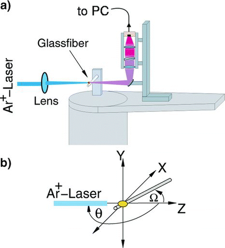
To avoid bleaching before the measurements, a HeNe-laser was used to place the pollen correctly in the scattering volume. The HeNe laser was adjusted so that its beam coincides with the beam of the Ar-Ion laser. The positioning of the pollen was guided by the observation of the scattering pattern. The angular distribution of the fluorescence of the sample excited by the focused laser beam was recorded by the detection unit. This unit consists of a collimating lens with f = 100 mm with an aperture diameter of d = 25 mm. A holographic notch-filter was placed behind this lens in the collimated beam to remove the elastically scattered light. A second lens focused the fluorescence light free from elastic scattered light onto a photo diode. The range of the detection angle θ was limited due to the construction of the detection unit to 5° < θ < 175°.
The detecting unit mounted on the goniometer was rotated computer controlled in steps of 6 degree while the laser was kept focused on the sample. The step size was chosen in accordance with the aperture angle of the detecting unit. In order to avoid bleaching of the dye the laser was shut-off while the goniometer moved from one measuring angle to the next. At each measuring position the shutter was opened and the laser beam illuminated the pollen. Ten successive measurements were made alternating the scanning direction (first measurement with increasing scattering angle, second measurement with decreasing scattering angle, and so on). After completing the scans the average for each scattering angle was calculated. By this procedure bleaching effects were minimized.
The samples were always fixed on the same silica fiber (d = 25 μm) simply by adhesive forces. The coating of the fiber was flame removed.
The main part of the experimental set-up is shown in . The orientation of the pollen and the scattering plane is sketched in . The laser beam was polarized in the y,z plane and propagated in positive z-direction.
RESULTS
To test the effect of the silica fiber, which was used as carrier for the pollen, we measured the angular distribution of the fluorescence emitted by a spherical polymer particle, d = 40 μm, doped with rhodamine 6G on its surface and placed on the fiber as shown in . The angle of incidence Ω as kept at 90°. Light scattered inelastically into the lower half plane (-x, z-plane) was recorded as function of scattering angle. A comparison of results from simulation with the measurements reproduced in shows the following: If the fiber is not hit by the laser beam before it hits the particle and if the fiber is not between the particle and the detector of the scattered light, the effect of the fiber on the inelastically scattered light is small, and the experimental results are close to the theoretical predictions. These conditions are fulfilled by the arrangement shown in .
FIG. 2 Angular distribution of the fluorescence light for a spherical dye doped polymer particle (d = 40 μm), numerical simulation for inelastic scattering of a spherical particles of the same size, with and without being placed on a fiber with different orientation to the laser beam (z-axis).
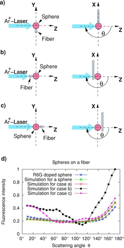
The measurements were compared with calculations for various arrangements of the sphere on a fiber and without the fiber. The calculations were made using geometrical optics. Details of the procedure can be found elsewhere (CitationSchulte et al. 2001; CitationWeigel et al. 2004). Results are shown in .
The numerical simulations were made for a plane incident wave and scattering on a sphere with d = 40 μm and refractive index n = 1,505 (SOMOS 3100) homogeneously filled with fluorescing molecules. As refractive index of the silica fiber n = 1,544 was used. Theoretical results are shown for the arrangements displayed in , , and and for a free sphere. The experiment agrees very well with the calculations. In addition, if a scattering arrangements as depicted in is used, the influence of the silica fiber on the lower scattering half plane is small. The angular distribution is nearly identical to scattering from a free sphere. This is predicted form the simulation and confirmed by experiment. In addition, a comparison of the experimental results with the calculations sows that scattering from a homogeneously doped sphere is not much different from scattering from the surface doped sphere. As expected, for the scattering arrangements shown in and the angular distribution of the scattered radiation differs remarkably from scattering on an isolated sphere.
For nonspherical particles the orientation of the particle in the laser beam is important. The scattered light was therefore recorded for different orientations. The orientation of the pollen is quantified by the angles Ω defined in .
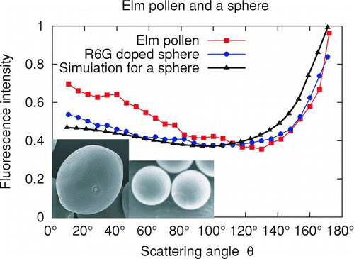
Figures 3–5 show results from measurements on various pollen and of SOMOS particles. All particles except the SOMOS particle, were immersed in a solution of DCM in alcohol for typically one and a half day before the measurements. SEM photographs show the shape and orientation of the particles investigated. Results from measurement on elm pollen (lat. ulmus), a spherical polymer particle, d = 40 μm, doped with rhodamine 6G, and the numerical simulation for a sphere homogeneously filled with fluorescing molecules are represented in . The angular distribution of the fluorescence from the elm pollen differs slightly from the results from the sphere due to the deviation of the pollen shape from a sphere.
FIG. 3 Angular distribution of the fluorescence light for a spherical dye doped polymer particle (d = 40 μm), numerical simulation for inelastic scattering of a spherical particle of the same size, and for an elm (lat. Ulmus) pollen (d ∼ 30 μm).
Due to the leak of interference effects in the scattered radiation, the scattering pattern of inelastic scattering is much simpler than for elastic scattering. A direct comparison for elastic and inelastic scattering for a d = 5.1 μm sphere can be found in (CitationLee et al. 1978). Nevertheless, a, b, and c indicate that inelastic scattered light for a hazel (lat. Corylus), willow (lat. Salix alba), and oak (lat. Quercus robur) pollen shows distinct differences. The characteristic features are also preserved if the orientation of the particles relative to the incident laser beam is varied. All three pollen were measured under different positions by adjusting the angle of incidence Ω of the laser beam.
FIG. 4 Angular distribution of the fluorescence light for three different orientations of the fiber with respect to the laser beam of incidence as indicated: (a) Hazel (lat. Corylus) pollen, (b) willow (lat. Salix alba) pollen, and (c) oak (lat. Quercus robur) pollen.
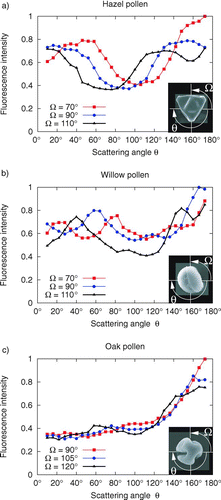
The hazel pollen has two threefold symmetry axes as shown in . We expect from the orientation of the pollen, shown in the inset, that the fluorescence light should therefore show similar features in the scattering range from 0 <θ< 90° as in the quadrant 90 <θ< 180°. This expectation is confirmed by the measurement. In addition, with increasing angle Ω the minimum in the scattered intensity shifts to smaller scattering angles in agreement with intuition. As expected, the graphs show that the angular distribution of the fluorescence depends on the angle of incidence of the laser beam. In all three figures, regions can be identified that shift with the angle of incidence.
The oak pollen () shows that notches in the pollen have no significant effect on the angular distribution of the fluorescence light. The scattering features are not much different from a spherical particle. This confirms that the inelastically scattered light is not very sensitive to details in the shape of the particles.
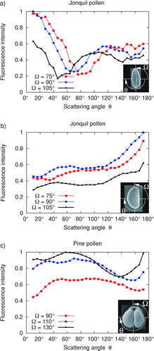
Results for measurements on jonquil (lat. Narcissus) and pine (lat. Pinus sylvestris) pollen are shown in . The jonquil pollen has a tortilla like shape. The scattering signature depends therefore strongly on the orientation of the pollen as a comparison of with clearly shows. The shape of the pine pollen is basically spherical and the angular distribution is therefore relatively insensitive to the angle of incidence. However, the grooves seems to be responsible for a pronounce difference of the scattering signature compared to spheres.
FIG. 5 Angular distribution of the fluorescence light for three different orientations of the fiber with respect to the laser beam of incidence as indicated: (a) and (b) jonquil (lat. Narcissus) pollen, (c) pine (lat. Pinus sylvestris) pollen.
Despite the effort to avoid photo bleaching of the laser dye the intensity of the fluorescent light decreased slightly when the same pollen was scanned twice. Nevertheless the signature of the inelastically scattered light of the pollen maintained the same.
CONCLUSION
We have shown that the angular distribution of the fluorescence emitted from dye doped pollen contains information on the shape of these particles. The pollen were deposited on a thin tapered glass fiber that may slightly disturb the incident light and the angular distributed fluorescent light. Nevertheless, the effect on the angular distribution of the fluorescence light was found to be small, if the pollen were directly exposed to the incident laser beam and the fiber was not between the particle and the detector. This for example was the case in the scattering arrangement shown in .
The shape of the pollen may deviate from the shape of freshly collected pollen. Nevertheless, we have shown that the angular distribution of inelastically scattered light contains sufficient specific shape information to assist in a rapid pollen identification. Our next investigation will be to abandon the doping with dye solutions and exploit the intrinsic fluorescence of the pollen.
Acknowledgments
The work was partly supported by the Deutsche Forschungsgemeinschaft (GRK 358).
REFERENCES
- Eversole , J. D. , Cary , Jr. W. K. , Scotto , C. S. , Pierson , R. , Spence , M. and Campillo , A. J. 2001 . Continuous Bioaerosol Monitoring using UV Excitation Fluorescence: Outdoor Test Results . Field Anal. Chem. Techol , 15 : 205 – 212 .
- Esen , C. and Schweiger , G. 1996 . Preparation of Monodisperse Polymer Particles by Photopolymerization . J. Colloid Interface Sci , 179 : 276 – 280 .
- Holler , S. , Pan , Y.-L. and Chang , R. K. 1998 . Two-dimensional Angular Optical Scattering for the Characterization of Airborne Microparticles . Opt. Lett , 23 : 1489 – 1491 .
- Hill , S. C. , Pinnick , R. G. , Niles , S. , Pan , Y.-L. , Holler , S. , Chang , R. K. , Bottiger , J. R. , Chen , B. T. , Orr , C-.S. and Feather , G. 1999 . Real-time Measurement of Fluorescence Spectra from Single Airborne Biological Particles . Field Anal. Chem. Technol , 3 : 221 – 239 .
- Hill , S. C. , Pinnick , R. G. , Niles , S. , Fell , N. F. , Pan , Y.-L. , Bottiger , J. , Bronk , B. V. , Holler , S. and Chang , R. K. 2001 . Fluorescence from Airborne Microparticles: Dependence on Size, Concentration of Fluorophores, and Illumination Intensity . Appl. Opt. , 40 : 3005 – 3013 .
- Hill , S. C. , Boutou , V. , Yu , J. , Ramstein , S. , Wolf , J.-P. , Pan , Y.-L. , Holler , S. and Chang , R. K. 2000 . Enhanced Backward-directed Multiphoton-excited Fluorescence from Dielectric Microcavities . Phys. Rev. Lett , 85 : 54 – 57 .
- Kaye , P. H. , Barton , J. E. , Hirst , E. and Clark , J. M. 2000 . Simultaneous light Scattering and Intrinsic Fluorescence Measurement for the Classification of Airborne Particles . Appl. Opt , 39 : 3738 – 3745 .
- Krathovil , J. P. , Lee , M. P. and Kerker , M. 1978 . Angular Distribution of Fluorescence from Small Particles . Appl. Opt , 17 : 1978 – 1980 .
- Laucks , M. L. , Roll , G. , Schweiger , G. and Davis , E. J. 2000 . Physical and Chemical (Raman) Characterization of Bioaerosols-Pollen . J. Aerosol. Sci , 31 : 307 – 319 .
- Lee , E.-H. , Benner , R. E. , Fenn , J. B. and Chang , R. K. 1978 . Angular Distribution of Fluorescence from Monodispersed Particles . Appl. Opt , 17 : 1980 – 1982 .
- Manoharan , R. , Ghiamati , E. , Britton , K. A. , Nelson , W. H. and Sperry , J. F. 1991 . Resonance Raman Spectra of Aqueous Pollen Suspensions with 22.5–242.4 nm Pulsed Laser Excitation . Appl. Spectrosc. , 45 : 307 – 311 .
- Miguel , A. G. , Taylor , P. E. , House , J. , Glovsky , M. M. and Flagan , R. C. 2006 . Meteorological Influence on Respirable Fragment Release from Chinese Elm Pollen . Aerosol. Sci. Technol , 40 : 690 – 696 .
- Mishchenko , M. I. , Travis , L. D. and Mackowski , D. W. 1996 . T-matrix Computations of Light Scattering by Nonspherical Particles: A Review . J. Quant. Spectrosc. Rad , 55 : 535 – 575 .
- Pan , Y.-L. , Holler , S. , Chang , R. K. , Hill , S. C. , Pinnick , R. G. , Niles , S. and Bottiger , J. R. 1999 . Single-shot Fluorescence Spectra of Individual Micrometer-sized Bioaerosols Illuminated by 351- or 266-nm Ultraviolet Laser . Opt. Lett , 24 : 116 – 118 .
- Pan , Y.-L. , Cobler , P. , Rhodes , S. , Potter , A. , Chou , T. , Holler , S. , Chang , R. K. , Pinnick , R. G. and Wolf , J.-P . 2001 . High-speed, High-sensitivity Aerosol Fluorescence Spectrum Detection using a 32-anode Photomultiplier Tube Detector . Rev. Sci. Instr , 72 : 1831 – 1836 .
- Pan , Y.-L. , Aptowicz , K. B. , Chang , R. K. , Hart , M. and Eversole , J. D. 2003a . Characterizing and Monitoring Respiratory Aerosols by Light Scattering . Opt. Lett , 28 : 589 – 591 .
- Pan , Y.-L. , Hartings , J. , Pinnick , R. G. , Hill , S. C. , Halverson , J. and Chang , R. K. 2003b . Single-particle Fluorescence Spectrometer for Ambient Aerosols . Aerosol. Sci. Technol. , 37 : 628 – 639 .
- Pan , Y.-L. , Surbek , M. , Aptowicz , K. B. and Chang , R. K. 2003c . Rapid Classification of Aerosols by Single-laser-shot Fluorescence Spectra and Elastic Scattering Patterns. CLEO'03 Conference on Lasers and Electro-Optics.
- Schulte , J. and Schweiger , G. 2001 . Inelastic Scattering on Spherical Microparticles with Inclusion . J. Opt. Soc. Am. , A18 : 117 – 123 .
- Schulte , J. , Weigel , T. and Schweiger , G. 2004 . Inelastic Scattering at Arbitrary Shaped Particles . J. Aerosol. Sci. , 35 : 485 – 486 .
- Secker , R. , Kaye , P. H. , Greenaway , R. S. , Hirst , E. , Bartley , D. and Videen , G. 2000 . Light Scattering from Deformed Droplets and Droplets with Inclusions. I. Experimental Results . Appl. Opt. , 39 : 5023 – 5030 .
- Sivaprakasam , V. , Huston , A. L. , Scotto , C. and Eversole , J. D. 2004 . Multiple UV Wavelength Excitation and Fluorescence of Bioaerosols . Opt. Express , 12 : 4457 – 4466 .
- Stowers , M. A. , van Wuijckhuijse , A. L. , Marijnissen , J. C. M. , Kientz , Ch. E. and Ciach , T. 2006 . Fluorescence Preselection of Bioaerosols for Single-particle Mass Spectrometry . Appl. Opt , 45 : 8531 – 8536 .
- Taylor , P. E. , Flagan , R. C. , Valenta , R. and Glovsky , M. M. 2002 . Release of Allergens as Respirable Aerosols: A Link Between Grass Pollen and Asthma . Allergy Clin. Immunol , 109 : 51 – 58 .
- Weigel , T. , Schulte , J. and Schweiger , G. 2004 . Inelastic Light Scattering on Nonspherical particles . J. Quant. Spectrosc. Radiat. , 89 : 365 – 369 .
- Weigel , T. , Schulte , J. and Schweiger , G. 2004 . Determination of the Composition and Shape of a Particle with Help of Inelastic Light Scattering . J. Aerosol. Sci. , 35 : 1171 – 1172 .
- Weigel , T. , Schulte , J. and Schweiger , G. 2006 . Inelastic Scattering by Particles of Arbitrary Shape . J. Opt. Soc. Am. A , 23 : 2797 – 2802 .