Abstract
Influenza viruses cause pandemics in humans. The aim of this study was therefore to evaluate filter/real-time qPCR to quickly and accurately determine and quantify the airborne influenza and avian influenza virus. In this study, the sampling stress of filtration to influenza virus and the storage effects for both the virus and extracted RNA were evaluated in the laboratory. Then, the collection efficiencies of open-face and closed-face filter cassettes were compared in a wet poultry market. From July 8 to August 19, 2006, a total of 36 air samples were quantified by filter/real-time qPCR.
The recovery rate of the virus on a filter stored at 4°C reached 0.94 after 3-day storage, whereas the RNA stored at –80°C was 100% after 3-month storage. In terms of collection efficiency, the closed-face filter cassette was superior to the open-face filter cassette in the field study. In the wet poultry market, it was revealed that both the positive rate and concentration of influenza A virus in the chicken pen were higher than that in the duck pen, possibly due to differences in ventilation type, climate factors, and avian characteristics. To our knowledge, this is the first report describing the quantification of airborne influenza virus in field samples. This quantitative technique should provide more insight into influenza/avian influenza virus transmissibility and epidemiology of influenza/avian influenza, as well as infection control.
INTRODUCTION
Influenza A and influenza B viruses cause epidemics in humans (CitationLamb and Krug 2001; CitationWright and Webster 2001). Each year as many as 40,000 people in America die due to influenza virus infection and its complications (CitationKrug 2003). In addition, these viruses mutate from year to year to cause even worldwide pandemics (CitationGhedin et al. 2005). In the 20th century, at least four pandemics of influenza occurred, which killed approximately 100 million people around the world at irregular intervals (CitationProença-Módena et al. 2007). In 1997, a highly pathogenic avian influenza virus (H5N1) emerged in Hong Kong, causing severe human disease. To date, 363 human cases of avian influenza (61% of them lethal) have been officially reported by the World Health Organization (CitationWHO 2008). Therefore, preparation for an influenza pandemic must include an early warning system to stop initial outbreaks is of extreme importance.
Human influenza viruses, including influenza A/H1, A/H2, A/H3, and B virus, are readily and rapidly transmitted from human to human by aerosol (CitationWright and Webster 2001). For avian influenza virus, more than 200 human cases of avian influenza virus infection due to A/H5, A/H7, and A/H9 subtypes, mainly a result of poultry-to-human transmission, have been reported (CitationWong et al. 2006). An experimental animal study recently revealed that airborne transmission of highly pathogenic avian influenza virus (H5N1) to chickens has occurred (CitationTsukamoto et al. 2007). Therefore, a sensitive yet specific method for monitoring airborne influenza virus is crucial for early warning and infection control of human influenza and avian influenza outbreaks.
Poultry markets are considered high risk areas of avian influenza virus infections, and based on surveillance, were apparently the source of H5N1 influenza viruses by early December 1997 (CitationShortridge et al. 1998). Although studies in Thailand and Vietnam found that the most statistically significant risk factor was recent exposure to sick or dead poultry (CitationAreechokchai et al. 2006; CitationDinh et al. 2006), some urban patients with influenza A (H5N1), who had no known direct contact with sick poultry or poultry that died of illness were reported in China (CitationYu et al. 2007). Among 6 confirmed influenza A (H5N1) patients who resided in urban areas of the People's Republic of China, only 1 had known exposure to sick poultry or poultry that had died from illness, but before becoming ill, all 6 had visited wet poultry markets (CitationYu et al. 2007). Therefore, monitoring airborne influenza virus in high risk areas such as wet poultry markets is critical for early awareness to stop initial outbreaks.
The most commonly used methods for quantifying airborne virus involve the capture of viruses directly either on solid media, as with an Andersen sampler, in liquid buffer, as with all-glass impingers (AGI), or through a filter. Because the culture method remains the dominant analytical method for bioaerosol measurements, many studies have investigated sampling efficiency using culturability and the preservation of culturability measurements as a function of the composition of the collection medium (CitationAgranovski et al. 2004; CitationHermann et al. 2006; CitationPyankov et al. 2007; CitationTseng and Li 2007). However, culture techniques tend to underestimate bioaerosol concentrations by two orders of magnitude (CitationHeidelberg et al. 1997; CitationChen and Li 2005). In addition, culture techniques have not successfully detected airborne infectious virus in field samples. Due to high sensitivity and high specificity, biological molecular techniques, however, might be superior tools for monitoring infectious bioaerosol in our environment.
Specific infectious viruses in field samples have only successfully been detected by filtration coupled with a PCR-based method. In hospitals, airborne varicella-zoster virus, human cytomegalovirus, respiratory synctial virus, and acute respiratory syndrome coronavirus have been detected by using filtration coupled with a PCR method in 1994, 1996, 1998, and 2006, respectively (CitationSawyer et al. 1994; CitationMcCluskey et al. 1996; CitationAintablian et al. 1998; CitationTsai et al. 2006). In 2004, Myatt et al. validated a filter/semi-PCR method for monitoring Rhinovirus inside and outside office buildings. For influenza virus, it was aerosolized and evaluated in chamber studies (CitationHermann et al. 2006); however, no concentration profile was investigated in the field. These studies revealed that filter/PCR was a powerful technique for specifically detecting infectious virus aerosol. However, this method is only qualitative or semi-quantitative, involving only positive or negative responses in a narrow dynamic range (less than four orders of magnitude) (CitationSchafer et al. 1998; CitationSchafer et al. 1999). In our previous study (CitationChen and Li 2005; CitationChen and Li 2008), we successfully demonstrated that the filter/real-time qPCR method is a sensitive and quick method to quantify another important infectious bioaerosol, M. tuberculosis, in hospitals. This newly established technique shows promise for quantifying infectious bioaerosols in field samples with sensitivity and specificity. Therefore, the aim of the current study was to develop a fast and sensitive method to detect airborne influenza virus and avian influenza virus by using filter/real-time qPCR method. We evaluated sampling stress of filtration to influenza virus, compared collection efficiency of open-face and closed-face filter cassettes and validated this method to detect and quantify influenza virus in a wet poultry market.
MATERIALS AND METHODS
Reference Viruses
Reference strains of influenza A and influenza B were kindly provided by the Laboratory for Virology, Kaohsiung Medical University Hospital (Kaohsiung, Taiwan). Prototype strains utilized to develop the real-time qPCR method were: A/New Caladonia/20/99 H1N1, A/Hiroshima/52/2005 H3N2, and B/Malaysia/2506/04. For safety concern, avian influenza A/H5 was provided as plasmid cDNA. In chamber study, only A/Hiroshima/52/2005 H3N2 was used to evaluate the sampling stress and storage effect.
Chamber Study
Sampling Stress
Sampling stress on influenza virus was evaluated in a chamber study. We applied medium that contains viral test strain on clean polytetrafluoroethylene (PTFE; “Teflon”) membrane filters (Pall Corporation, New York, USA). Then, 37 mm diameter filters with 0.2 μ m pore size supported by cellulose pads were loaded into open-face and closed-face plastic cassettes, respectively. Both cassettes were simultaneously evaluated by passing clean air through the filters at 20 L/min at sampling times of 0, 2, 8, 24, and 48 h, respectively. The Teflon filters were then removed from each cassette immediately for viral RNA extraction and further real-time qPCR analysis. The sampling stress on the virus was defined by the ratio Qt/Q0, where Qt and Q0 are the quantity of the simultaneously collected samples subjected to airflow for t hours and 0 hours, respectively.
Storage Effect on Virus and RNA
During transport of the virus between collection and analysis, the concentration of the collected virus, and thus the extracted RNA (an analysis target of the one-step real-time aPCR), might change due to storage time. The effect of storage on both the virus and extracted RNA concentration was therefore evaluated as follows. Equal volumes of virus medium were loaded separately on clean Teflon filters. Each Teflon filter was stored at 4°C and then removed for viral RNA extraction and real-time qPCR analysis at 0, 1, 3, 7, and 14 days to obtain a kinetic curve of the concentration variation up to 14-day storage. Extracted RNA samples were stored at −80°C and then removed for real-time qPCR analysis at 0, 7, 30, and 90 days to obtain a kinetic curve of concentration variation up to 3 month storage. The effect of storage time on extracted RNA was determined by the ratio Qt/Q0, where Qt and Q0 are the quantity of the simultaneously collected samples stored for t days and 0 days, respectively.
Field Study
Sampling Location
The filter/real-time qPCR technique was validated by using it to determine and quantify airborne influenza virus in both a chicken pen and duck pen in an actual wet poultry market in an urban area of Taipei, Taiwan. The chicken pen was a semi-open space (6.4 m long and 5.3 m wide) with 4 industrial fans blowing from inside to outside, and housed about 50,000 chickens during the field study. The duck pen was an open space (7.2 m long and 3.6 m wide), and housed about 4,000 ducks.
Collection of Samples
Airborne influenza virus was collected on a 0.2 μ m pore size Teflon membrane filter in a disposable plastic cassette by using a sampling pump operating at 20 L/min with a sampling time of 4 h. Filters and support pads had been autoclaved and the plastic cassettes had been sterilized with ethylene oxide. The sampling efficiency of both the closed-face and open-face filter cassettes were simultaneously evaluated 1 m above the floor at the center of the chicken pen and at the center of the duck pen on 9 different days. A total of 36 air samples were collected from July 8 to August 19, 2006. By using direct-reading instruments (Sekunden-Hygrometer 601; Testo, Tirana, Albania), the temperature and relative humidity were recorded at the same two locations as the sampling. The samples were then transported under refrigeration to our laboratory within 1 day. For quality control, trip blank and field blank controls were also evaluated. Results confirmed no detectable influenza virus RNA in either the trip blank or field blank control (results not shown). In addition, side-by-side duplicate field samples yielded comparable results (with relative difference of 11%).
Viral Genomic RNA Isolation RNA in the samples was isolated using commercially available kits, QIAamp Viral RNA Mini Kit (Qiagen Gmbh, Hilden, Germany). A single RNA preparation was prepared from each chamber and field sample. RNA extraction from laboratory-prepared samples and field samples was performed essentially as recommended by the manufacturer, except that in Step 2, “the sampled Teflon filter was folded into quadrants and put into the Buffer AVL-carrier RNA in the microcentrifuge tube.” All manipulations of samples were performed in a biological safety cabinet. The viral RNA was stored at –80°C within 1 month before further analysis.
Real-Time Quantitative PCR Assay
Primers and Probes
The primers and probes for influenza A virus and influenza B virus were based on genomic regions highly conserved in various subtypes and genotypes of influenza A virus (matrix protein gene) and influenza B virus (hemagglutinin gene segment) (Citationvan Elden et al. 2001). The primers and probe for influenza A virus can detect all types of influenza A virus, including human influenza virus and avian influenza virus. For influenza A virus subtype H5, the primer and probe were primarily targeted to conserved regions of North American H5 influenza viruses (CitationSpackman et al. 2002). The specificity, evaluated in the previous study, of these three sets of primers and probes were 100% for influenza A, B, and A/H5 viruses, respectively (Citationvan Elden et al. 2001; CitationSpackman et al. 2002). The sequences were shown in . In our study, samples were firstly analyzed for influenza A and B virus. The analysis for A/H5 was only done for those samples in which influenza virus A was detected.
TABLE 1 Primers and probes of influenza A virus, influenza B virus, and avian influenza A/H5 virus
Amplification and Detection
The PCR assay was performed in 25 μ l volume using MicroAmp Optical 96-well reaction plates and MicroAmp Optical Caps (PE Biosystems, Forster City, CA) for each well. TaqMan One-step Reverse Transcriptase PCR Master Mix Reagents Kit (PE Applied Biosystems) was used for cDNA synthesis and ABI Prism 7500 sequence detection system was used for Real-time qPCR. The PCR mixture was as follows. For influenza A virus detection, the PCR mixture was 5 μ l of viral RNA, 1 μ l of primer INFA-1 of 150 nM, 1 μ l of primer INFA-2 of 150 nM, 1 μ l of INFA-3 of 150 nM, 1μ l of INFA probe of 22.5 μ M, 12.5 μ l of 2X Master Mix without UNG, 0.625 μ l of 40X MultiScribe and RNase Inhibitor Mix and 2.875 μ l of DEPC water. For influenza B virus detection, the mixture was 5 μ l of viral RNA, 1 μ l of primer INFB-1 of 300 nM, 1 μ l of primer INFB-2 of 300 nM, 1 μ l of INFB probe of 22.5 μ M, 12.5 μ l of 2X Master Mix without UNG, 0.625 μ l of 40X MultiScribe and RNase Inhibitor Mix and 3.875 μ l of DEPC water. For avian influenza A/H5 virus detection, the mixture was 5 μ l of viral RNA, 1 μ l of primer H5-1 of 200 nM, 1 μ l of primer H5-2 of 200 nM, 1μ l of INFA probe of 22.5 μ M, 12.5 μ l of 2X Master Mix without UNG, 0.625 μ l of 40X MultiScribe and RNase Inhibitor Mix and 3.875 μ l of DEPC water. Amplification and detection were performed under the following conditions: 30 min at 48°C for reverse transcription, 10 min at 95°C for activation of AmpliTaq Gold DNA polymerase, 40 cycles of 15 s at 95°C for cDNA denaturation, and 1 min at 60°C for annealing and extension. All samples analyzed using the real-time qPCR were done in triplicate.
Standard Curve
The target cDNA standard solution of influenza A, B, and A/H5 virus from cloning plasmid was purchased from Mission Biotech (Taipei, Taiwan). For standard curve preparation, the standard solution was serial diluted from 107 to 100 copy/ul. The sensitivity of the real-time qPCR assay was evaluated using 10-fold dilutions of plasmid cDNA run in triplicate reactions. The threshold was set at 10 times the standard deviation of the mean baseline emission calculated for PCR cycles 3 to 10. The amount of product in a given field sample was determined by interpolation from a standard curve of cycle threshold (C t ) values based on the known concentration of cDNA (C0) at the same run. The positive (purified dilutions of plasmid cDNA) and negative controls were analyzed with each run in triplicate. All samples were analyzed in triplicate (coefficients of variation were less than 6%).
Inhibitory Effect
In field studies, possible interference from different mediums must be carefully considered. Because collected airborne samples might contain compounds that inhibit amplification assay, environmental interference must be determined to assess the utility of PCR under different environments. In a previous study, air samples containing 103 to 104 CFU/m3of environmental bacteria and fungi inhibited PCR amplification, whereas a 1/10 dilution of these samples did not (CitationAlvarez et al. 1995), suggesting that interference could be corrected by serial dilution of samples. In the present study, the interference was corrected by 1/10, 1/100, and 1/1000 dilutions of all the samples. If cDNA was quantified in 1/10, 1/100, or 1/1000 diluted solution, the true concentration was corrected by multiplying the detected concentration by 10, 100, or 1000, respectively. Inhibitory samples that were initially negative, became positive after dilution.
Statistical Methods
Software used for data analysis included Excel for Windows XP, version 7.0 (Microsoft, Redmond, WA) and SigmaPlot for Windows, version 3.06 (SPSS, Chicago, IL). The differences were examined using the student's t test.
RESULTS AND DISCUSSION
This study evaluated a filter/real-time qPCR method for quick and accurate quantification of airborne influenza virus. First, the sampling stress of open-face and closed-face filter cassettes and the storage effects of both the virus and extracted RNA were evaluated in a chamber study. Then, the collection efficiencies of open-face and closed-face filter cassettes were compared in a field study. Then, the filter/real-time qPCR method was validated as an early warning system to accurately assess the concentration of airborne influenza virus and avian influenza virus in a chicken pen and duck pen in an actual wet poultry market.
Dynamic Range and Analytical Sensitivity of Real-Time qPCR
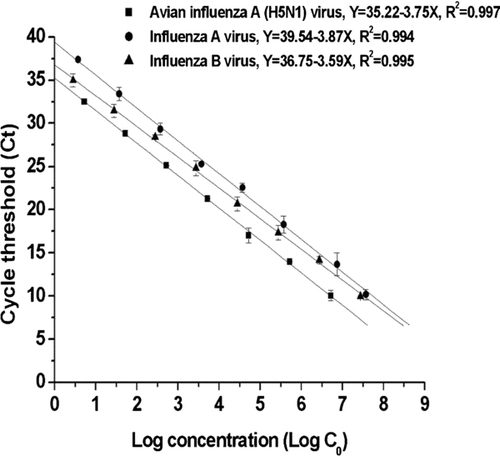
shows the dynamic range of the standard curve for influenza A, B, and A/H5 virus, respectively. For influenza A, the range was 3.75 × 100∼ 3.75 × 10⁷ copy/μl with r = 0.988. For influenza B, the range was 2.79 × 100∼ 2.79 × 10⁷ copy/μ l with r = 0.995. For influenza A/H5, the range was 5.27 × 100∼ 5.27 × 10⁶ copy/μ l with r value of 0.995, respectively. The calibration curve was linear over 7 orders of magnitude with r > 0.988. Because the filters were eluted into 1.12 ml, the detection limit of the filter/real-time qPCR method for influenza A, B, and A/H5 virus was 886 copy/m3, 653 copy/m3, and 1236 copy/m3, respectively. If the positive results observed in the 1/1000 dilution samples were due to the inhibitory effect, then the actual detection limit corrected for this effect for influenza A, B, and A/H5 virus was 0.8 copy/m3, 0.65 copy/m3, and 1.23 copy/m3, respectively.
FIG. 1 Standard curve of known cDNA and threshold cycle (C t ) measured using a real-time qPCR for influenza A virus, influenza B virus, and influenza A/H5 virus.

In comparison with the previous study, only the detection limit of real-time qPCR was reported. In previous studies, the detection limit for influenza A virus and influenza B virus was 5.1 × 10⁴ copies/reaction and 1.1 × 10⁵ copies/reaction, respectively, with the TaqMan assay (CitationKrafft et al. 2005). In another study, the detection limit for influenza A virus and influenza B virus was 13 copies/μ l, and 11 copies/μ l, respectively, with the TaqMan assay (Citationvan Elden et al. 2001). For avian influenza virus (A/H5), the reported detection limit was 103 copies (CitationSpackman et al. 2002). Because the concentration of airborne influenza virus in field samples might be low, high sensitivity of the analytical method is crucial for detecting airborne influenza virus in a wet poultry market. The filter/real-time qPCR method developed here could be useful to rapidly identify and quantify airborne influenza virus in field samples.
Storage Effect
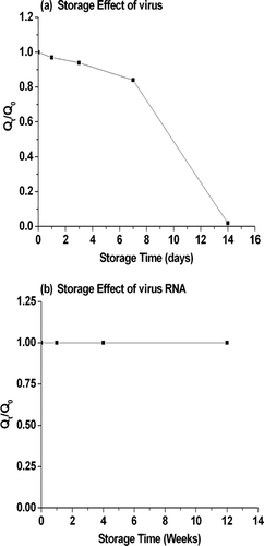
shows the storage effect of a virus on a filter at 4°C. The concentration of virus decreased as storage time increased, and the recovery rate reached 0.94 within 3 day storage. Therefore, to ensure a high recovery rate in the field test, the RNA isolation in the further analysis was done within 3 days of sampling. For RNA storage at −80°C, the recovery rate of RNA was 100% after 3-month storage. In our field study, the RNA samples were therefore analyzed by using real-time qPCR within 1 month after isolation. In clinical samples, the quantity of influenza A virus detected using real-time qPCR assay was nearly double that detected using the culture method when samples were collected and sent through postal mail and thus in transit between 1 and 4 days at 4°C (CitationSchweiger et al. 2000; Citationvan Elden et al. 2001). To date, no data on the storage effect of filtered influenza virus has been reported. By using the culture method to evaluate filtered samples, only 1/3 of the yeast colony of a cell-type fungus was detected after 24 h storage, and no colony recovered after 4 day storage (CitationLin and Li 2003). For cell-type bacteria, filtered E. coli cells were found to lose more than 70% of the colony after 1 h storage (CitationLi and Lin 2001). Because a virus is more fragile than E. coli and yeast in terms of sampling stress in different samplers and inactivation by ultraviolet germicidal irradiation and ozone (Tseng and Li 2005a; Tseng and Li 2005b; CitationTseng and Li 2006), the effect of storage on filtered virus analyzed using the culture method should be much stronger than on virus analyzed using real-time qPCR. Therefore, the filter/real-time qPCR technique should successfully detect and quantify airborne influenza virus in field studies.
Sampling Stress on Open-Face and Closed-Face Filter Cassettes
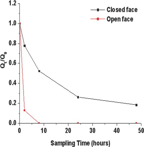
shows that, in the chamber study, the sampling stress of open-face filter cassettes was higher than that of closed-face filter cassettes (). After pure air was passed through the filter for 2 h, Q t /Q0 decreased to 0.78 and 0.14 by using the closed-face and open-face filter cassettes, respectively. After 8 h sampling, Q t /Q0 decreased to 0.52 and 0 by using closed-face and open-face filter cassettes, respectively. Therefore, the recovery rate of a virus could exceed 0.5 when a closed-face filter cassette was used and when the sampling time was less than 8 hours.
FIG. 3 Sampling stress of open-face filter cassette and closed-face filter cassette on influenza virus.

shows the collection efficiency of open-face and closed-face filter cassettes was compared by simultaneously collecting parallel samples in both the chicken pen and duck pen on 9 different days (n = 36). When an open-face filter cassette was used to assess airborne influenza A virus in the pens, the average concentration of the virus was 2.9 × 10³ copy/m3 (concentration range: below the lower detection limit to 2.8 × 10⁴ copy/m3) and the positive rate was 33.3%. When a closed-face filter cassette was used, the average concentration was 4.5 × 10³ copy/m3 (below the lower detection limit to 3.7 × 10⁴ copy/m3) and the positive rate was 55.5%. The samples collected using a closed-face filter cassette had a higher positive rate and higher concentration of influenza virus compared with those collected using an open-face filter cassette. Same trend was also observed when samples from chicken pen and duck pen were analyzed independently. These results are consistent with the sampling stress results from our chamber study (see preceding section). In summary, a closed-face filter cassette is superior to an open-face filter cassette in evaluating the concentration of airborne influenza virus in a field study.
TABLE 2 The concentration of airborne influenza virus in wet poultry market
Concentration of Airborne Influenza Virus in an Actual Wet Poultry Market
In our field study, the filter/real-time qPCR method successfully quantified the concentrations of airborne influenza virus in an actual wet poultry market. To our knowledge, this is the first report describing the quantification of airborne influenza virus in field samples. First, all air samples were analyzed for influenza A virus and influenza B virus. Then, samples in which influenza A virus was detected were further analyzed for avian influenza virus (A/H5). However, neither influenza B virus nor avian influenza virus (A/H5) was detected in the samples. No influenza B virus and avian influenza virus (A/H5) existed in that environment during sampling period might be the reason. lists the measured concentration of airborne influenza A virus collected using open-face and closed-face filter cassettes in the two pens in our field study. Both the concentration and positive rate of influenza A virus in the chicken pen were higher than those in the duck pen. Overall, the filter/real-time qPCR method revealed airborne influenza A virus in about 55.5% of the air samples in the wet poultry market, and the concentrations of airborne influenza virus varied widely from below the lower detection limit to 3.7 × 10⁴ copies/m3 ().
The climate factors in the wet poultry market were also measured. The temperature in the chicken pen was significantly lower than that in the duck pen (p < 0.01), whereas the relative humidity was significantly higher (p < 0.01). In the chicken pen, the average temperature was 29.6°C (range from 26.9°C to 31.4°C), and the average relative humidity was 78.8% (range from 68.8°C to 88.4°C). In the duck pen, the average temperature was 35.8°C (range from 31.7°C to 38.2°C) and the average relative humidity was 51.6% (range from 32.8°C to 69.7°C). The differences of climate factors between chicken pen and duck pen may affect the concentrations of airborne influenza virus.
Three mechanisms might explain the higher positive rate and concentration of influenza A virus in the chicken pen compared with the duck pen: namely, ventilation type, climate factors, and avian characteristics. In terms of ventilation, the duck pen was an open space, whereas the chicken pen was a semi-open space. The duck pen therefore had more fresh air to dilute the airborne influenza virus, and thus the measured airborne virus concentration was lower for the duck pen. In terms of climate factors, the transmission of influenza virus under different temperature and relative humidity has been evaluated using animal studies (CitationHarper 1961; CitationHemmes et al. 1960; CitationLowen et al. 2007; CitationSchaffer et al. 1976). One study reported that the transmission of influenza virus decreased as the temperature increased from 5°C to 30°C (CitationLowen et al. 2007), and another study reported that the stability of airborne influenza virus depended on the relative humidity (CitationHarper 1961; CitationHemmes et al. 1960; CitationSchaffer et al. 1976). The most recent report showed viral stability to be minimal at intermediate relative humidity (50%), and high at elevated relative humidity (60% to 80%) (CitationSchaffer et al. 1976). In the present study, lower virus concentration was measured in the duck pen with average temperature of 35.8°C and relative humidity of 51.6% (range: 32.8°C to 69.7°C). Our results are consistent with previously reported results. In terms of avian characteristics, chickens and ducks might exhibit different responses after being infected with avian influenza virus. Such avian-specific response characteristics need further investigation.
In conclusion, we successfully demonstrated that the filter/real-time qPCR technique with closed-face filter cassettes is a sensitive and fast (< 5 h) method to quantify airborne influenza virus in field samples. This quantitative method should provide deeper insight into influenza/avian influenza virus transmissibility and epidemiology of influenza/avian influenza, as well as infection control. Quick and accurate measurement of the concentration of airborne influenza/avian influenza virus will be crucial for the early warning of an outbreak. Further study is needed, however, on background influenza virus levels within high-risk microenvironments, and on the potentially infectious influenza dose for susceptibility groups and the general public. Furthermore, the profile of subtypes, including human influenza virus (H1, H2, and H3) and avian influenza (H5, H7, and, H9), should be investigated to identify high risk areas for the reassortment of human and avian influenza viruses.
This work was supported by grant NSC-95-EPA-Z-002-004 and NSC96-EPA-Z002-005 from the National Science Council, Republic of China.
REFERENCES
- Agranovski , I. E. , Safatov , A. S. , Borodulin , A. I. , Pyankov , O. V. , Petrishchenko , V. A. , Sergeev , A. N. , Agafonov , A. P. , Ignatiev , G. M. , Sergeev , A. A. and Agranovski , V. 2004 . Inactivation of Viruses in Bubbling Processes Utilized for Personal Bioaerosol Monitoring . Appl. Environ. Microbio , 70 ( 12 ) : 6963 – 6967 .
- Aintablian , N. , Walpita , P. and Sawyer , M. H. 1998 . Detection of Boedetella Pertussis and Respiratory Synctial Virus in Air Samplers from Hospital Rooms . Infect Control Hosp Epidemiol , 19 : 918 – 923 .
- Alvarez , A. J. , Buttner , M. P. and Stetzenbach , L. D. 1995 . PCR for Bioaerosol Monitoring: Sensitivity and Environmental Interference . Applied and Environmental Microbiology , 61 : 3639 – 3644 .
- Areechokchai , D. , Jiraphongsa , C. , Laosiritaworn , Y. , Hanshaoworakul , W. and O'Reilly , M. 2006 . Investigation of Avian Influenza (H5N1) Outbreak in Humans—Thailand 2004 . MMWR Morb Mortal Wkly Re , 55 (Suppl 1) : 3 – 6 .
- Chen , P. S. and Li , C. S. 2005 . Concentration of Airborne Mycobacterium Tuberculosis in Patient Rooms Measured using Real-Time qPCR Coupled to an Air-sampling Filter Method . Aerosol Sci. Technol. , 39 : 371 – 376 .
- Chen , P. S. and Li , C. S. 2005 . Sampling Performance for Bioaerosols by Flow Cytometry with Fluorochrome . Aerosol Sci. Technol. , 39 : 231 – 237 .
- Chen , P. S. and Li , C. S. 2008 . Concentration Profiles of Airborne Mycobacterium Tuberculosis in a Hospital . Aerosol Sci. Technol. , 42 ( 3 ) : 194 – 200 .
- Chen , P. S. and Li , C. S. 2005 . Quantification of Airborne Mycobacterium Tuberculosis in Health Care Setting using Real-Time qPCR Coupled to an Air-Sampling Filter Method . Aerosol Sci. Technol. , 39 ( 4 ) : 371 – 376 .
- Dinh , P. N. , Long , H. T. , Tien , N. T. , Hien , N. T. , Maie , T. Q. , Phong , le H. , Tuan , le V. , Van , T. H. , Nguyen , N. B. , Van , T. P. and Phuong , N. T. 2006 . Risk Factors for Human Infection with Avian Influenza A H5N1,Vietnam 2004 . Emerg Infect Dis. , 12 : 1841 – 1847 .
- Ghedin , E. , Sengamalay , N. A. , Shumway , M. , Zaborsky , J. , Feldblyum , T. , Subbu , V. , Spiro , D. J. , Sitz , J. , Koo , H. , Bolotov , P. , Dernovoy , D. , Tatusova , T. , Bao , Y. , St George , K. , Taylor , J. , Lipman , D. J. , Fraser , C. M. , Taubenberger , J. K. and Salzberg , S. L. 2005 . Large-Scale Sequencing of Human Influenza Reveals the Dynamic Nature of Viral Genome Evolution . Nature. , 437 ( 20 ) : 1162 – 1166 .
- Harper , G. J. 1961 . Airborne Micro-Organisms: Survival Tests with Four Viruses . J. Hyg. (Lond.) , 59 : 479 – 486 .
- Heidelberg , J. F. , Shahamat , M. , Rahman , I. , Stelma , G. , Grim , C. and Colwell , R. R. 1997 . Effect of Aerosolization on Culturability and Viability of Gram-Negative Bacteria . Appl. Environ. Microbio. , 63 : 3585 – 3588 .
- Hemmes , J. H. , Winkler , K. C. and Kool , S. M. 1960 . Virus Survival as a Seasonal Factor in Influenza and Poliomyelitis . Nature. , 188 : 430 – 431 .
- Hermann , J. R. , Hoff , S. J. , Yoon , K. J. , Burkhardt , A. C. , Evans , R. B. and Zimmerman , J. J. 2006 . Optimization of a Sampling Systems for Recovery and Detection of Airborne Porcine Reproductive and Respiratory Syndrome Virus and Swine Influenza Virus . Appl. Environ. Microbiol. , 72 : 4811 – 4818 .
- Proença-Módena , José , Luiz , Santos , Macedo , Izolete , Arruda and Eurico . 2007 . H5N1 Avian Influenza Virus: An Overview . The Brazilian J. Infect. Dis. , 11 ( 1 ) : 125 – 133 .
- Krafft , A. E. , Russell , K. L. , Hawksworth , A. W. , McCall , S. , Irvine , M. , Daum , L. T. , Connoly , J. L. , Reid , A. H. , Gaydos , J. C. and Taubenberger , J. K. 2005 . Evaluation of PCR Testing of Ethanol-Fixed Nasal Swab Specimens as an Augmented Surveillance Strategy for Influenza Virus and Adenovirus Identification . J. Clin. Microbiol. , 43 : 1768 – 1775 .
- Krug , R. M. 2003 . The Potential Use of Influenza Virus as an Agent for Bioterrorism . Antiviral Research , 57 : 147 – 150 .
- Lamb , R. A. and Krug , R. M. 2001 . “ Orthomyxoviridae: The Viruses and Their Replication ” . In Fields Virology, , 4th ed , Edited by: Knipe , D. M. and Howley , P. M. 1487 – 1532 . Philadelphia : Lippincott Williams & Wilkins .
- Lin , W. H. and Li , C. S. 2003 . Influence of Storage on the Fungal Concentration Determination of Impinger and Filter Samples . AIHA J (Fairfax, Va). , 64 : 102 – 107 .
- Li , C. S. and Lin , Y. C. 2001 . Storage Effects on Bacterial Concentration: Determination of Impinger and Filter Samples . Science Total Environ. , 278 : 231 – 237 .
- Lowen , A. C. , Mubareka , S. and Palese , P. 2007 . Influenza Virus Transmission is Dependent on Relative Humidity and Temperature . PLoS Pathog. , 3 ( 10 ) : 1470 – 1476 .
- McCluskey , R. , Sandin , R. and Greene , J. 1996 . Detection of Airborne Cytomegalovirus in Hospital Rooms of Immunocompromised Patients . J. Virol. Meth. , 56 ( 1 ) : 115 – 118 .
- Pyankov , O. V. , Agranovski , I. E. , Pyankova , O. , Mokhonova , E. , Mokhonov , V. , Safatov , A. S. and Khromykh , A. A. 2007 . Using a Bioaerosol Personal Sampler in Combination with Real-Time PCR Analysis for Rapid Detection of Airborne Viruses . Environ. Microbiol. , 9 ( 4 ) : 992 – 1000 .
- Reid , A. H. , Fanning , T. G. , Slemons , R. D. , Janczewski , T. A. , Dean , J. and Taubenberger , J. K. 2003 . Relationship of Pre-1918 Avian Influenza HA and NP Sequences to Subsequent Avian Influenza Strains . Avian Dis. , 47 (3 Suppl) : 921 – 925 .
- Sawyer , M. H. , Chamberlin , C. J. , Wu , Y. N. , Aintablian , N. and Wallace , M. R. 1994 . Detection of Varicella-Zoster Virus DNA in Air Samplers from Hospital Rooms . J Infect Dis. , 169 : 91 – 94 .
- Schafer , M. P. and Fernback , J. E. 1999 . Detection and Characterization of Airborne Mycobacterium Tuberculosis H37Ra Particles, a Surrogate for Airborne Pathogen M. Tuberculosis . Aerosol Sci. Technol , 30 : 161 – 173 .
- Schafer , M. P. , Fernback , J. E. and Jensen , P. A. 1998 . Sampling and Analytical Method Development for Qualitative Assessment of Airborne Mycobacterial Species of Mycobacterium Tuberculosis Complex . AIHA J. , 59 : 540 – 546 .
- Schaffer , F. L. , Soergel , M. E. and Straube , D. C. 1976 . Survival of Airborne Influenza Virus: Effects of Propagating Host, Relative Humidity, and Composition of Spray Fluids . Arch. Virol. , 51 ( 4 ) : 263 – 273 .
- Schweiger , B. , Zadow , I. , Heckler , R. , Timm , H. and Pauli , G. 2000 . Application of a Fluorogenic PCR Assay for Typing and Subtyping of Influenza Viruses in Respiratory Samples . J. Clin. Microbiol. , 38 : 1552 – 1558 .
- Shortridge , K. F. , Zhou , N. N. , Guan , Y. , Gao , P. , Ito , T. , Kawaoka , Y. , Kodihalli , S. , Krauss , S. , Markwell , D. , Murti , K. G. , Norwood , M. , Senne , D. , Sims , L. , Takada , A. and Webster , R. G. 1998 . Characterization of Avian H5N1 Influenza Viruses from Poultry in Hong Kong . Virology. , 252 : 331 – 342 .
- Spackman , E. , Senne , D. A. , Myers , T. J. , Bulaga , L. L. , Garber , L. P. , Perdue , M. L. , Lohman , K. , Daum , L. T. and Suarez , D. L. 2002 . Development of a Real-Time Reverse Transcriptase PCR Assay for Type A Influenza Virus and the Avian H5 and H7 Hemagglutinin Subtypes . J. Clin. Microbiol , 40 : 3256 – 3260 .
- Tsai , Y. H. , Wan , G. H. , Wu , Y. K. and Tsao , K. C. 2006 . Airborne Severe Acute Respiratory Syndrome Coronavirus Concentrations in a Negative-Pressure Isolation Room . Infect. Control Hosp. Epidemiol. , 27 : 523 – 525 .
- Tseng , C. C. and Li , C. S. 2007 . Inactivation of Viruses on Surface by Ultraviolet Germicidal Irradiation . J. Occup. Environ. Hyg. , 40 : 400 – 405 .
- Tseng , C. C. and Li , C. S. 2006 . Ozone for Inactivation of Aerosolized Bacteriophages . Aerosol Sci. Technol. , 40 : 683 – 689 .
- Tseng , C. C. and Li , C. S. 2005 . Inactivation of Virus-Containing Aerosols by Ultraviolet Germicidal Irradiation . Aerosol Sci. Tech. , 39 : 1136 – 1142 .
- Tseng , C. C. and Li , C. S. 2005 . Collection Efficiencies of Aerosol Samplers for Virus-Containing Aerosols . J. Aerosol Sci. , 36 : 593 – 607 .
- Tsukamoto , K. , Imada , T. , Tanimura , N. , Okamatsu , M. , Mase , M. , Mizuhara , T. , Swayne , D. and Yamaguchi , S. 2007 . Impact of Different Husbandry Conditions on Contact and Airborne Transmission of H5N1 Highly Pathogenic Avian Influenza Virus to Chickens . Avian Dis. , 51 : 129 – 132 .
- van Elden , L. J. , Nijhuis , M. , Schipper , P. , Schuurman , R. and van Loon , A. M. 2001 . Simultaneous Detection of Influenza Viruses A and B Using Real-Time Quantitative PCR . J. Clin. Microbiol. , 39 : 196 – 200 .
- WHO . (2008) . Cumulative Number of Confirmed Human Cases of Avian Influenza A/(H5N1) Reported to WHO 2008 [accessed Jan. 14, 2008]
- Wong , S. S. and Yuen , K. Y. 2006 . Avian Influenza Virus Infections in Humans . Chest. , 129 ( 1 ) : 156 – 168 .
- Wright , P. F. and Webster , R. G. 2001 . “ Orthomyxoviruses ” . In Fields Virology , 4th ed , Edited by: Knipe , D. M. and Howley , P. M. 1533 – 1579 . Philadelphia : Lippincott Williams & Wilkins .
- Yu , H. , Feng , Z. , Zhang , X. , Xiang , N. , Huai , Y. , Zhou , L. , Li , Z. , Xu , C. , Luo , H. , He , J. , Guan , X. , Yuan , Z. , Li , Y. , Xu , L. , Hong , R. , Liu , X. , Zhou , X. , Yin , W. , Zhang , S. , Shu , Y. , Wang , M. , Wang , Y. , Lee , C. K. , Uyeki , T. M. , Yang , W. and Avian Influenza H5N1 Study Group2 . 2007 . Human Influenza A (H5N1) Cases . Emerg. Infect. Dis. , 13 ( 7 ) : 1061 – 1064 . Urban Areas of People's Republic of China 2005–2006 www.cdc.gov/eid
