Abstract
Thermal inactivation of viruses has been studied in relevance to food sterilization, water purification, and other “non-aerosol” applications, in which heat treatment is applied for a relatively long time. No data are available on the inactivation of airborne viruses exposed to dry heat for a short time, although this is relevant to bio-defense and indoor air quality control. In this study, we investigated inactivation of aerosolized MS2 viruses in a continuous air flow chamber with axial heating resulted from exposures during ∼ 0.1–1 s. For an airborne virion, the characteristic exposure temperature, T e , was defined utilizing the air temperature profiles in the chamber. The tests were conducted at two air flow rates, Q, which allowed for establishing different thermal flow regimes and exposure time intervals. The experimentally determined inactivation factor, IF, was subjected to correction to account for the temperature profiles. At T e up to ∼ 90°C (Q = 18 L/min) and up to ∼ 140°C (Q = 36 L/min), the loss of viral infectivity was relatively modest (≤ 10). However, IF increased exponentially as T e rose from ∼ 90°C to ∼ 160°C (for 18 L/min) or from ∼ 140°C to ∼ 230°C (for 36 L/min). Under specific thermal exposure conditions (∼ 170°C and ∼ 250°C, respectively), IF exceeded ∼ 2.4 × 10⁴ (∼ 99.996% infectivity loss)—the maximum quantifiable in this study. The airborne MS2 virions exposed to hot air for < 1 s were found to have survived much higher temperatures than those subjected to thermal treatment in liquid for minutes or hours. The findings are significant for establishing limitations of the heat-based bioaerosol control methods.
INTRODUCTION
Different methods are available for protecting air environments from bioaerosol hazards including, but not limited to, pathogenic aerosol agents. While some methods achieve this goal by reducing the total aerosol concentration, others specifically target viable bioaerosol particles. Heat treatment has been found efficient for inactivating viable bacteria (CitationNorthrop and Slepecky 1967; CitationSetlow 1995; CitationSetlow and Setlow 1995; CitationStumbo 1973) and viruses (CitationJohnson et al. 2007; CitationStonehouse and Stockley 1993). However, most of the previous studies have been primarily motivated by “non-aerosol” applications, such as the sterilization of food products, water purification, and surface decontamination. Consequently, these studies involved microorganisms in aqueous media or on solid surfaces. Few investigations on heat-induced microbial inactivation have dealt with aerosolized biological particles—bacterial spores (CitationGrinshpun et al. 2010; CitationMullican et al. 1971), vegetative cells (CitationJung et al. 2009a; CitationLee and Lee 2006), or fungal spores (CitationJung et al. 2009b). To our knowledge, no data are currently available on the inactivation of aerosolized viruses by their exposure to hot air flow.
It is anticipated that aerosolized microorganisms exhibit different responses to heat stress than those suspended in liquid or attached to solid surfaces (CitationNicholson et al. 2000). One parameter that promotes the crucial difference in stress responses is the time of exposure because viruses in aerosol phase may be exposed to heat over relatively short period of time as compared to those subjected to the heat treatment in liquids or on surfaces.
The quantification of the thermal inactivation of airborne virions is particularly important for situations when the exposure time is as short as ∼ 0.1 s (or even shorter) and the air temperature ranges from ∼ 100°C to ∼ 1000°C. Bio-defense applications serve as a good example of such bioaerosol exposure. It is presently unknown whether viral warfare agents aerosolized under high-temperature conditions (e.g., in a bio-agent defeat scenario) are likely to lose 100% of their infectivity during a short-term exposure to dry heat. Should some virions survive the exposure, their presence in the atmosphere may pose a serious public health threat. Inactivation of airborne virions in an air flow drawn through a heating unit is also relevant to indoor air quality. Heat treatment is believed to be an effective, safe and environment-friendly method for purifying air contaminated with bioaerosol particles in continuous-flow settings, e.g., heating, ventilation, and air-conditioning systems (CitationJung et al. 2009a, Citation2009b).
In this study, we investigated inactivation of aerosolized MS2 virions in a continuous air flow with axial heating. In the absence of moisture, the dry heat induced inactivation was evaluated for the characteristic air temperatures ranging from < 50°C to > 300°C and exposure time intervals of ∼ 0.1–1 s. MS2 bacteriophage, approx. 0.03 μ m tailless non-enveloped icosahedral RNA-coliphage, is relatively stable against environmental stress, and has been used in the past as a simulant of mammalian and other viruses (CitationJones et al. 1991; CitationVerreault et al. 2008). It is also known as an indicator for enteric viruses (CitationHavelaar et al. 1993).
MATERIALS AND METHODS
Preparation of Viral Suspension and Experimental Protocol
Stock suspension of MS2 virus was prepared as described in our previous publication (CitationBalazy et al. 2006). In brief, a volume of 9 mL of Tryptone Yeast extract Glucose (TYG) broth with ultrafiltered water was added to a freeze-dried bacteriophage MS2 vial (ATCC 15597-B1) obtained from the American Type Culture Collection (ATCC, Manassas, VA). The aliquot was filtered and serially diluted to achieve 108–109 PFU (plaque forming units) per mL of the suspension. The titer was determined with a modified plaque assay (ISO 2000) using the Escherichia coli (ATCC 15597, strain C3000) as the host organism.
While the airborne stability of bacteriophages depends on multiple factors (CitationDubovi and Akers 1970; de Jong et al. 1975), MS2 virus has been shown to maintain its infectivity in a nebulizer suspension and in aerosol phase with variability < 25% for at least 90 min (CitationTseng and Li 2005). More recent study conducted with MS2 virions dispersed in a large chamber at air temperatures 24–26°C and relative humidity of 21–30% revealed that the aerosol concentration of active virions remains stable (within 20% tolerance) over 60 min (CitationGrinshpun et al. 2007). The virion can also maintain its infectivity for long time after it is collected on conventional filters (CitationRengasamy et al. 2010).
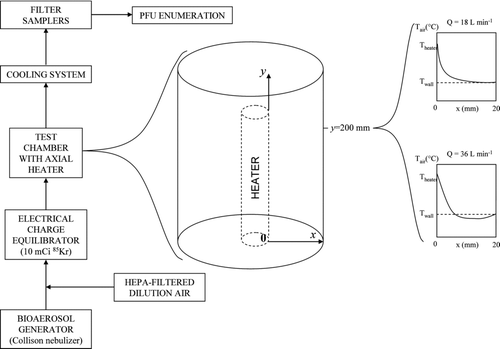
The experimental facility and test design were described in detail in our earlier paper (CitationGrinshpun et al. 2010). The setup was operated inside a class II biosafety cabinet (Model 6TX, Baker Co., Inc., Sanford, ME). provides the general schematics of the setup. MS2 virions were aerosolized by a Collison nebulizer (BGI Inc., Waltham, MA) operated at 6 L/min. Dry dilution air was supplied through a HEPA-filter at two flow rates of 12 and 30 L/min producing the total flows of Q = 18 L/min and Q = 36 L/min, respectively, which represented two different thermal flow regimes and exposure times. Charge-equilibrated by a 10 mCi 85Kr source (model 3012, TSI Inc., St. Paul, MN), the bioaerosol was dispersed in a vertically oriented, cylindrical test chamber of 50 mm in inner diameter and 320 mm in height. The chamber as well as other sections of the experimental setup was made of stainless steel. A cylindrical electric heating element (Mighty-Watt Heater, Gordo Sales, Inc., Layton, UT) of 10 mm in diameter and 230 mm in height was installed in the test chamber along its axis on a flat holder 16 mm above the chamber entrance. The axial air flow heating design was utilized due to its relevance to another laboratory study conducted in parallel to investigate the selected bio-agent defeat during explosion and combustion. This configuration is also being explored for thermal air purification of biocontaminated indoor air (CitationJung et al. 2009a, Citation2009b; CitationLee and Lee 2006). Connected to a variable voltage source (Staco Energy Products, Co., Dayton, OH) through a general purpose transformer (Acme Electric Corp., Lumberton, NC), the heating element was operated at voltages of 50 to 160 V, producing a wide range of temperatures T heater . Depending on the voltage applied to the heating element and the air flow rate, it took approximately 30–60 min to achieve a stable thermal condition represented by a specific target temperature of the test chamber wall, T wall , which was measured using thermocouple probes (Model 5TC-GG-20, Omega Engineering, Inc., Stamford, CT) with a digital thermometer (Model HH12A, Omega Engineering, Inc.).
After passing through the test chamber and a 500-mm long transportation line, the aerosol was isoaxially sampled into three identical probes and transported through a cooling system that cooled the air down to room temperature. The viruses were collected from each probe downstream of the cooling system on a sterile 25-mm diameter gelatin filter (SKC Inc., Eighty Four, PA). Gelatin filters are appropriate for collecting airborne viruses as viral infectivity is not critically impacted during collection (CitationVerreault et al. 2008). The sampling was conducted at a flow rate of 5 L/min over a period of 1 min. The filters were subsequently removed, dissolved in sterile, filtered deionized water, and analyzed using a modified plaque assay (ISO 2000). The PFU count was performed on TYG agar plates from 1 mL aliquots of the original or diluted filter extracts. Dilution ratios of 101, 102, 103, and 104 were used. The concentration of plaque forming viruses was determined as an average from the three filter samples obtained in each trial. At least two trials were conducted for each set of conditions. The lowest countable number was 1 PFU per plate. Some plates (representing the samples obtained under high thermal exposure conditions) showed zero count. For those, we assigned a value of 0.5 PFU that was input when determining an average count from several replicates—the approach conventionally used in other studies (CitationHeinrich et al. 2003). The limit of detection (LOD) was calculated using 1 PFU per sample and a total sample volume of 1 mL. Available analytical guidelines recommend using the Practical Quantification Limit (PQL), which is 5-fold greater than LOD. It represents the lowest level that can be reliably achieved within specific precision and accuracy limitations provided by routine laboratory operating conditions (CitationBenjamin and Belluck 2001; CitationMcBean and Rovers 1998; CitationWayman et al. 1999). Following these guidelines, we adopted PQL = 5 × LOD as the quantification limit for our analysis. We acknowledge that in quantitative microbiology credible estimates are usually based on colony/plaque counts higher than 5 CFU or PFU; however, as this test protocol aimed at achieving conditions causing the maximum possible inactivation, we accepted 5 PFU as a reasonable limit.
The experiments were conducted with no voltage applied to the heating element, i.e., at a room temperature, T 0 = 20°C (regarded as control), and at various voltages representing specific thermal conditions to which the virions were exposed in the chamber (temperature T). The inactivation factor (IF) can be quantified through the test-control comparison as follows:Footnote 1
Characteristic Exposure Temperature and Conservative Estimate of Thermal Inactivation in Axially Heated Air Flow
In accordance to the approach developed in our earlier study (CitationGrinshpun et al. 2010), the characteristic exposure temperature was defined based on the following considerations:
-
The longitudinal air temperature profile is non-monotonic. For any specific flow streamline (x = const), the air temperature T air increases as the air moves along the heater and then decreases. Based on our previous measurements, depending on the target temperature and the radial coordinate, the peak (T y, max x = const ) occurs at different points, mostly around y = 200 mm. For a specific particle trajectory, the peak temperature largely determines the airborne virus inactivation.
-
While being very high for the air streamlines near the heater (small x), the peak values of air temperature are much lower for streamlines near the wall (large x). T y, max decreases radially following a monotonic pattern (mostly for lower flow rates) or a non-monotonic pattern (for higher flow rates; ). The difference is associated with the thermal development of the air flow (CitationHolman 2002; CitationThomas 1992). At any flow rate, an annular area can be identified between x = x* and x = 20 mm (wall), in which the air flow is heated the least. In our earlier study involving the same experimental set-up, the entire cross-section was divided in four annuli, x = 0–5 mm, x = 5–10 mm, x = 10–15 mm, and x = 15–20 mm, and the “low-heat zone” was identified as the last two annuli (CitationGrinshpun et al. 2010). It was postulated that if the microorganisms moving through this zone did not survive, those moving through a hotter area (closer to the heater) would not survive either. According to this conservative approach, the zone of x > x* was selected for characterizing the exposure causing inactivation, and the maximum integral temperature achieved in this zone was regarded as the characteristic air temperature, T air .
-
There is a certain time needed for an aerosol particle to get adjusted to a specific surrounding air temperature. Thus, in an air flow with constantly changing T air , the particle temperature at a point (x, y) may differ from the air temperature measured at the same point. However, for the test conditions of this study, the thermal equilibration time for particles as small as MS2 virions is estimated to fall below 1 millisecond, based on the assessment provided in CitationGrinshpun et al. (2010). This suggests that the airborne virions adapted the air temperature practically instantly, i.e., the characteristic air temperature defines the characteristic virion exposure temperature: T air = T e .
Similar to our previous investigation (CitationGrinshpun et al. 2010), the air temperature profiles inside the test chamber were determined with non-sheathed thermocouple probes (Type J, Model TJ36-ICIN-116, Omega Engineering) positioned at a specific points (x, y); all thermocouple-measured air temperature values were subjected to correction for radiation. In the previous study, the measurement precision was Δ T = ± 15°C, representing variations associated with positioning the thermocouple inside the chamber. In this study, we improved the spatial resolution as compared to the earlier measurements by decreasing the radial increment from Δ x = 5 mm to Δ x = 1 mm. The air temperatures determined at x = 1, 2, 3, …, 19 mm from the heater were combined with the surface temperature data assuming T air x = 0 = T heater and T air x = 20 mm = T wall . For each of the four 5-mm thick annuli, the average temperature value was calculated by integrating over the corresponding cross-section. The average air temperature values of adjacent annuli were compared. If the difference fell below 30°C (twice the precision-based uncertainty, Δ T = 15°C), no differentiation was made between the annuli with respect to the heat exposure. These annuli were combined to establish the characteristic exposure zone (x* to 20 mm) for the conservative determination of the heat-induced viral inactivation. The interval of air temperatures, [ttilde] air , measured across this zone with a 1-mm increment and the average value, T air , calculated by integrating over the zone's cross-section, were recorded. The former represented the temperature variation within the characteristic exposure zone, and the latter allowed assigning a single characteristic exposure temperature for which the IF-value was experimentally determined.
The control sample accounted for all viable virions arrived from the test chamber, i.e., those which moved through the entire cross-section (x = 0 to 20 mm). As explained above, the test sample accounted for the surviving virions arrived from the characteristic exposure zone (x = x* to 20 mm), because others were postulated to be 100% inactivated by the more intense heat. The relative difference between the corresponding cross-sectional areas, expressed as
Data Analysis
The mean IF-value and the geometric standard deviation were calculated for every test. The upper limit of the reportable IF was derived from PQL = 5 × LOD and referred to as the highest practical quantification limit (HPQL).
RESULTS
According to the criteria presented in the Materials and Methods section, the combination of the third (x = 10–15 mm) and the fourth (x = 15–20 mm) annuli was established as the characteristic exposure zone for determining the inactivation factor. presents IF corrected = 0.667 × IF measured plotted against the characteristic exposure temperature for two exposure/flow conditions: Q = 18 and 36 L/min. The horizontal bars represent the above-defined range of temperatures [Ttilde] air . The vertical error bars represent the geometric standard deviations (GSD) for the corrected values of the inactivation factor.
FIG. 2 Effect of the characteristic exposure temperature and the air flow rate through the test chamber on the inactivation of aerosolized MS2 virions. Points: experimentally obtained values of IF corrected (T e ); horizontal bars: range of [ttilde] air ; vertical bars: GSD of IF corrected . HPQL = Highest Practical Quantification Limit.
![FIG. 2 Effect of the characteristic exposure temperature and the air flow rate through the test chamber on the inactivation of aerosolized MS2 virions. Points: experimentally obtained values of IF corrected (T e ); horizontal bars: range of [ttilde] air ; vertical bars: GSD of IF corrected . HPQL = Highest Practical Quantification Limit.](/cms/asset/21534fb4-eec9-4221-aa52-c7ca45985117/uast_a_509119_o_f0002g.gif)
It is seen that a short-term exposure to temperatures of up to ∼ 90°C (Q = 18 L/min) or up to ∼ 140°C (Q = 36 L/min) produced a steady MS2 virus inactivation at relatively modest levels of IF ∼ 10 or lower. Further increase in T e resulted in a rapid infectivity decline demonstrated by an exponential increase of the inactivation factor. The latter reached ∼ 103 (∼ 99.9% viruses killed) at T e ∼ 150°C as a result of a longer exposure (Q = 18 L/min) or at T e ∼ 230°C after shorter exposure (Q = 36 L/min). Further—even moderate—increase of T e beyond the above-indicated values generated a rapid jump of the efficiency of MS2 inactivation with IF exceeding HPQL. This was confirmed by multiple measurements performed at T e > 170°C (Q = 18 L/min) and T e > 250°C (Q = 36 L/min), as shown in by vertical arrows at specific temperatures corresponding to counts of zero or few PFUs per sample that produced IF > HPQL. These IF levels translate into a virus inactivation rate of ∼ 99.996% or greater.
DISCUSSION
The time of a specific thermal exposure was not explicitly defined by this study protocol because a virion moving along a specific trajectory was exposed to different air temperatures at different points between the entrance and the exit of the test chamber. Thus, in spite of experimental advantages and practical relevance, the axial design presents a clear limitation for establishing relationship between the exposure temperature and exposure time. In this context, a more accurate characterization of thermal inactivation of bioaerosol particles in air flows may require a more pertinent experimental system. Nonetheless, the exposure time was estimated by applying the time-of-flight concept to a distance hypothetically characterized by a constant temperature [Ttilde] air . Based on the review of the longitudinal air temperature profiles, we estimated this distance to be approximately one quarter of the heater's length. Consequently, the time of flight along this distance (i.e., 230/4 = 57.5 mm) was assigned as a reasonable estimate of the characteristic exposure time. Accounting for the dependence of air velocity on air temperature (through its effect on the air density), the estimated characteristic exposure time ranged approximately from 0.10 to 0.15 s at Q = 36 L/min (calculated for the tested range of T e = 90 – 250°C; the higher temperature corresponds to the shorter exposure time). At 18 L/min, it ranged from 0.24 to 0.33 s (calculated for the tested T e = 45 – 170°C).
For each flow rate, the results show that the initial temperature increase caused only a moderate inactivation with IF exhibiting a weak or no dependence on T e . This is due to insufficient exposure to heat that resulted in relatively low heat energy transfer. Generally, thermal inactivation is quantitatively characterized by the probability of the virus colliding with a molecule of sufficient energy to cause lethal damage or a number of such collisions. With further increase in T e , this probability is expected to increase exponentially (CitationHiatt 1964), which is evident from the experimentally determined exponential increase of the inactivation factor from ∼ 101 to ∼ 103 (). The final phase is portrayed by a sudden boost in IF above the reportable limit of the study protocol as the temperature further increased by a small margin. This may possibly indicate that a different, or an additional, mechanism is responsible for inactivation at extreme temperatures.
The data sets obtained at the two flow rates show the same trends but quantitatively are remarkably different. The 2-fold difference between the tested flow rates (corresponding to a relatively modest difference in exposure time) produced a sizable change in the virus inactivation. The assessment described in the online Supplemental Information section demonstrates that it is possible.
Little is known about physicochemical and biochemical mechanisms that lead to thermal inactivation of aerosolized viruses. However, some information generated in studies with thermal inactivation of the MS2 bacteriophage in liquids seems relevant and is utilized in the discussion below.
The full virion of MS2 consists of a non-enveloped icosahedral protein capsid surrounding an RNA genome. The protein coat is formed from 180 identical subunits through self-assembly. In addition, this bacteriophage features the assembly protein A, which is an important component required for the phage attachment to the side of the pilus of the host bacterium containing the F plasmid (F+ or male E. coli) (CitationBrock et al. 1994). Although the location of protein A in the MS2 protein shell structure is not well characterized (e.g., it has not yet been resolved by the three-dimensional X-ray crystallographic analysis), chemical labeling and antibody-binding experiments suggest that protein A is at least partially exposed on the capsid surface (CitationCurtiss and Krueger 1974; CitationO'Callaghan et al. 1973). Thus, heat treatment may damage not only the coat protein but potentially also protein A. Heat may also damage the single-stranded RNA inside the capsid. This damage is represented by effects such as denaturation, mutation, and strand breaks, or their combination. Each of the effects or all of them combined can inactivate an MS2 virion and make it non-infectious to the host bacterium, which ultimately reduces the fraction of viruses forming plaques. While the exponential decrease in infectivity with the temperature observed in our experiments is consistent with the conventionally accepted first-order concept (CitationHiatt 1964), the same concept does not explain the rapid enhancement of viral inactivation observed at T e ∼ 170°C for Q = 18 L/min and at ∼ 250°C for Q = 36 L/min. Additional experiments are needed to identify possible mechanisms associated with the final abrupt drop in the phage survival.
CitationStonehouse and Stockley (1993) investigated the damage of MS2 coat protein by exposing aliquots of assembled coat protein (from empty capsid lacking RNA) to specific temperatures for a 10-min period. Once heated to 70.5°C (in liquid), reassembly of coat proteins was found no longer possible. CitationJohnson et al. (2007) compared the recovery of the capsid proteins after heating for 2 h using sodium dodecyl sulfate polyacrylamide gel electrophoresis (SDS–PAGE) and reported that no capsids were recovered at temperatures above 70°C due to precipitation of the denatured protein. It is acknowledged that the temperature and exposure time in the above-quoted studies were different than in ours and, therefore, the results may not be directly comparable. Nevertheless, these studies provide evidence that disassembly and reassembly of capsid proteins may take place until a certain “threshold” temperature is reached, which makes the disassembly of coat proteins irreversible.
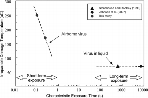
From the practical perspective, it seems meaningful to define the “threshold” exposure temperature which inflicts irreparable damage to all viruses within the quantification limit of the test method. schematically demonstrates this “threshold” plotted against the thermal exposure time. The left part of the scale represents short-term treatments, which typically take place in aerosol systems, while the right part features much longer thermal exposures occurring in liquid suspensions. In this figure, we plotted the actual experimental data obtained in the present study with aerosolized MS2 virions (circles) as well as the data reported by CitationStonehouse and Stockley (1993) and CitationJohnson et al. (2007) from their tests on the inactivation of MS2 bacteriophage in liquid (triangle and diamond).Footnote 2 For longer exposures, the threshold temperature corresponding to the virus irreparable damage is not expected to show an appreciable dependence on time. In contrast, manifestation of the temperature-time dependence is anticipated when virions are exposed to heat over sub-second time intervals, which is sufficient for adopting the surrounding air temperature but may fall short of providing sufficient time for the expected lethal response. Thus, the airborne virions exposed to hot air for less than one second may survive much higher temperatures than those suspended in hot liquid media for minutes or hours. In addition, the data points representing viruses in liquid may signify individual dispersed MS2 virions. In contrast, the MS2 aerosols generated by the Collison nebulizer are likely contained aggregates and/or particles covered by residue from the nebulization suspension (CitationEninger et al. 2009; CitationHogan et al. 2005; CitationRiemenschneider et al. 2010). These factors, in principle, can enhance resistance of aerosolized viruses to environmental stress, including heat. Consequently, irreparable damage may occur at higher temperatures.
CONCLUSION
A short-term (∼ 0.1–1 s) thermal inactivation of aerosolized MS2 virions began at a certain level of heat exposure and continued gradually as T e increased from ∼ 90°C to ∼ 160°C (for Q = 18 L/min) or ∼ 140°C to ∼ 230°C (for Q = 36 L/min) exhibiting an exponential pattern. Longer thermal exposure produced greater inactivation. Under certain conditions (T e ∼ 170°C for Q = 18 L/min and ∼ 250°C for Q = 36 L/min), the experimentally determined IF-values exceeded ∼ 2.4 × 10⁴, indicating that over 99.996% of viable virions lost their infectivity—the maximum fraction quantifiable by this study protocol. The reported IF represents the lower (i.e., conservative) approximation of the actual inactivation. It was found that airborne MS2 virions exposed to hot air for sub-second time intervals may survive much higher temperatures than those subjected to thermal treatment in liquid suspensions for minutes or hours. We postulate that the findings may be attributed to effects such as denaturation of coat proteins and protein A, mutation, and strand breaks, or their combination. Further studies are necessary to identify the mechanisms of inactivation.
uast_a_509119_sup_15215268.zip
Download Zip (18.1 KB)The authors gratefully acknowledge the support from the Defense Threat Reduction Agency (US Department of Defense) through grant HDTRA-1-08-1-0012. The authors are also thankful to Dr. S. Mohan for assisting in the temperature analysis.
Chunlei Li is on leave from Fudan University, Shanghai, People's Republic of China.
Notes
1 It is common in microbiology to use the “log inactivation” by quantifying log 10(IF).
2 Straight lines in are drawn through the data points and do not aim at representing specific linear trends.
REFERENCES
- Balazy , A. , Toivola , M. , Adhikari , A. , Sivasubramani , S. K. , Reponen , T. and Grinshpun , S. A. 2006 . Do N95 Respirators Provide 95% Protection Level Against Airborne Viruses and How Adequate are Surgical Masks? . Amer. J. Infect. Control , 34 : 51 – 57 .
- Benjamin , S. and Belluck , D. 2001 . A Practical Guide to Understanding, Managing, and Reviewing Environmental Risk Assessment Reports , Boca Raton, FL : CRC Press .
- Brock , T. D. , Madigan , M. T. , Markinko , J. M. and Parker , J. 1994 . Biology of Microorganisms, , 7th ed. , Englewood Cliffs, NJ : Prentice-Hall .
- Curtiss , L. K. and Krueger , R. G. 1974 . Localization of Coliphage MS2 A-protein . J. Virol. , 14 : 503 – 508 .
- de Jong , J. C. , Harmsen , M. and Trouwborst , T. 1975 . Factors in the Inactivation of Encephalomyocarditis Virus in Aerosols . Infect. Immun. , 12 : 29 – 35 .
- Dubovi , E. J. and Akers , T. G. 1970 . Airborne Stability of Tailless Bacterial Viruses S–13 and MS–2 . Appl. Microbiol. , 19 : 624 – 628 .
- Eninger , R. , Hogan , C. J. , Biswas , P. , Adhikari , A. , Reponen , T. and Grinshpun , S. A. 2009 . Electrospray Versus Nebulization for Aerosolization and Filter Testing with Virus Particles . Aerosol Sci. Technol. , 43 : 298 – 304 .
- Grinshpun , S. A. , Adhikari , A. , Honda , T. , Kim , K. Y. , Toivola , M. , Rao , R. K. S. and Reponen , T. 2007 . Control of Aerosol Contaminants in Indoor Air: Combining the Particle Concentration Reduction with Microbial Inactivation . Environ. Sci. Technol. , 41 : 606 – 612 .
- Grinshpun , S. A. , Adhikari , A. , Li , C. , Reponen , T. , Yermakov , M. , Schoenitz , M. , Dreizin , E. , Trunov , M. and Mohan , S. 2010 . Thermal Inactivation of Airborne Viable Bacillus Subtilis Spores by a Short-Term Exposure in Axially Heated Air Flow . J. Aerosol Sci. , 41 : 352 – 363 .
- Havelaar , A. H. , van Olphen , M. and Drost , Y. C. 1993 . F-specific RNA Bacteriophages are Adequate Model Organisms for Enteric Viruses in Fresh Water . Appl. Environ. Microbiol. , 59 : 2956 – 2962 .
- Heinrich , J. , Hölscher , B. , Douwes , J. , Richter , K. , Koch , A. , Bischof , W. , Fahlbusch , B. , Kinne , R. W. and Wichmann , H. E. 2003 . INGA Study Group, Reproducibility of Allergen, Endotoxin and Fungi Measurements in the Indoor Environment . J. Expos. Anal. Environ. Epidemiol. , 13 : 152 – 160 .
- Hiatt , C. W. 1964 . Kinetics of the Inactivation of Viruses . Bacteriol. Rev. , 28 : 150 – 163 .
- Hogan , C. J. , Kettleson , E. M. , Lee , M.-H. , Ramaswami , B. , Angenent , L. T. and Biswas , P. 2005 . Sampling Methodologies and Dosage Assessment Techniques for Submicrometre and Ultrafine Virus Aerosol Particles . J. Appl. Microbiol. , 99 : 1422 – 1434 .
- Holman , J. P. 2002 . Heat Transfer, , 9th ed. , Boston : McGraw-Hill .
- 2000 . Water Quality—Detection and Enumeration of Bacteriophages—Part 2: Enumeration of Somatic Coliphages ISO—International Organization for Standardization 10705–2:2000
- Johnson , H. R. , Hooker , J. M. , Francis , M. B. and Clark , D. S. 2007 . Solubilization and Stabilization of Bacteriophage MS2 in Organic Solvents . Biotechnol. Bioengin. , 97 : 224 – 234 .
- Jones , M. V. , Bellamy , K. , Alcock , R. and Hudson , R. 1991 . The Use of Bacteriophage MS2 as a Model System to Evaluate Virucidal Hand Disinfectants . J. Hosp. Infect. , 17 : 279 – 285 .
- Jung , J. H. , Lee , E. L. and Kim , S. S. 2009a . Thermal Effects on Bacterial Bioaerosols in Continuous Air Flow . Sci. Total Environ. , 407 : 4723 – 4730 .
- Jung , J. H. , Lee , J. E. , Lee , C. H. , Kim , S. S. and Lee , B. U. 2009b . Treatment of Fungal Bioaerosols by a High-Temperature, Short-Time Process in a Continuous Flow System . Appl. Environ. Microbiol. , 75 : 2742 – 2749 .
- Lee , Y. H. and Lee , B. U. 2006 . Inactivation of Airborne E. coli B. subtilis Bioaerosols Utilizing Thermal Energy . J. Microbiol. Biotechnol. , 16 : 1684 – 1689 .
- McBean , E. A. and Rovers , F. A. 1998 . Statistical Procedures for Analysis of Environmental Monitoring Data and Risk Assessment , Upper Saddle River, NJ : Prentice Hall .
- Mullican , C. L. , Buchanan , L. M. and Hoffman , R. K. 1971 . Thermal Inactivation of Aerosolized Bacillus subtilis var. niger Spores . Appl. Microbiol. , 22 : 557 – 559 .
- Nicholson , W. L. , Munakata , N. , Horneck , G. , Melosh , H. J. and Setlow , P. 2000 . Resistance of Bacillus Endospores to Extreme Terrestrial and Extraterrestrial Environments . Microbiol. Mol. Biol. Rev. , 64 : 548 – 572 .
- Northrop , J. and Slepecky , R. A. 1967 . Sporulation Mutations Induced by Heat in Bacillus subtilis. . Science , 155 : 838 – 839 .
- O'Callaghan , R. , Bradley , R. and Paranchych , W. 1973 . Controlled Alterations in the Physical and Biological Properties of R17 Bacteriophage Induced by Gunaidine Hydrochloride . Virology , 54 : 476 – 494 .
- Rengasamy , S. , Fisher , E. and Shaffer , R. E. 2010 . Evaluation of the Survivability of MS2 Viral Aerosols Deposited on Filtering Face Piece Respirator Samples Incorporating Antimicrobial Technologies . Amer. J. Infect. Control , 38 : 9 – 17 .
- Riemenschneider , L. , Woo , M.-H. , Wu , C.-Y. , Lundgren , D. , Wander , J. , Lee , J.-H. , Li , H.-W. and Heimbuch , B. 2010 . Characterization of Reaerosolization from Impingers in an Effort to Improve Airborne Virus Sampling . J. Appl. Microbiol. , 108 : 315 – 324 .
- Setlow , P. 1995 . Mechanisms for the Prevention of Damage to the DNA in Spores of Bacillus Species . Annual Rev. Microbiol. , 49 : 29 – 54 .
- Setlow , B. and Setlow , P. 1995 . Small, Acid-Soluble Proteins Bound to DNA Protect Bacillus subtilis Spores from Killing by Dry Heat . Appl. Environ. Microbiol. , 61 : 2787 – 2790 .
- Stonehouse , N. J. and Stockley , P. G. 1993 . Effects of Amino Acid Substitution on the Thermal Stability of MS2 Capsids Lacking Genomic RNA . FEBS Lett. , 334 : 355 – 359 .
- Stumbo , C. R. 1973 . Thermobacteriology in Food Processing , London : Academic Press .
- Thomas , L. C. 1992 . Heat Transfer, , 2nd ed. , Englewood Cliffs, NJ : Prentice Hall .
- Tseng , C.-C. and Li , C.-S. 2005 . Collection Efficiencies of Aerosol Samplers for Virus-Containing Aerosols . J. Aerosol Sci , 36 : 593 – 607 .
- Verreault , D. , Moineau , S. and Duchaine , C. 2008 . Methods for Sampling of Airborne Viruses . Microbiol. Mol. Biol. Rev. , 72 : 413 – 444 .
- Wayman , C. , Gordon , E. and King , J. 1999 . The Method Detection Limit and Practical Quantitation Level: Their Derivations and Regulatory Implications Proceedings of Waste Management Conference (WM1999, Tucson, AZ, February 28–March 4, 1999)

