Abstract
Cough generated infectious aerosols are of interest while developing strategies for the mitigation of disease risks ranging from the common cold to SARS. In this work, the velocity field of human cough was measured using particle image velocimetry (PIV). The project subjects (total 29) coughed into an enclosure seeded with stage fog. Cough flow velocity profiles, average widths of the cough jet, and maximum cough velocities were measured. Maximum cough velocities ranged from 1.5 m/s to 28.8 m/s. The average width of all coughs ranged between 35 to 45 mm. Wide variability in the data suggests that future cough simulations consider a range of conditions.
1. Introduction
Many diseases can be transmitted by inhalation of airborne microorganisms. In the early part of the last century, CitationWells and Wells (1938) showed that during coughing and sneezing, small microorganism-containing droplets were released into the air and that these droplets could evaporate and remain suspended in the air. Transmission of tuberculosis by inhalation of airborne Mycobacterium tuberculosis aerosolized by coughing has been well documented (CitationBarnes et al. 1996; CitationValway et al. 1998). More recently, attention has focused on virally-transmitted diseases. CitationCouch et al. (1966) maintains that coughing is capable of producing recoverable viruses 40% of the time. Severe Acute Respiratory Syndrome (SARS) is a virus that is suspected of being transmitted by direct inhalation of viral particles released from infected individuals. Many healthcare workers that were infected during the 2003 SARS outbreak were present during particle generating procedures conducted in the hospitals, such as sputum induction, suggesting that these particle generating procedures were not adequately controlled and may have increased the risk of SARS infection (Centers for Disease Control 2004).
Airborne transmission of infectious diseases occurs by dissemination of either airborne droplet nuclei or droplets in the respirable size range containing infectious agents that remain infective over time and distance (CitationSiegel et al. 2007). Respiratory droplets are generated when an infected person coughs, sneezes, or talks or during respiratory procedures. Droplets are defined as being greater than 5 μm in size; droplet nuclei are particles arising from desiccation of suspended droplets and are defined as less than or equal to 5 μm (CitationSiegel et al. 2007; CitationXie et al. 2009). Nicas and colleagues (2005) show by modeling that emitted droplets will evaporate to 50% of their initial value and that if the initial diameter is <20 μm this process will happen instantaneously.
Despite the obvious importance of infectious aerosols in disease transmission, limited information is available on the flow fields of the primary generation mechanisms, human cough and sneeze. Specifically, there has been little research conducted on the velocities of particles released during a cough or on the velocity field of a cough. Most related research in this area has studied the volumetric flow rate, or particle sizes. Other studies have investigated the sounds of cough (CitationPiirilä and Sovijärvi 1989), the physiological function of cough, and the effectiveness of different asthma medications.
CitationZhu et al. (2006) measured the velocity of coughs from three males using particle image velocimetry (PIV) with flour as the tracer particle. The initial cough velocity ranged from 6–22 m/s with an average velocity of 11.2 m/s, and an estimated standard deviation of 2.85 m/s. An average of 6.7 mg of saliva was expelled from the coughs. The affected area extended as far as 2 m or more. CitationChao et al. (2009) reported particle size data from 11 subjects and PIV data from one male and one female during coughing and speaking, using a saline fog as tracer. The average velocity of 50 coughs from each subject was 11.7 m/s.
Two studies measured flow rates of a voluntary cough as a function of time with a tussometer (CitationMahajan et al. 1994; CitationSingh et al. 1995). Mahajan et al. found that peak flow rates ranged from 200 L/min to 950 L/min with a mean peak flow rate of 300 L/min and the time to reach those flow rates ranged from 15 ms to 40 m/s. Singh et al. found the peak flow rate of women reached 750 L/min and the peak flow rate of men reached 1300 L/min.
CitationWells (1955), referring to work by CitationChaussé and Magne (1916), reported that the peak velocity from sneezing or coughing approaches 100 m/s and 16–48 m/s, respectively, and that the majority of droplets expelled by expiratory processes at these velocities are 10 microns or less in diameter.
CitationPapineni and Rosenthal (1997) measured particle diameters of aerosols generated while a human breathed (mouth and nose), coughed, and talked in situ using a Climet CI7300 optical particle counter that is capable of measuring particles in five size bins with diameters in the range 0.3 μm – >10 μm. They also used electron microscopy to estimate the droplet sizes from the residue after the aerosols had dried on copper substrates coated with carbon film. From these experiments, droplet optical diameters were determined to range from <0.6 μm to 2.5 μm. The highest droplet counts were for coughing, and most of the particles were less than 2 μm in diameter.
In another study of cough, CitationFennelly et al. (2004) isolated cough-generated particles of Mycobacterium tuberculosis from sixteen participants that had tested positive for tuberculosis. Particles were collected from voluntary cough using a cough aerosol sampling system based on two six-stage cascade impactors. Particles ranged in size from 0.65 μm to 3.3 μm.
CitationPiirilä and Sovijärvi (1989) studied the acoustics of a cough compared to the cough flow in people with asthma and bronchitis. They measured the volume flow rate to and from the lungs with a pneumotachograph and simultaneously recorded the sound spectra of spontaneous cough. This study illustrated that the cough sound starts at the beginning of the cough flow.
Detailed measurements of in vitro cough flow fields are reported by CitationAfshari et al. (2002), who used PIV to map the flow of a cough flow simulator, which is a hydraulic pump producing oil droplets less than 5 μm in diameter. One measurement of each simulated cough flow was taken at 0.4 s. The cough simulator was programmed using airflow waveforms developed from two volunteers, one with asthma, and one without. CitationBadeau et al. (2002) later used Afshari's data in a numerical CFD simulation of the particle and flow field trajectories in a closed environment. CitationZhao et al. (2005) used Badeau's cough conditions in their CFD simulation of indoor human generated aerosol transport. On a related topic, CitationMarr et al. (2005) used PIV to map the flow from a thermal mannequin with simulated breathing and showed that there was a threefold increase in local turbulence intensity during the exhalation part of the waveform due to the unsteady flow characteristics.
In the context of fluid mechanics, human cough can be classified as a transient turbulent jet, and likely an impulsively started one. In their studies of transient jets, CitationJohari et al. (1997) and CitationBajpai and Tirumkudulu (2008) use a nondimensional time scale based on steady-state jet speed Vj and diameter D of tV j /D. This can be interpreted as a nondimensionalized equivalent piston stroke of length L, L/D. For human cough, an estimate of this parameter can be made from CitationMahajan et al.'s (1994) data. They report an average cough volume of roughly 2.5 liters. Assuming a mouth diameter D of 30 mm, this gives a stroke length L of 3.5 m, and an L/D of 118. This places the cough in the regime of short-duration jets, but far from the very short duration regime where much of the cough would be entrained in a single vortex ring (CitationRosenfeld et al. 1998). Transient turbulent jets are generally modeled as a quasi-steady state jet with a vortex ring at the head following CitationTurner (1962). Although the starting jet is acknowledged to be not self-similar, particularly in its initial stages and close to the head of the jet, experimental and theoretical analyses often explore the application of steady-state turbulent jet scalings. Several such recent works have had success in predicting the evolution of the tip of the jet (CitationJohari et al. 1997; CitationHill and Ouellette 1999; CitationSong and Abraham 2003; CitationCossali et al. 2004; CitationAi et al. 2005; CitationBajpai and Tirumkudulu 2008).
The objective of the present work is to increase our understanding of cough flow fields by making PIV measurements of actual cough flow fields produced by human subjects. The flow field of human cough was measured by first filling an enclosure with theatrical fog and then having 29 healthy subjects cough into the enclosure. PIV measurements were made, using a cross correlation algorithm to determine the velocities of the fog particles. We report the maximum velocities, maximum velocity location, waveform characteristics, average velocity profile, width of the cough flow, and turbulent scaling for the flow. The immediate use of this data will be to improve the design of mechanical simulators such as those described above, and in numerical simulations of cough, which require appropriate initial and boundary condition data. Ultimately, the information gathered from this research may be helpful to health care workers in estimating the distances infectious particles may spread, and to aid in the design of infectious aerosol control technologies and strategies.
2. Materials and Methods
2.1. Apparatus
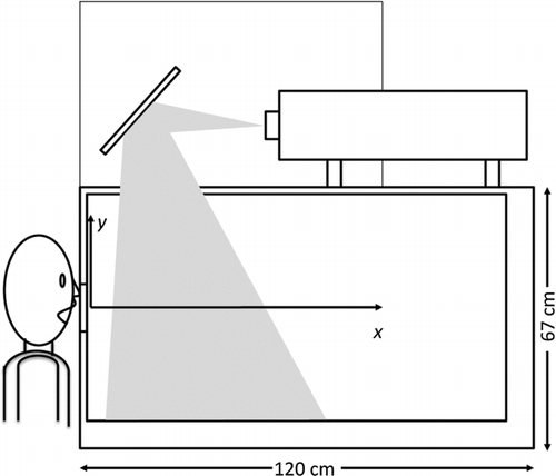
Volunteer human subjects coughed into an enclosure 120 cm in length, 76 cm in width, and 67 cm height. The enclosure, shown in , was lined with black plastic sheeting. The enclosure represents a limitation of the study. The average cough jet volume to enclosure volume ratio is roughly 240, thus little indirect influence of recirculating flow can be expected. However, at an average speed of 10 m/s, the cough would likely impinge on the far wall at 0.12 s. Thus care must be taken when interpreting the data. The front of the enclosure was a pane of glass to allow imaging of the cough flow field. The left side, adjacent to the subject, was plywood with a pear-shaped hole, 10 cm high, 9 cm wide at the base, and 5 cm at the top. This hole was large enough to accommodate the subject's nose and mouth only and the plywood protected the subject from stray laser light. The laser sheet entered through a window, 25 cm by 15 cm on the top of the enclosure. The laser rested on top of the enclosure and was directed downward by a flat mirror. The laser and the mirror were further encased in shielding. The apparatus and procedures were approved by the University of Colorado's Institutional Review Board.
FIG. 1 Diagram of apparatus.
Healthy volunteers produced an insufficient quantity of particles from a cough for determination of the velocity field, so the air in the enclosure was seeded as described below. Illumination of the seed particles was provided by a New Wave Solo 120 mJ Nd:YAG laser at a wavelength of 532 nm (New Wave Research Inc., Fremont, CA). The laser sheet was oriented so that it would provide a cross section of the center of the cough as the cough flow traveled away from the volunteer. A spherical lens of focal length = 1000 mm was used to focus the sheet to a thickness of 0.15 mm in the region of the measurement. A high power setting was used, with a Q-switch delay of 170 μs, resulting in 117 mJ/pulse. Two diverging lenses were used to give a combined focal length of –9.4 mm and high power was used. Images were recorded by a TSI PIVCam 138 Model 630047 camera with 1K × 1K resolution (TSI Inc., Minneapolis, MN) with a Nikkor 60 mm focal length lens.
TSI's Insight PIV control and analysis program was used to capture and process two digital images (frames) of the cough flow field with a known pulse separation time (tps) of 100 μs between the frames. A recursive Nyquist grid was used with a central difference offset scheme, beginning with a spot size of 64 × 64 pixels, and ending with 32 × 32. Each spot was preconditioned by subtraction of the average spot intensity (employing the ‘zero pad mask’). The image pairs were correlated with an FFT algorithm, and peak locations in the correlation maps were detected to subpixel accuracy with a bilinear fit. Each correlation was validated in two ways. First the average image intensity in the spot must be at least 1/10 of the image average. Second, the height of the maximum correlation peak (signal) in each spot was compared to the next highest peak (presumed to be noise) and correlations with a ratio less than 1.5 were rejected. Typically, less than 4% of vectors were rejected from an image.
2.2. Methods
Twenty-nine nonsmoking volunteers were enrolled in this study: ten male and nineteen female volunteers. All participants were healthy normal subjects with ages ranging from 20 to 55. The median height was 1.70 m (range: 1.57 to 1.85 m) and the median weight was 61.9 kg (range: 47.6 to 61.9 kg). Two volunteers had mild, athletic-induced asthma, but were not prescribed an inhaler or any other medication for their asthma. Each participant was asked to produce a voluntary single cough as they normally would and typically three measurements were made during each cough. The volunteers were then asked to repeat this single voluntary cough five to eight times. A total of 195 coughs (130 female, 65 male) were recorded.
It was determined initially that healthy coughing subjects did not produce enough particles to be imaged by the PIV system. Subjects were then asked to fill their mouth with water and then cough, causing water particles to be expelled from the mouth with the cough flow. This method also did not produce meaningful results. Subsequently theatrical fog generated by an evaporation/condensation type fog generator (model G150, Le Maitre Ltd., Surrey, England) was used to seed the volume of the enclosure. Fog fluid is composed of a proprietary blend of glycols and water, has a specific gravity of 1.037, and is non-toxic for volunteers to inhale (Le Maitre Special Effects, Inc., 2006). This fog was injected into the enclosure in low concentrations, fanned to create uniformity, and then allowed to stagnate for between 30 seconds and one minute. The subject then breathed the fog from the enclosure before coughing into the enclosure. The mean diameter of the aerosol is 1μm for this type of generator (CitationSomers et al. 1991). While it is probable that the use of theatrical fog affected the real cough air flow, these small particles more accurately represent the particles typically expelled from a human's mouth during coughing or breathing, and also represent the flow better.
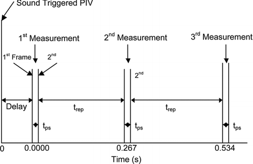
The sound of the volunteer's cough triggered the PIV system. A microphone was placed inside the enclosure just above the hole for the volunteer's mouth. When the volunteer coughed, cough signal was transmitted through the microphone and pre-amplifier, and then the voltage was sent to a low-powered quad op amp comparator circuit, which then triggered the PIV. Details are provided in CitationVanSciver (2005).
Three measurements (comprised of two frames each) were taken of each cough with a pre-determined delay time between each measurement. Once the sound of the cough triggered the PIV, the first measurement was taken after a delay of 20 μs. Successive measurements of the same cough were taken after a delay of 0.267 s. In summary, the first measurement was taken at 20 μs, the second at 0.267 s and the third at 0.534 s after the cough triggered the PIV, as illustrated in .
FIG. 2 Diagram depicting timing of image frames for PIV measurements.
3. Results and Discussion
3.1. Maximum Velocities
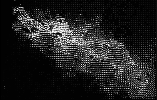
An image of a typical cough flow can be seen in . Each volunteer tended to cough in a different direction; in this case, the volunteer coughed downward.
FIG. 3 PIV of cough flow using theatrical fog.
Maximum velocities ranged from 1.15 to 28.8 m/s for all coughs (n= 195), with Reynolds numbers ranging from 2,300 to 57,600, based on an estimated mouth diameter of 30 mm and a kinematic viscosity of 1.5 10–5m2/s. The velocities of the surrounding stagnant air were low and on the order of 10–2 m/s, which allows the cough flow to be readily identified. The overall average maximum cough velocity was 10.2 m/s (standard deviation (SD) = 6.7 m/s) and the median was 8.1 m/s. The corresponding average Reynolds number Re = 2.04 × 10⁴. The maximum cough velocities had a log-normal distribution (based on Kolmogorov's goodness-of-fit test) with a geometric mean of 8.4 m/s and a geometric standard deviation of 1.9.
The data was evaluated for associations between sex and weight. A single-factor analysis of variance (ANOVA) was performed on natural log-transformed data. No significant difference was found in maximum cough velocity between sexes (p value = 0.67) or between weights less or greater than 61.7 kg (p value = 0.25).
Flow rate measurements by CitationSingh et al. (1995) found a difference between the peak flow rates of the sexes, with the peak flow rates of women 60% of the men's values. While the maximum velocities from the present study did not differ between sexes, the discrepancy could be caused by Singh et al. asking each participant to cough with maximum effort while here the participants were asked to cough as they normally would.
The velocities derived in this study are comparable to velocities reported in the literature. CitationZhu et al. (2006) reported an average velocity of 11.2 m/s. CitationMahajan et al. (1994) measured peak cough volumetric flow rates to be between 200 L/min to 950 L/min with a mean peak flow rate of 300 L/min. CitationSingh et al. (1995) measured peak flow rates of women to reach 750 L/min and the peak flow rate of men to reach 1300 L/min. Using these flow rates, approximate cough velocities were estimated by dividing by the area of the mouth assuming a circle with a diameter of 30 mm. details these estimated velocities, which are consistent with the data from this study. The data are also comparable to the range of cough velocities reported by CitationWells (1955): 16–48 m/s.
TABLE 1 Characteristic cough volumetric flow rates from CitationMahajan et al. (1994) and CitationSingh et al. (1995); corresponding average velocities based on diameter D= 30 mm
3.2. Maximum Velocity Location
The location (in the horizontal (x) direction) of the maximum velocity was also determined for each cough. The location of the mouth was defined by the enclosure's wall. The location of the maximum velocity ranged from 3.9 mm to 309 mm. The median location of the maximum velocity was 35.3 mm downstream (n= 175), while the average location was at 67.8 mm from the subject's mouth with a standard deviation of 78 mm. The data were log-normally distributed (using Kolmogorov's goodness-of-fit test), with a geometric mean of 36.6 mm and a geometric standard deviation of 3.19. The development of the maximum velocity away from the orifice is consistent with a vena cava effect, suggesting that the front of the mouth and the lips form a constriction.
3.3. Waveform
There were three velocity measurements taken during each cough: one at 20 μs, one at 0.267 s, and the third at 0.534 s (). details the distribution parameters for each measurement and shows the decrease in cough velocity as a function of time., The maximum velocity usually occurred in the first measurement, at 20 μs, and the velocity decreased exponentially by 74% at 0.267 s, and by 86% at 0.534 s compared to the initial measurement. The cough velocity measurements at each time had a log-normal distribution (using Kolmogorov's goodness-of-fit test).
FIG. 4 Average waveform of a cough. Error bars are the standard deviation of the measurement (n= 145). Distribution statistics and an exponential fit to the data are given.
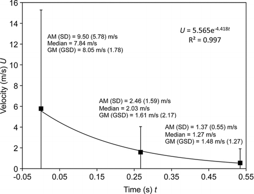
Typical air flow waveforms were mapped using a tussometer by CitationMahajan et al. (1994) and are similar to the current results: the flow rate in their experiment also drops to less than 30% by 0.25 s.
3.4. Average Velocity Profile
Initial analysis of the cough velocity fields revealed, unsurprisingly, that humans exhibit great variability in the direction and strength of cough, even from one subject. Here we attempt to mitigate this effect by scaling and offsetting the profiles vertically so that an average profile can be estimated. The subsequent analysis was performed only on the first measurement (at 20 μs) of each cough. The velocity profile as a function of y (vertical direction) of each cough was normalized by the maximum velocity of that cough found at that downstream distance (U/U max). The maximum cough velocities occurring at different distances for each cough can be seen in . At this measurement time, the highest velocities are found closest to the mouth, although the distribution is broad and peaks at 6 m/s. High velocity coughs might be expected to have penetrated farther at this time, but in general the decay of jet speed with distance dominates. This is consistent with studies of transient turbulent jets (CitationJohari et al. 1997; CitationCossali et al. 2004). The maximum normalized velocity is thus equal to 1 for each cough at each downstream distance. Each of the velocity profiles was then aligned so that the maximum for each profile occurred at the same y location: yc = 0, which defines the center of these normalized and offset profiles. The variation of cough profile shapes for five representative coughs at x= 50 mm can be seen in .
FIG. 5 Distribution of maximum velocity of each cough at specified locations in x.
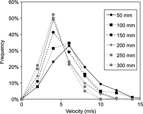
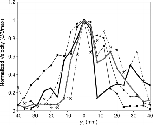
FIG. 6 Offset velocity profiles for five representative coughs at x= 50 mm.
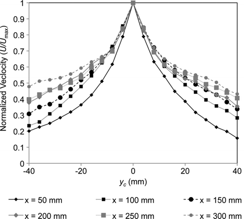
An average velocity profile was determined for each downstream distance (). The cough spreads with increasing downstream location, although the central portion of the profile maintains a similar steepness into the far field. The typical Gaussian profile is not observed, but this may be due to the ensemble averaged nature of the data. shows that the profiles tended to be asymmetric, with the peak velocity occurring towards either the top or bottom edge. When offset to align the peak velocities and averaged, the resulting profile was steepest at the centerline.
FIG. 7 Normalized velocity profiles for each x location, averaged over all coughs.
3.5. Width of the Cough Flow
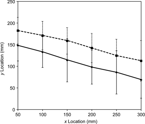
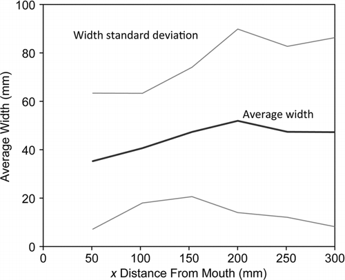
Normalized profiles occurring at each x location (U/U max) were used to determine the average width of the cough jet produced from subjects. The width was determined for each normalized (but not offset) velocity profile by interpolating to find the width at half velocity, i.e., the upper and lower y locations with a U/U max of 0.5. These locations were averaged over all coughs to give the jet spread shown in . The width varied greatly per person and per cough. The error bars indicate a large variance in the widths of cough flows, with the standard deviation ranging from 30 mm to 46 mm. Note the average downward direction and slow growth in the width of the jet.
FIG. 8 Average lower y and upper y locations for 1/2 of centerline velocity. The width, d, was determined from the difference between the upper and lower half velocity locations, then averaged over all coughs (). Error bars are the standard deviation.
represents the average width d of a cough as the horizontal distance xincreases. The width varied greatly per cough as can be seen from the lines representing the standard deviation of the width. The cough width data were lognormally distributed for the x= 50 mm position only (Kolmogorov test), with a median of 30.8 mm, GM of 30.0 mm (GSD = 1.8). The average cough width was statistically significantly different for the first three positions (x= 50, 100, 150 mm), based on a two sample t-test with unequal variances, α=0.05, with averages of 35.8 (SD = 27.9) mm, 41.4 (22.2) mm, and 48.2 (26.2) mm, respectively. From x= 150 mm and farther, the cough width was not statistically significantly different with an average of 49.3 mm (34.8 mm). Although mouth openings were not measured, an average diameter can be estimated from this data by extrapolating the near field data (x= 50 to 200 mm, where the jet growth is linear) to x= 0, which results in a value D= 29.4 mm. This value is used in subsequent scaling efforts. Further extrapolation provides an estimate of the virtual origin a of the jet to be at 8.8 diameters upstream of x= 0. Care must be taken in the interpretation of this value since the data are derived from an ensemble of measurements made at a specific point in time, early in each cough, and do not represent the time averaged behavior.
FIG. 9 Average and standard deviation of the cough flow width as a function of the X distance from mouth.
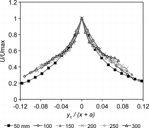
3.6. Scaling
The average over all coughs of the normalized and offset velocity profiles at each X location is shown in as a function the width of the cough jet (d), as determined above. For typical steady turbulent jets, the normalized velocity profile can be collapsed by scaling the width by the downstream (x) location, including an offset due to the virtual origin a (CitationHinze 1975). Transient jets are observed to attain self-similarity after some development time and some distance downstream (L/D> 6) (CitationCossali et al. 2004). However, at the early time and in the near field analyzed here, the average cough jet does not collapse well with this scaling, using the virtual origin defined by the half-height width.
FIG. 10 Normalized velocity profiles scaled by x + a.
4. Conclusion
Twenty-nine nonsmoking healthy volunteers produced voluntary coughs of average effort with velocities ranging from 1.5 m/s to 28.8 m/s. The overall average maximum cough velocity was 10.2 m/s (SD = 6.7 m/s). No correlation was found for sex and weight.
Based on an ensemble average of measurements early in the cough, the width of the cough was found to expand linearly initially, and then remain constant at distances farther from the mouth. This affected the normalized velocity profiles, which were found to partially collapse when scaled with distance from a virtual origin.
This study has demonstrated that PIV can be used to map the flow of a human cough in vivo. With this data, it is now possible to refine the flow characteristics of a simulated cough, and to more accurately model cough flows. This study shows that human cough cannot be reduced to a well-defined flow field. In particular, the high degree of variance in the velocity data, including the width of the jet, the maximum velocities, and the direction of the jet demonstrates that studies of human cough based on numerical or in vitro simulations should consider incorporating a wide range of conditions, rather than focus on a single “typical” cough flow.
Future work may include a second trial. During this trial, each volunteer would cough with maximum force while their heads were placed to control the direction of the cough flow. The cough flow would also be mapped at greater distances from the mouth to evaluate the far field velocities and widths to better determine the cough's penetration into a room. Ensemble averages throughout the period of the cough are also needed to fully characterize the turbulence quantities in this unsteady, short-duration jet.
REFERENCES
- Afshari , A. , Azadi , S. , Ebeling , T. , Badeau , A. , Goldsmith , W. , Weber , K. and Frazer , D. 2002 . “ Evaluation of Cough Using Digital Particle Image Velocimetry ” . In Conference Proceedings. Second Joint EMBS-BMES Conference 2002 24th Annual International Conference of the Engineering in Medicine and Biology Society. Annual Fall Meeting of the Biomedical Engineering Society (Cat. No. 02CH37392) , 975 – 976 . Piscataway , NJ : IEEE .
- Ai , J. J. , Yu , S. C. M. , Law , A. W. and Chua , L. P. 2005 . Vortex Dynamics in Starting Square Water Jets. . Phys. Fluids. , 17 ( 1 ) : 014106 – 12 .
- Badeau , A. , Afshari , A. , Goldsmith , T. and Frazer , D. 2002 . “ Preliminary Prediction of Flow and Particulate Concentration Produced from Normal Human Cough Dispersion ” . In Proceedings of the 2002 IEEE Engineering in Medicine and Biology 24th Annual Conference and the 2002 Fall Meeting of the Biomedical Engineering Society (BMES / EMBS), October 23, 2002 - October 26, 2002 , 246 – 247 . Houston , TX : Institute of Electrical and Electronics Engineers Inc. .
- Bajpai , S. and Tirumkudulu , M. 2008 . An Experimental Study of Impulsively Started Turbulent Axisymmetric Jets. . European Physical Journal B: Eur. Phys. J. B. (France). , 61 : 293 – 7 .
- Barnes , P. F. , El-Hajj , H. , Preston-Martin , S. , Cave , M. D. , Jones , B. E. , Otaya , M. , Pogoda , J. and Eisenach , K. D. 1996 . Transmission of Tuberculosis among the Urban Homeless. . JAMA. , 275 : 305 – 307 .
- CDC . 2004 . CDC | Public Health Guidance for Community-Level Preparedness and Response to Severe Acute Respiratory Syndrome (SARS) Version 2 Retrieved from http://www.cdc.gov/ncidod/sars/guidance.
- Chao , C. , Wan , M. , Morawska , L. , Johnson , G. , Ristovski , Z. , Hargreaves , M. , Mengersen , K. , Corbett , S. , Li , Y. , Xie , X. and Katoshevski , D. 2009 . Characterization of Expiration Air Jets and Droplet Size Distributions Immediately at the Mouth Opening. . J. Aerosol Sci. , 40 : 122 – 133 .
- Chaussé , P. and Magne , H. 1916 . Contribution à l'étude de la Toux et de Quelques Actes Expulsifs Analogues; Pression et Eitesse de l'Air; Conséquences Relatives à l'Etologie de Certaines Affections de Voies Respiratoires. . Arch. de méd. experiméntale et d'anat. path. , 27 : 213
- Cossali , G. , Marengo , M. , Coghe , A. and Zhdanov , S. 2004 . The Role of Time in Single Drop Splash on Thin Film. . Experiments in Fluids, Exp. Fluids (Germany). , 36 : 888 – 900 .
- Couch , R. B. , Cate , T. R. , Douglas , R. G. , Gerone , P. J. and Knight , V. 1966 . Effect of Route of Inoculation on Experimental Respiratory Viral Disease in Volunteers and Evidance for Airborne Transmission. . Bacterilogical Reviews , 30 ( 3 ) : 517 – 529 .
- Fennelly , K. P. , Martyny , J. W. , Fulton , K. E. , Orme , I. M. , Cave , D. M. and Heifets , L. B. 2004 . Cough-Generated Aerosols of Mycobacterium Tuberculosis: A New Method to Study Infectiousness. . Am. J. Respiratory Critical Care Med. , 169 : 604
- Hill , P. G. and Ouellette , P. 1999 . Transient Turbulent Gaseous Fuel Jets for Diesel Engines. . J. Fluids Engng, Transactions of the ASME. , 121 : 93 – 101 .
- Hinze , J. O. 1975 . Turbulence (2nd ed.) , 520 – 540 . New York : McGraw-Hill Companies .
- Johari , H. , Zhang , Q. , Rose , M. and Bourque , S. 1997 . Impulsively Started Turbulent Jets . AIAA J., AIAA J. (USA). , 35 : 657 – 62 .
- Le Maitre Special Effects, Inc. 2006 . Material Safety Data Sheet Retrieved from http://www.lemaitrefx.com/attachments/282.pdf.
- Mahajan , R. P. , Singh , P. , Murty , G. E. and Aitkenhead , A. R. 1994 . Relationship between Expired Lung Volume, Peak Flow Rate and Peak Velocity Time during a Voluntary Cough Manoeuvre. . Br. J. Anaesth. , 72 : 298 – 301 .
- Marr , D. , Khan , T. , Glauser PhD , M. , Higuchi , H. and Zhang , J. 2005 . On Particle Image Velocimetry (PIV) Measurements in the Breathing Zone of a Thermal Breathing Manikin. . ASHRAE Transactions. , 111 : 299 – 305 .
- Nicas , M. , Nazaroff , W. W. and Hubbard , A. 2005 . Toward Understanding the Risk of Secondary Airborne Infection: Emission of Respirable Pathogens. . J. Occup. Environ. Hygiene. , 2 : 143
- Papineni , R. S. and Rosenthal , F. S. 1997 . The Size Distribution of Droplets in the Exhaled Breath of Healthy Human Subjects. . J Aerosol Med. , 10 : 105 – 116 .
- Piirilä , P. and Sovijärvi , A. R. 1989 . Differences in Acoustic and Dynamic Characteristics of Spontaneous Cough in Pulmonary Diseases. . Chest. , 96 : 46 – 53 .
- Rosenfeld , M. , Rambod , E. and Gharib , M. 1998 . Circulation and Formation Number of Laminar Vortex Rings. . J. Fluid Mech. , 376 : 297 – 318 .
- Siegel , J. D. , Rhinehart , E. , Jackson , M. , Chiarello , L. and Healthcare Infection Control Practices Advisory Committee . 2007 . Guideline for Isolation Precautions: Preventing Transmission of Infectious Agents in Healthcare Settings 2007. , Atlanta : Centers for Disease Control and Prevention .
- Singh , P. , Mahajan , R. P. , Murty , G. E. and Aitkenhead , A. R. 1995 . Relationship of Peak Flow Rate and Peak Velocity Time during Voluntary Coughing. . Br. J. Anaesth. , 74 : 714 – 716 .
- Somers , J. , Magill , J. , Richter , K. , Fourcaudot , S. , Lajarge , P. and Barraux , P. 1991 . Acoustic Agglomeration of Liquid and Solid Aerosols: A Comparison of a Glycol Fog and Titanium Dioxide. . J. Aerosol Sci. , 22 : S109 – S112 .
- Song , L. and Abraham , J. 2003 . Entrainment Characteristics of Transient Turbulent Round, Radial, and Wall-Impinging Jets: Theoretical Deductions. . J. Fluids Eng. , 125 : 605 – 612 .
- Turner , J. 1962 . Starting Plume' in Neutral Surroundings. . J. Fluid Mech. , 13 : 356 – 368 .
- Valway , S. E. , Sanchez , M. P. , Shinnick , T. F. , Orme , I. , Agerton , T. , Hoy , D. , Jones , J. S. , Westmoreland , H. and Onorato , I. M. 1998 . An Outbreak Involving Extensive Transmission of a Virulent Strain of Mycobacterium Tuberculosis. . N. Engl. J. Med. , 338 : 633 – 639 .
- VanSciver , M. 2005 . Flow Field Measurements of Human Generated Infectious Aerosols , 12 Boulder , Co : MS Thesis, University of Colorodo .
- Wells , W. F. 1955 . Airborne Contagion and Air Hygiene: An Ecological Study of Droplet Infection , 2 – 3 . Cambridge , MA : Harvard University Press for the Commonwealth Fund .
- Wells , W. F. and Wells , M. W. 1938 . Measurement of Sanitary Ventilation. . Am. J. Public Health Nations Health. , 28 : 343 – 350 .
- Xie , X. , Li , Y. , Sun , H. and Liu , L. 2009 . Exhaled Droplets Due to Talking and Coughing. . J. R. Soc. Interface. , 6 : S703 – S714 .
- Zhao , B. , Zhang , Z. and Li , X. 2005 . Numerical Study of the Transport of Droplets or Particles Generated by Respiratory System Indoors. . Building Environ. , 40 : 1032 – 1039 .
- Zhu , S. , Kato , S. and Yang , J. 2006 . Study on Transport Characteristics of Saliva Droplets Produced by Coughing in a Calm Indoor Environment. . Building Environ. , 41 : 1691 – 1702 .