Abstract
The aerodynamic size of pathogen-laden expiratory aerosols plays an important role in their dispersion in air and deposition onto surfaces, both of which are related to the spread of infectious respiratory diseases. The size of bacterial cells is on a similar scale to the size of expiratory aerosols, but because some bacterial cells are nonspherical, bacterium-laden expiratory aerosols often have irregular shapes and highly variable aerodynamic sizes. An algorithm that can estimate their aerodynamic sizes is highly desirable in studying their physical transport and to assess the subsequent exposure level and infection risk. In this study, an algorithm based on stochastic modeling was developed to predict the distribution of the aerodynamic size of bacterium-laden expiratory aerosols. The applicability of the algorithm was tested experimentally by conducting biological air sampling using a multi-stage impactor in a test facility. The proposed algorithm was used to predict the size profile of simulated expiratory aerosols encasing a strain of benign rod-shaped bacterium. Simulated bacterium-laden expiratory aerosols were generated using a cough machine with a solution containing the bacteria. Air at three different positions was then sampled to obtain the size profile of bacterium-laden aerosols at each position. The results were compared to the prediction by the algorithm and by another method, which simply considers the evaporative shrinkage of the expiratory aerosols and neglects the inclusion of the pathogen. It was found that the prediction by the proposed algorithm generally matched the measured results much better than the method that neglects the inclusion of the bacterium. Limitations of the current algorithm and further research and development are also discussed in this article.
Copyright 2012 American Association for Aerosol Research
1. INTRODUCTION
Aerosols exhaled by infectors of respiratory diseases, such as tuberculosis (TB), influenza, and severe acute respiratory syndrome (SARS), may carry pathogens. These polydispersed aerosols consist of about 91% of water and 9% of nonvolatile content (Effros et al. Citation2002). Once the expiratory aerosols are introduced into the air, the water content will evaporate and the aerosols will shrink. Most of these droplet-based aerosols will reach an equilibrium state, when they become what is “droplet nuclei,” in less than a second (Wan and Chao Citation2007).
Many studies have shown that smaller expiratory aerosols can be dispersed farther away from the source than larger ones, and the gravitational settling and deposition of expiratory aerosols also depend on their sizes (Wan and Chao Citation2007; Xie et al. Citation2007; Wan et al. Citation2009). Other than the physical transport, the size of these infectious particles will also affect their infectivity and the intake into the respiratory tract (Sze To and Chao Citation2010). Therefore, the size of pathogen-laden expiratory aerosols is an important parameter to consider when studying the spread of infectious respiratory diseases.
FIG. 1 Illustration of expiratory droplet nucleus shrinkage from (a) a droplet free of pathogen, (b) virus-laden droplet, (c) a bacterium-laden droplet, (d) a droplet containing a rod-shaped bacterium, and (e) a droplet containing multiple bacteria.
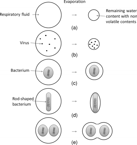
The initial size profile of expiratory droplets has been studied by numerous researchers using different techniques (Duguid Citation1946; Loudon and Roberts Citation1967; Fairchild and Stamper Citation1987; Nicas et al. Citation2005; Yang et al. Citation2007; Chao et al. Citation2009; Morawska et al. Citation2009). To determine the size profile of expiratory droplet nuclei, one commonly used approach is to consider both the expiratory droplet and the droplet nucleus as spheres and estimate the shrinkage in size of the droplet due to the evaporation of water content. Knowing the amount of nonvolatile content in the respiratory fluid and the humidity of air, the ratio of the size of the droplet nucleus to the initial droplet size can be estimated (Nicas et al. Citation2005; Xie et al. Citation2007). This approach is reasonable in estimating the size of droplet nucleus size of an expiratory droplet that is free of pathogens, which is illustrated in . However, the size of the nucleus of a droplet containing one or more pathogens is expected to be larger than that estimated by this approach, since microorganisms consist of more nonvolatile content than the respiratory fluid. The average dry mass to total mass of bacterial cells is about 20% (Luria 1960) and in some cases up to 57% (Bratbak and Dundas Citation1984). Possibly due to the complexity of the problem and the associated experimental risk involved in characterizing expiratory aerosols exhaled by infected persons, studies investigating the size profiles of pathogen-laden expiratory droplet nuclei have been few and usually only reported small samples. Some examples are the work of Fennelly et al. (Citation2004), who studied the size profile of the cough-generated expiratory aerosols of humans infected with TB, and Fabian et al. (Citation2008), who studied the size profile of aerosols exhaled by humans infected with influenza. Another study by Sze To et al. (Citation2008) discussed the scenario of a relatively small expiratory droplet (2 μm) containing 10 relatively large (300 nm) virus particles, corresponding to an extremely high pathogen concentration in the respiratory fluid. The increase in the size of the droplet nucleus on account of the presence of the virus particles was only about 1%. This suggests that the approach of evaporative shrinkage can be applied to estimate the size of a virus-laden expiratory droplet nucleus with insignificant error, since the nanometer size virus particle has a negligible volume compared with the expiratory droplet, which mostly has micrometer size. This is illustrated in .
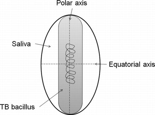
Bacteria, however, have sizes in the micrometer scale, comparable to expiratory droplets. As illustrated in , the nucleus size of a bacterium-laden expiratory droplet may be larger than that estimated from the evaporative shrinkage of a clean droplet. In addition, some bacterial cells are nonspherical. For example, TB bacillus is a slim rod-shaped bacterium that causes TB and ranges from 2 to 10 μm in length (Sherris et al. Citation1990). As illustrated in , when a rod-shaped bacterium is encased in an expiratory droplet, the bacterium-laden droplet nucleus may not be spherical. A similar situation can be found in the case where one droplet contains multiple bacterial cells, as illustrated in . These properties add to the complexity of estimating the aerodynamic size of the bacteria-laden expiratory droplet nucleus.
The variability of the expiratory droplet size, bacterial cell size, and the number of bacterial cells in a droplet also adds to the complexity. Expiratory droplets are polydispersed in nature. Bacterial cells also have variable sizes. These two size parameters exist in the form of size distributions rather than as single values. Also, not all droplets contain the same number of bacterial cells. Some droplets may contain no bacterial cells at all. The number of bacterial cells encased in a droplet should also be described by a distribution instead of a single value. Stochastic modeling can be used to address these parametric variability. The Monte Carlo simulation is a viable tool for providing the numerical estimation of statistical results (Tung and Yen Citation2005). The Monte Carlo method computes the result by drawing random samples from the specified distributions to obtain a set of input parameters. The parameters are then input into the algorithm as defined by the user to calculate the quantity under investigation. A single calculation process is regarded as one iteration. With a large number of iterations, the distribution of the calculated values will be close to the actual distribution of the quantity under investigation.
The objective of this study was to develop an algorithm to estimate the aerodynamic size profile of bacterium-laden expiratory droplet nuclei. Stochastic modeling based on the Monte Carlo method was used in the algorithm. The algorithm focuses on rod-shaped bacteria under a low bacterial concentration in the respiratory fluid that allows a reasonable degree of complexity. Assumptions made in the algorithm are discussed in the article. Microbiological sampling was performed to test the applicability of the algorithm in a realistic scenario. A strain of benign Escherichia coli (E. coli), also a rod-shaped bacterium, was used to generate simulated bacterium-laden expiratory droplets in an isolation ward testing facility. The aerodynamic size profiles measured at various positions were compared to the estimation by the algorithm and also by the method of evaporative shrinkage. Further researches to improve the algorithm were also discussed.
2. METHODS
2.1. The Algorithm
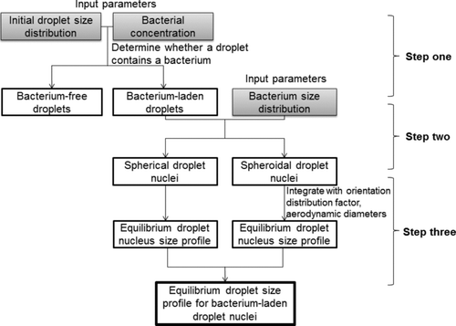
An algorithm was developed to estimate the aerodynamic size profile of rod-shaped bacterium-laden droplet nuclei in three major steps. The flow of the algorithm is shown in . Each step is discussed in detail in this section.
2.1.1. Step One: Identifying the Bacterium-Laden Droplets
The first step of this algorithm is to determine whether or not a droplet contains a bacterium according to the initial droplet size and the bacterial concentration in the respiratory fluid. Initial droplet size profile is defined as the size distribution of expiratory droplets without evaporation loss of water content. This profile can be obtained from the literature (Chao et al. Citation2009; Morawska et al. Citation2009). A sample, a droplet of a particular size, is drawn from this distribution. Then the expected number of bacteria encased in that droplet, E b, can be determined by multiplying the droplet volume, calculated as a sphere, with the bacterial concentration in the respiratory fluid:
Under the situation of low bacterial concentration, it is reasonable to assume that most droplets will either contain one or no bacterium. On the basis of this assumption, a yes-no distribution (Bernoulli distribution) is used to identify the bacterium-laden droplets from the bacterium-free droplets by setting the value of E b as the probability of outcome “yes.” A sample is then drawn from this distribution, and if the outcome is “yes,” the droplet contains one bacterium and the algorithm moves on to Step two; otherwise the droplet is bacterium free. The aerodynamic size of the bacterium-free nucleus is then calculated using Equation (2) by considering the evaporative shrinkage:
2.1.2. Step Two: Distinguishing Between the Spherical and the Spheroidal Droplet Nuclei
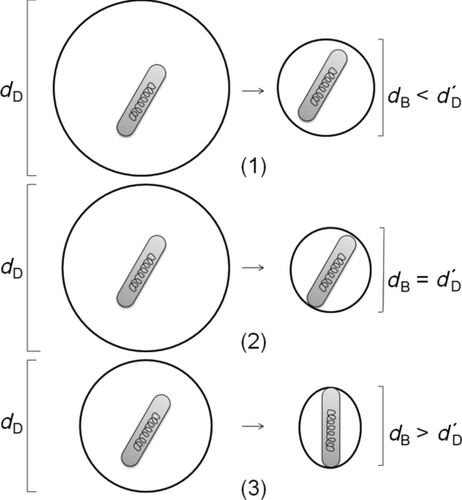
The nuclei of some bacterium-laden droplets may not be spherical but are spheroidal depending on the sizes of the droplets and the bacteria inside them. When an initial droplet has a comparable size with the encased bacterium, the liquid with encased bacterium will form a droplet nucleus with a minimum total surface area due to the surface tension, as illustrated in . A different set of equations is used to estimate the aerodynamic size of the nucleus in each case. Therefore, the second step is to distinguish spheroidal bacterium-laden nuclei from the spherical ones. A sample is drawn from the bacterial size distribution to determine the size of the bacterium inside the droplet. The iteration is considered as invalid and is not counted into the result if the bacterium is larger than the droplet, since a droplet cannot contain a bacterium larger than itself.
For those valid iterations, the ratio of the size of the initial droplet to the length of the bacterium, d B, is defined. It is assumed that a bacterium maintains the same length after the evaporation of water content. When this ratio is larger than or equal to a certain critical value (see scenario 1 and 2 in ), the shape of the nucleus is considered to be spherical. When the ratio is smaller than this critical value, the shape of the nucleus is considered to be spheroidal (see scenario 3 in ). The value of this critical ratio can be determined by considering scenario 2 in , where the diameter of the nucleus is exactly equal to the length of the encased bacterium. The critical ratio can then be calculated with the following equation:
2.1.3. Step Three: Calculating the Aerodynamic Diameter
The aerodynamic diameter of the bacterium-laden droplet nucleus is calculated using two sets of equations according to the shape of the droplet nucleus determined in Step two.
TABLE 1 Average shape factors for various axial ratios of the spheroid droplets
For a spherical bacterium-laden droplet nucleus, the volume shrinkage of water content is determined by the amount of water evaporating from the droplet. The residual volume of liquid can be determined by evaporative shrinkage, assuming that there is no change in the bacterial volume. Then, the residual droplet volume can be determined by summing the liquid volume and the bacterial volume as shown in Equation (4):
The aerodynamic diameter is defined as the diameter of the spherical particle with a density of 1000 kg/m3 that has the same settling velocity as the particle under study. For a spheroidal particle, its diameter can be calculated according to the density of the particle as well as its shape factor as follows:
The shape factor of a spheroidal aerosol also depends on the orientation of the aerosol to the oncoming airflow (Kasper Citation1982). For instance, when the polar axis is perpendicular to the oncoming airflow, the shape factor is the largest since the drag force is at the maximum. In contrast, when the polar axis is parallel to the oncoming airflow, the shape factor is the smallest since the drag force is at the minimum. Time-averaged random orientation in the case of Brownian rotation is considered to determine the mean shape factor (Fuchs Citation1964):
TABLE 2 Aerosol size ranges collected on different stages of the impactor
2.2. Experimental Validation of the Predicted Size Profile of E. coli-Laden Droplet Nuclei
A set of experiments was performed to test the applicability of the proposed algorithm. The idea was to evaluate how close the size profile of bacteria-laden droplet nuclei estimated by the proposed algorithm is to the actual size profile of bacteria-laden droplet nuclei measured by microbiological air sampling using a multistage impactor. This is because the laser spectrometer can provide the size profile of droplet nuclei but cannot distinguish the bacteria-laden droplet nuclei from bacteria-free droplet nuclei. shows the aerosol size ranges collected on different stages of the impactor and the size channels defined for each stage. Aerosols collected on stages 1–5 were analyzed in this study.
The experiments were conducted in an isolation facility to examine the proposed algorithm under a realistic situation. The isolation facility has dimensions of 5 × 4 × 3 m (W × L × H). The wall material used in the isolation facility was concrete coated by paint. A downward flow system was installed in the facility and ventilation air was supplied by two four-way spread-type ceiling supply diffusers (0.7 × 0.7 m). Two exhaust vents (0.7 × 0.15 m) were placed on one sidewall next to the floor. The isolation ward has a constant supply airflow rate of 648 m3/h and has an air exchange rate of 12.5 h−1. Due to the high air exchange rate, the room temperature and the relative humidity could be maintained at around 22°C and 60%, during the experiments.
A strain of benign E. coli (ACCT number E1121) was aerosolized to generate bacterium-laden droplets in the isolation ward. E. coli is a rod-shaped bacterium and is normally 2.0–6.0 μm in length (Prescott et al. Citation1996). These characteristics make it a suitable choice for the experiments. A broth solution containing 30 g of tryptone soya broth powder (OXOID CM129) dissolved in a liter of distilled water was prepared for bacteria propagation. The solution was sterilized by autoclaving at 121°C for 15 min. Biologically active E. coli were transferred into the broth solution and propagated for 8 h at 37°C. After the 8 h, the E. coli solution was diluted 109 times using simulated human respiratory fluid. One liter of simulated human respiratory fluid was produced by mixing 86 g of glycerin with distilled water to simulate the nonvolatile content weighting in the human respiratory fluid as reported by Effros et al. (Citation2002). The presence of nonvolatile solutes in the simulated respiratory droplet causes a depression of the vapor pressure relative to that above pure water. Some water will remain in the droplet when they reach hygroscopic equilibrium states. With this composition, the ratio of the residual droplet nucleus diameter to initial droplet diameter was about 0.39 (Wan et al. Citation2009). An E. coli concentration of 8.4 × 10⁶ cfu/mL was used in the experiments. Under this concentration, droplets with an initial size of up to 61 μm would have contained no more than one bacterium on average as determined from Equation (1), while the largest collectable aerosols of interest was 7 μm in size, corresponding to a droplet with an initial size of 18 μm. Hence, the assumption to be presented in section 2.1.1 is reasonable in this situation.
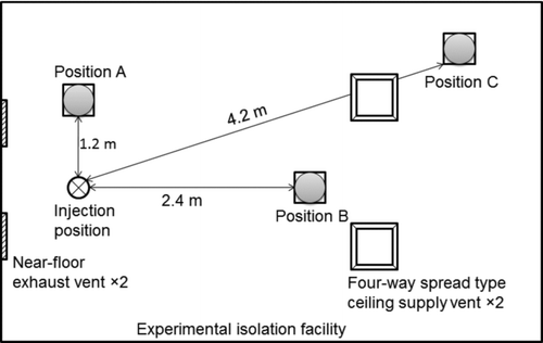
An in-house-built droplet generator was used to produce test droplets from the simulated human respiratory fluid. The generator is composed of liquid and gas supply systems and a pneumatic nozzle (Spraying Systems Co. B1/4JAU-SS) with a similar configuration to the one used by Wan et al. (Citation2007) and Sze To et al. (Citation2008). The generator consists of two flow-rate controllers (Cole-Parmer Instrument Co. 32907-43), two solenoid valves (SMC Pneumatics Ltd. VT317-4G-02), and two pressure regulators (SMC Pneumatics Ltd. AR20-02). This droplet generator can produce polydispersed droplets with a size profile similar to human expiratory droplets generated from a cough (Wan et al. Citation2009). The nozzle was installed on the mouth of a manikin 0.8 m above the floor, which is about the breathing height of a patient lying down. A puff of test aerosols was injected vertically upward. Each simulated cough lasted for 1 s and about 0.072 mL of simulated human respiratory fluid was injected into the isolation ward with 0.4 L of air. This amount of solution roughly corresponded to the amount of expiratory fluid expelled by 10 coughs (Zhu et al. Citation2006). The background concentration level in the ward was measured using an isokinetic aerosol spectrometer that recorded data every 6 s (GRIMM model 1.108, measurable range: 0.3–20 μm in 15 channels) for 1 min before each injection to obtain the average background concentration. The detailed size classes for the aerosol spectrometer were shown in , while the smaller size classes were neglected. The background concentration was then subtracted from the aerosol counting results. The background levels were around fifty times lower than the measured concentration levels during test aerosol injections. After an injection, the aerosol spectrometer was used to monitor the aerosol concentration level to make sure it returned to the background level before another round of injection and this process normally took 5 min. Measurements were made at 1.2, 2.4, and 4.2 m away from the nozzle (all at 1.6 m above the ground) as shown in . Each measurement was repeated five times. The nozzle expelled the droplet toward the ceiling and the spectrometer and impactor were located at sufficient distance horizontally to make sure the droplets reached equilibrium state before they approached the measurement equipment.
TABLE 3 Predicted aerodynamic size profile of E. coli-laden droplet nuclei
2.2.1. Prediction of the Size Profile of E. coli-Laden Droplet Nuclei Using the Algorithm
The proposed algorithm was used in a realistic case to predict the size profile of E. coli-laden droplet nuclei. Direct measurement to obtain the size profiles was used rather then calculating them using the well-mixed assumption. The algorithm was defined in a Monte Carlo modeling software (CRYSTAL BALL, Version 11.1.1.1.00) following the three steps described in section 2.1. A set of input values was generated by drawing random values based on the defined distributions. The values were substituted into the algorithm to calculate the aerodynamic diameter of a droplet nucleus. This process is regarded as iteration. When a large number of iterations are performed, the aerodynamic size distribution of the droplet nuclei can be determined. The accuracy of the estimation obtained through the Monte Carlo approach is a function of the number of iterations performed. However, a rule for determining the minimum number of iterations is unavailable. Thus, the replication of the Monte Carlo simulation under different numbers of iterations is the only way to check the accuracy (Melching 1995). In the current study, it was found that going beyond five million iterations does not change the results significantly. Therefore, five million iterations were performed to determine the aerodynamic size profile of the bacterium-laden droplet nuclei. Three required parameters were input into the algorithm, which were the initial droplet size profile, bacterial size profile, and the bacterial concentration in the simulated human respiratory fluid. As described in section 2.2, the bacterial concentration was 8.4 × 10⁶ cfu/mL. The other two parameters are described below.
2.2.1.1. Estimation of the initial droplet size profile. The size profile of droplet nuclei at each position was determined using the aerosol spectrometer described before. Since the aerosols measured were droplet nuclei, the initial droplet size profile at each measurement position was determined by the method of evaporative shrinkage, represented by Equation (2). In this part of the validation study, after the test droplets were injected, the aerosol spectrometer was set to count the number concentration of the aerosols every 6 s for 5 min after the injection, since microbiological air sampling also lasted for 5 min. The cumulative concentration count within the 5-min period was used to calculate the percentage size profile of droplet nuclei at that position. Then, the measured size profile of droplet nuclei was back adjusted to the initial droplet size profile by the method of evaporative shrinkage. According to Equation (1) and considering the low concentration of E. coli in the simulated respiratory fluid, most of the droplet nuclei (>99.8%) will be bacteria free. Therefore, the profiles can be taken as the initial droplet size profiles of the simulated cough.
2.2.1.2. Approximation of the E. coli length profile. The E. coli size profile was one of the input parameters. There is currently no study directly investigating the E. coli size profile. Thus, the volume distribution profile of E. coli (Kubitschek Citation1969) was used to calculate the E. coli bacterial size distribution. The length of E. coli ranges from 2.0 to 6.0 μm and the cross-section diameter ranges from 1.1 to 1.5 μm (Prescott et al. Citation1996). On the basis of these values, the mean ratio of the bacterial length to the cross-section diameter was calculated as 2.91, which was used to adjust the volume distribution profile to the bacterial length distribution profile as shown in Equation (11):
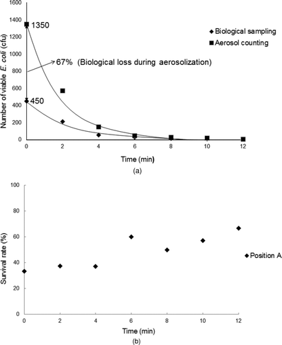
2.2.1.3. Survivability measurement. During the aerosolization process, a significant amount of bacteria lost viability (Sze to et al. Citation2008). Since the impactor can only reveal the number of viable E. coli, the viability loss of E. coli in the airborne state and due to the aerosolization process needs to be incorporated into the algorithm. Since only viable E. coli are counted and compared to the simulation result, the bacterial concentration used in the algorithm was factored by the survival rate. The method of measuring the survivability of the E. coli was adopted from Sze To et al. (Citation2008). The viability function of E. coli in droplets, f(t), was determined by biological sampling with the one-stage Andersen viable impactor (measurable size range: 0.65–23 μm) and by aerosol counting using an aerosol spectrometer. Again, the size of the droplet nuclei measured using the spectrometer was back adjusted to their initial sizes using Equation (2). Knowing the flow rate of the impactor, the total volume of droplets collected on the impactor, as measured using the aerosol spectrometer, was multiplied with the bacterial concentration used to calculate the total number of E. coli (both viable and unviable) collected on the impactor. Comparing this value to the count of viable E. coli collected on the impactor, its percentage viability was then determined. In order to be compatible with the size range of the Andersen impactor, only the count data between 0.65 and 20 μm in size were used in the calculation. After a 1-s injection of simulated expiratory droplets, six 1-min biological air samples were collected every 2 min sequentially onto six culture plates at position A. The aerosol spectrometer was operated simultaneously during this period at the same position. After incubation, the number of colonies formed by the viable E. coli on each of the six plates was adjusted by performing the positive hole correction and the calculated total number of E. coli collected on each plate were obtained and plotted against time. The viability loss due to aerosolization was determined by extrapolating the curves to t = 0. The viability of the bacteria in the subsequent airborne state was also determined by comparing the two curves.
2.2.2. Measurement of the Size Profile of E. coli-Laden Droplet Nuclei
The biological sampler (Thermo Scientific Six-stage Viable, Andersen Cascade Impactor) was used to collect the E. coli-laden droplet nuclei with different aerodynamic diameters. The measurable aerodynamic droplet diameter ranges from 0.65 to 7 μm and above. The six-stage biological air sampler sampled for 5 min once the simulated expiratory droplets were injected. The working mechanism of the six-stage biological sampler is such that the sampled droplets are deposited on culture plates according to the aerodynamic droplet diameter at each stage. After sampling, the agar plates were sealed and then cultured under a constant temperature of 37°C until the E. coli colonies became visible and countable. By counting the colonies formed by the E. coli bacteria in the six culture plates with subsequent positive hole correction, the size profile of the E. coli-laden droplet nuclei was obtained. Five sets of measurements were taken at each position. After each set of measurements, the aerosol concentration level in air was monitored by the aerosol spectrometer to ensure that it returned to the original background droplet concentration level before another set of injections and measurements. This directly measured size profile was then compared with the predicted size profile of the E. coli-laden droplet nuclei estimated by the proposed algorithm.
2.2.3. The Size Profile of Bacterium-Laden Droplet Nucleus Measured by Evaporative Shrinkage
A set of parallel measurements was performed to investigate the size profiles of droplet nuclei using evaporative shrinkage for comparison. Clean simulated respiratory fluid was used in this set of measurements. Measurements were conducted at the same selected positions following the same procedures as described in section 2.2.1.1. The aerosol generator injected clean droplets into the isolation ward for 1 s and then the aerosol spectrometer counted the droplets for 5 min after the injection. The background aerosol level was also measured for 1 min ahead of the simulated expiratory droplets injection and was subtracted from the droplets counting results. Five sets of measurements were taken at each selected position and the average droplet counts were estimated. Considering that the droplets were bacteria free, there was no impact on the residual droplet volume due to the presence of bacteria. Therefore, the measured size profile of clean droplet nuclei is equivalent to the profile estimated by evaporative shrinkage.
3. RESULTS AND DISCUSSIONS
3.1. The E. coli Survival Rate During the Aerosolization Process
The E. coli counts estimated from the aerosol counting data is represented by the upper curve in and the measured results using air sampling is represented by the lower curve. The upper curve represents physical aerosol reduction (dilution by ventilation and deposition), and the lower one represents a combination of physical reduction and biological loss (aerosolization loss and loss of viability; Sze to et al. Citation2008). The two curves were extrapolated to t = 0 using exponential form of equation to work out the number of E. coli at that time. A large portion (around 67%) of E. coli was lost at t = 0, which represented the aerosolization loss. The viability loss of E. coli during the subsequent airborne state after aerosolization can be determined by comparing the two curves in . The E. coli survival rate during the first few minutes was around 33% (33%–37%) as shown in . This is because the bacteria were surrounded and protected by the external liquid from inactivation. Similar results were also reported by others (Ehrlich et al. Citation1970; Sze To et al. Citation2008). The measured survival rate increased to over 40% after 4 min. The bacteria should not have replicated so significantly in the aerosols during this short time interval. Since the concentration of bacterium-laden aerosols became very low after 4 min, the observation was likely to be due to measurement error. The survival rate of E. coli, 33%, was used to adjust the E. coli concentration in the estimation by the proposed algorithm.
3.2. The Predicted Size Profile of E. coli-Laden Droplet Nuclei
shows the size profile of E. coli-laden droplet nuclei at three positions estimated by the proposed algorithm in eight size channels of the aerosol spectrometer. To obtain a more reliable result by using the Monte Carlo method, a larger number of sampling is essential. The algorithm was set to take five million iterations. It is tedious and time consuming to collect five millions aerosols in the measurement. For comparison purpose, the results are shown in terms of the percentage of the total number of droplets. The estimated droplet size profile ranged between 1.6 and 20 μm. Droplets not within this range were neglected considering that the smaller they are, the more difficult it is for them to contain a larger volume of bacteria, and the larger they are, the less likely it is for them to cause airborne infections due to their stronger gravitational settling ability. The size profile for the 2.5-μm size channel had a much larger value than that for the 1.8-μm size channel for all the measured positions. The droplet number peaked for the size channel of 2.5 μm for position A, while it peaked for the 6.25-μm size channel for positions B and C. From the schematic diagram of the experimental setup shown in , one can see that both positions B and C were close to the ceiling supply vents. This enhanced the ventilation dilution of the droplets. Also, position A was located much closer to the cough machine compared with the other two positions. As a result, the total droplet counts at positions B and C were significantly lower than the one at position A. The average particle concentration at position A was 83,440 particle/L, while it was 17,083 particle/L and 18,236 particle/L at position B and C, respectively.
3.3. The Measured Size Profiles of E. coli-Laden Droplet Nuclei
The E. coli-laden droplet size profiles were measured using the six-stage biological air sampler. Five sets of measurements were taken at each position. The averaged E. coli counts for the different stages are shown in together with the droplet size profile predicted by the proposed algorithm. The maximum amounts of collected viable E. coli-laden droplet nuclei ranged from 2.1 to 3.3 μm (the fourth collection stage of the six-stage biological sampler) for all three measured positions. Very few E. coli-laden droplets were collected at stage six at the three measured positions.
FIG. 7 Comparison between the size profiles of bacterial droplet nuclei obtained from the method of evaporative shrinkage, the proposed algorithm, and actual measurement. (a) Position A. (b) Position B. (c) Position C.
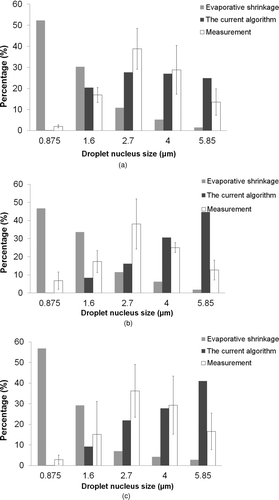
At position A, both the measured result and the predicted one peaked at the size channel of 3.33 μm. Other predicted results generally matched the measured ones well for all the size channels at position A. The predicted size profiles at positions B and C matched the measured ones reasonably well for small size channels, while more significant differences were observed for large size channel. Therefore, it can be concluded that the proposed algorithm can reasonably predict the size profile for most of the size channels.
It was observed that the predicted profiles were slightly larger than the measured ones. In the current algorithm, it was assumed that the encased rod-shaped bacterium maintains its original shape after the evaporation of the water content. However, in real situations, the bacterium may shrink or deform. The shrinkage/deformation of the bacterium due to the evaporation of water content may affect the aerodynamic size of the droplet nucleus. When a droplet nucleus is holding a bent/shrunk bacterium, the nucleus resembles more of a sphere than a spheroid, which will further reduce the aerodynamic droplet diameter. Further studies on this possibility are needed.
The mean size profiles of droplet nuclei estimated by the method of evaporative shrinkage are shown in together with the size profiles obtained from the proposed algorithm and measurement. Substantially different size profiles were obtained from these two methods. For the method of evaporative shrinkage, most of the droplets concentrated around 0.875 μm, while the ones from our algorithm peaked at a larger size for all three positions. Although the measured droplet size profiles of E. coli-laden droplets were smaller than sizes from the predicted results of the proposed algorithm (compared with positions B and C in ), the proposed algorithm provides a more accurate means of estimating the size profile of bacterium-laden droplet nucleus compared to the method of evaporative shrinkage.
3.4. The Number of Bacteria Enclosed in One Droplet
The algorithm proposed by the current study was designed for cases where only one single bacterium is encased in each droplet. Thus in this study, the assumption that no more than one bacterium is encased in each droplet was made. In the case of TB, a single bacillus is enough to cause infection (Wells Citation1955; Williams et al. Citation2005; Bowong and Tewa Citation2009). Therefore, the current algorithm describes the situation for TB infection well. On the basis of this assumption, all the droplets were treated as either bacteria free or have at most one bacterium encased in each of them. Scenarios where two or more bacteria are contained in one expiratory droplet were not discussed in the current study. However, in order for a droplet to be able to contain more than one bacterium, either the droplet has to be large or the bacterial concentration in the respiratory fluid has to be very high. Large droplets and nuclei will settle on a surface quickly due to gravity and cannot act as effective airborne pathogen carriers. The bacterial concentration for one droplet containing more than one bacterium can be calculated using Equation (1). A droplet with an initial diameter of 10 μm can be used as an example. In order for the expected number of bacteria encased in this droplet to be equal to two, the bacterial concentration needs to be 3.8 × 10⁹ cfu/mL. The TB bacillus concentration of TB patients ranges between 6.6 × 10⁴ and 3.4 × 10⁷ cfu/mL (Yeager et al. Citation1967). A concentration of 3.8 × 10⁹ cfu/mL is considered to be extremely high. Therefore, the assumption of one droplet containing no more than one bacterium is reasonable and applicable to realistic scenarios. With high bacterial concentration, the droplet nuclei may contain more than one bacterium (Lighthart and Kim Citation1989) and this scenario needs to be further studied. When considering a high bacterial concentration, a Poisson-based function can be incorporated into the algorithm to form a distribution describing the count of droplets containing different numbers of bacteria. The current study only considers rod-shaped bacteria. Further study was also planned to extend the algorithm to be suitable for various cases of irregular shaped bacteria encased.
3.5. Applications to Droplet Transport Study, Exposure and Infection Risk Assessments
Expiratory droplets are potential carriers of pathogenic bacteria spread through airborne route or indirect contact route. Various physical and biological mechanisms are involved in infectious disease transmission in indoor environments (Cole and Cook Citation1998). The transport and deposition characteristics of expiratory droplets are highly dependent on the droplet size (Chao and Wan Citation2006; Wan and Chao Citation2007; Wan et al. Citation2007; Chao et al. Citation2008). By applying the developed algorithm, the size profile of bacteria-laden droplet nuclei can be more accurately estimated. The motions of bacteria-laden droplet nuclei can then be tracked more accurately in numerical models.
The aerodynamic size of infectious particles is also an important parameter governing the intake and infection processes. Droplets with smaller aerodynamic diameters can penetrate deeper into the respiratory system, and the deposition of aerosols onto the respiratory tract is dependent on their aerodynamic sizes (Inthavong et al. Citation2006). In addition, different pathogen infectivity is observed when the pathogens are carried by droplets of different sizes (Wells Citation1955; Day and Berendt Citation1972). In order to assess the exposure and infection risk of airborne bacteria more accurately, it is important to incorporate a more accurate size profile of bacteria-laden droplet nuclei into the assessment.
4. CONCLUSIONS
An algorithm was developed in this study to estimate the size profile of bacterium-laden droplet nuclei. It is based on a number of assumptions and stochastic modeling techniques. Furthermore, the proposed algorithm was applied to predict the size profile of E. coli-laden droplet nuclei. Experiments were designed to validate the predicted results. Comparison between the results obtained from the prediction and those from the experiments indicated that the proposed algorithm can satisfactorily predict the size profiles of bacterium-laden droplet nuclei with reasonable accuracy. Results from the method of evaporative shrinkage were also compared with the results from the developed algorithm. It was found that the proposed algorithm has advantages in estimating the size profile of droplet nuclei when the bacteria were involved. Although submicron droplets were not covered in the current study, it does not affect the objective of this study which was to investigate the size profile of bacterium-laden droplet as the size of a droplet containing one bacterium is generally larger than a micron.
This algorithm can be used to study the transport and exposure of expiratory aerosols in risk assessment studies. This study was the first to use stochastic modeling to estimate the aerodynamic particle diameters of bacterium-laden expiratory aerosols. The Monte Carlo approach requires a large number of calculations. This study took five million trials. This approach in estimating aerodynamic particle size profiles requires multiple parametric inputs. For each input, individual variation may be substantial. Even so, this approach provides a valuable framework for studying the aerodynamic size and the subsequent transport and exposure of bacterial infectious particles.
Further research can extend the current algorithm to include the estimation of the size profile of droplet nuclei containing more than one bacterium or containing a bacterium of a different shape. Studies on the deformation/shrinkage of bacteria and bacteria of various shape profiles can also be considered in modifying the algorithm. The developed algorithm can be applied to the study of transport and exposure of TB-laden droplet nuclei as well as the study of infection risk assessment.
Acknowledgments
This research was financially supported by the Research Grants Council of the Hong Kong SAR Government through the GRF 611509 grant.
REFERENCES
- Bowong , S. and Tewa , J. J. 2009 . Mathematical Analysis of a Tuberculosis Model with Differential Infectivity . Comm. Nonlinear Sci. Number. Simulat. , 14 : 4010 – 4021 .
- Bratbak , G. and Dundas , I. 1984 . Bacterial Dry Matter Content and Biomass Estimations . Appl. Environ. Microbiol. , 48 ( 4 ) : 755 – 757 .
- Chao , C. Y. H. and Wan , M. P. 2006 . A Study of the Dispersion of Expiratory Aerosols in Uni-Directional Downward and Ceiling-Return Type Airflows Using Multiphase Approach . Indoor Air. , 16 ( 4 ) : 296 – 312 .
- Chao , C. Y. H. , Wan , M. P. , Morawska , L. , Johnson , G. R. , Ristovski , Z. D. Hargreaves , M. 2009 . Characterization of Expiration Air Jets and Droplet Size Distributions Immediately at the Mouth Opening . J. Aerosol Sci , 40 : 122 – 133 .
- Chao , C. Y. H. , Wan , M. P. and Sze To , G. N. 2008 . Transport and Removal of Expiratory Droplets in Hospital Ward Environment . Aerosol Sci. Tech. , 42 : 377 – 394 .
- Cole , E. C. and Cook , C. E. 1998 . Characterization of Infectious Aerosols in Health Care Facilities: An Aid to Effective Engineering Controls and Preventive Strategies . Am. J. of Infect. Control , 26 : 471 – 464 .
- Day , W. C. and Berendt , R. F. 1972 . Experimental Tularemia in Macaca Mulatta: Relationship of Aerosol Particle Size to the Infectivity of Aairborne Pasteurella Tularensis . Infect. Immun. , 5 : 77 – 82 .
- Duguid , J. P. 1946 . The Size and the Duration of Air-Carriage of Respiratory Droplets and Droplet-Nuclei . J. Hyg. , 44 : 471 – 479 .
- Effros , R. M. , Wahlen , K. and Bosbous , M. 2002 . Dilution of Respiratory Solutes on Exhaled Condensates . Am. J. Resp. Crit. Care Med. , 165 : 663 – 669 .
- Ehrlich , R. , Miller , S. and Walker , R. L. 1970 . Relationship Between Atmospheric Temperature and Survival of Airborne Bacteria . Am. Soc. Microbiol. , 19 : 245 – 249 .
- Fabian , P. , McDevitt , J. , DeHaan , W. H. , Fung , R. O. P. , Cowling , B. J. Chan , K. H. 2008 . Influenza Virus in Human Exhaled Breath: An Observational Study . PLoS ONE , 3 ( 7 ) : e2691 doi: 10.1371/journal.pone.0002691
- Fairchild , C. I. and Stamper , J. F. 1987 . Particle Concentration in Exhaled Breath . Am. Ind. Hyg. Assoc. J. , 48 : 948 – 949 .
- Fennelly , K. P. , Martyny , J. W. , Fulton , K. E. , Orme , I. M. , Cave , D. M. and Heifets , L. B. 2004 . Cough-Generated Aerosols of Mycobacterium Tuberculosis: A New Method to Study Infectiousness . Am. J. Respir. Crit. Med. , 169 : 604 – 609 .
- Fuchs , N. A. 1964 . The Mechanics of Aerosols , 38 New York : Pergamon .
- Inthavong , K. , Tian , Z. F. , Li , H. F. , Tu , J. Y. , Yang , W. Xue , C. L. 2006 . A Numerical Study of Spray Particle Deposition in a Human Nasal Cavity . Aerosol Sci. Tech. , 40 : 1034 – 1045 .
- Kasper , G. 1982 . Dynamics and Measurement of Smokes. I Size Characterization of Nonspherical Particles . Aerosol Sci. Tech. , 1 : 187 – 199 .
- Kubitschek , H. E. 1969 . Growth During the Bacterial Cell Cycle: Analysis of Cell Size Distribution . Biophys J. , 9 : 792 – 809 .
- Lighthart , B. and Kim , J. 1989 . Simulation of Airborne Microbial Droplet Transport . Appl. Environ. Microbiol. , 55 : 2349 – 2355 .
- Loudon , R. G. and Roberts , R. M. 1967 . Droplet Expulsion from the Respiratory Tract . Am. Rev. Resp. Dis. , 95 : 435 – 442 .
- Luria , S. E. 1960 . “ The Bacterial Protoplasm: Composition and Organization, in The Bacteria ” . Edited by: Gunsalus , I. C. and Stainer , R. Y. Vol. 1 , 1 – 34 . New York : Academic Press .
- Melching , C. S. 1995 . “ Reliability Estimation ” . In Computer Models of Watershed Hydrology , Edited by: Singh , V. P. 69 – 118 . Littleton , CO : Water Resources Publications .
- Morawska , L. , Johnson , G. R. , Ristovski , Z. D. , Hargreaves , M. , Mengersen , K. Corbett , S. 2009 . Size Distribution and Sites of Origin of Droplets Expelled from the Human Respiratory Tract During Expiratory Activities . J. Aerosol Sci. , 40 : 256 – 269 .
- Nicas , M. , Nazaroff , W. W. and Hubbard , A. 2005 . Toward Understanding the Risk of Secondary Airborne Infection: Emission of Respirable Pathogens . J. Occup. Environ. Hyg. , 2 : 143 – 154 .
- Prescott , L. M. , Harley , J. P. and Klein , D. A. 1996 . “ XXX ” . In Microbiology , 2nd ed. , Edited by: Sievers , E. M. 38 – 42 . Dubuque, IA, USA : Wm. C. Brown Publishers .
- Sherris , J. C. , Champoux , J. J. , Corey , L. , Neidhardt , F. C. , Plorde , J. J. Ray , C. G. 1990 . “ Mycobacteria ” . In Medical Microbiology an Introduction to Infectious Diseases , 2nd ed. , Edited by: Sherris , J. C. 443 – 444 . New York : Elsevier Science Publishers .
- Stober , W. 1972 . “ Dynamic Shape Factors of Nonspherical Aerosol Particles ” . In Assessment of Airborne Particles: Fundamentals, Applications, and Implications to Inhalation Toxicity , Edited by: Mercer , T. T. , Morrow , P. E. and Stober , W. 540 Springfield, IL, USA : Thomas Publishers .
- Sze To , G. N. and Chao , C. Y. H. 2010 . Review and Comparison Between the Wells—Riley and Dose-Response Approaches to Risk Assessment of Infectious Respiratory Diseases . Indoor Air , 20 : 2 – 16 .
- Sze To , G. N. , Wan , M. P. , Chao , C. Y. H. , Wei , F. , Yu , S. C. Y. and Kwan , J. K. C. 2008 . A Methodology for Estimating Airborne Virus Exposures in Indoor Environments Using the Spatial Distribution of Expiratory Aerosols and Virus Viability Characteristics . Indoor Air , 18 : 425 – 438 .
- Tung , Y. K. and Yen , B. C. 2005 . Hydrosystems Engineering Uncertainty Analysis , 213 New York : The McGraw-Hill Companies .
- Wan , M. P. and Chao , C. Y. H. 2007 . Transport Characteristics of Expiratory Droplets and Droplet Nuclei in Indoor Environments with Different Ventilation Air Flow Patterns . J. Biomech. Eng. , 129 ( 3 ) : 341 – 353 .
- Wan , M. P. , Chao , C. Y. H. , Ng , Y. D. , Sze To , G. N. and Yu , W. C. 2007 . Dis- persion of Expiratory Droplets in a General Hospital Ward with Ceiling Mixing Type Mechanical Ventilation System . Aerosol Sci. Technol. , 41 : 244 – 258 .
- Wan , M. P. , Sze To , G. N. , Chao , C. Y. H. , Fang , L. and Melikov , A. 2009 . Modeling the Fate of Expiratory Aerosols and the Associated Infection Risk in an Aircraft Cabin Environment . Aerosol Sci. Tech. , 43 : 322 – 343 .
- Wells , W. F. 1955 . Airborne Contagion and Air Hygiene , 2378 Cambridge , MA : Harvard University Press .
- Williams , A. , James , B. W. , Bacon , J. , Hatch , K. A. , Hatch , G. J. Hall , G. A. 2005 . An Assay to Compare the Infectivity of Mycobacterium Tuberculosis Isolates Based on Aerosol Infection of Guinea Pigs and Assessment of Bacteriology . Tuberculosis , 85 : 177 – 184 .
- Xie , X. , Li , Y. , Chwang , A. T. Y. , Ho , P. L. and Seto , W. H. 2007 . How Far Droplets Can Move in Indoor Environments—Revisiting the Wells Evaporation-Falling Curve . Indoor Air , 17 : 211 – 225 .
- Yang , S. H. , Lee , G. W. M. , Chen , C. M. , Wu , C. C. and Yu , K. P. 2007 . The Size and Concentration of Droplets Generated by Coughing in Human Subjects . J. Aerosol Med. , 20 : 484 – 494 .
- Yeager , H. Jr. , Lacy , J. , Smith , L. R. and LeMaistre , C. A. 1967 . Quantitative Studies of Mycobacterial Populations in Sputum and Saliva . Am. Rev. Resp. Dis. , 95 : 908 – 1004 .
- Zhu , S. , Kato , S. and Yang , J. H. 2006 . Study on Transport Characteristics of Saliva Droplets Produced by Coughing in a Calm Indoor Environment . Build. Environ. , 41 : 1691 – 1702 .
