Abstract
Accurate and time-resolved measurement of the size of water droplets is a prerequisite for the study of the microphysical processes of cloud formation by using an expansion chamber. We developed a droplet sizing method that uses color images of coronae observed under illumination of a white-light beam, also known as the corona-imaging colorimetry (CIC) method. In the CIC method, RGB data from images obtained by a commercial digital camera are converted into standard colorimetric parameters. The droplet size is estimated by optimizing the agreement of the measured colorimetric parameters with those estimated from Mie theory. For polystyrene latex spheres suspended in water, the particles size estimated by the CIC method agrees to within 2% of the predetermined value. We apply this method to the time-resolved measurement of the size of water droplets formed in an expansion chamber. The CIC method is technically simple and enables accurate and instantaneous measurements of the size of droplets with diameters larger than about 10 μm, which is the typical size of cloud droplets that form in the atmosphere.
Copyright 2013 American Association for Aerosol Research
1. INTRODUCTION
Atmospheric aerosols act as cloud condensation nuclei (CCN), which lead to the formation of cloud droplets under conditions of appropriate supersaturation of water vapor in the Earth's atmosphere. Among the important problems in obtaining a detailed understanding of the formation of cloud droplets from aerosols are the uncertainties in the condensation and evaporation rates of water vapor on existing droplets under supersaturation conditions (Chuang Citation2002). The condensation rate of water vapor on pure water droplets has been measured in the laboratory by using an expansion chamber (Winkler et al. Citation2006) and a laminar flow tube (Stratmann et al. Citation2004). In the expansion chamber methods, the condensation rate is determined directly from the measured time-dependent droplet size under known supersaturation conditions. For this purpose, the constant angle Mie scattering (CAMS) technique has been used for measuring the time-dependent droplet size in the chamber (Wagner Citation1985). In the CAMS method, the droplet diameter is estimated by fitting the measured time series of scattering intensity with that expected from Mie theory under the assumption of monotonic droplet growth. Therefore, determination of droplet size at any time requires a complete time series of scattering intensity, beginning from the time of droplet formation. The fitting procedure is unavoidably complicated for large size parameters (>30) because of the large numbers of maxima and minima in the time series data. The CAMS method is applicable only to the experimental situation in which droplet size increases monotonically as a function of time. Our droplet sizing method is characterized by measurements (1) with higher temporal resolution and (2) of diameters larger than those possible by the CAMS technique.
We developed a method for measuring time-dependent droplet size in an expansion chamber by observing the rings of the coronae produced by the droplets. On slightly overcast nights one can occasionally observe coronae composed of brilliantly colored rings around the moon. Coronae are caused by the wavelength and angular dependencies of diffraction (forward scattering) of light by spherical droplets with a monodisperse size distribution, which can be predicted rigorously by Mie theory (Bohren and Huffman Citation2004; Ward et al. Citation2008). We show that it is possible to estimate the droplet size based on the wavelength and angular dependencies of the diffraction image under illumination by a light beam. We derived the dependencies of the diffracted light intensity on wavelength and scattering angle from color images of the corona and compare them with theoretical calculations to determine the droplet size, a technique we call the corona-imaging colorimetry (CIC) method. Microdroplets size estimation methods that use Mie scattering theory (Grassmann and Peters Citation2004) and particle size measurements based on the angular distribution of scattered light (Kerker Citation1997; König et al. Citation1986; Han et al. Citation1998; Semyanov et al. Citation2004; Davies et al. Citation2012) have been reported. Similar techniques to study atmospheric coronae have been developed (Sassen Citation2003; Shaw and Neiman Citation2003). In the CIC method for droplet sizing, we deal with color quantitatively by using colorimetric parameters (Fairman et al. Citation1997).
The CIC method can accurately determine the size of droplets at any time from color images of coronae, without the use of time series of the scattering data of the assumption of monotonic droplet growth, which are needed for the CAMS method. In the CIC method, the accuracy of the measurement of the droplet size is high even for size parameters larger than ∼100. In this article, we describe the theoretical and technical details of the CIC method and a systematic calibration procedure that uses polystyrene latex spheres with known sizes. We also show an example of the growth of cloud droplets in an expansion chamber measured by the CIC method.
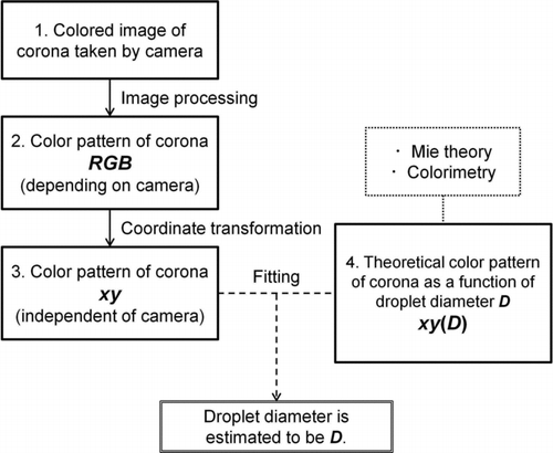
FIG. 1 Schematic diagram of the CIC method used to estimate droplet diameter. The left-hand side (1–3) starts from a color image of a corona taken by a camera and leads to experimental xy standard colorimetric parameters. The right-hand side (4) is the theoretical calculation for xy as a function of particle diameter. Finally, the particle diameter is estimated by fitting experimental and theoretical xy.
2. METHODOLOGY
Droplet size determination in the CIC method follows the procedure shown in . In the left and right columns are the experimental and theoretical procedures, respectively. First, a color image of a corona under illumination by a white-light source is obtained by a digital camera (step 1 in ). Second, the RGB values of individual pixels in the image are converted to standard colorimetric parameters in xy-color coordinates (steps 2 and 3), so as to be independent of characteristics of the image acquisition device and the absolute light intensity, which are not relevant to the present analysis. Finally, the droplet size is determined by fitting the experimental colorimetric parameters in xy-color coordinates to those calculated theoretically (step 4). Details of these procedures are described in the following subsections. As examples, color images of coronae are shown in .
FIG. 2 Color images of the corona (a) produced by PSL particles with a diameter of 7.088 μm suspended in water and (b) produced by cloud droplets growing in the expansion chamber at a pressure of about 790 hPa. Data in the scattering angles of (a) 4.25°–9.64° and (b) 3.52°–8.59° are shown. The left half shows experimentally obtained RGB values. A background image was subtracted and RGB values were circularly averaged. The right half shows theoretically calculated RGB values.
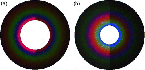
FIG. 3 Instrumental setup of the experiment for (a) a water tank and (b) a flask (expansion chamber). Dotted lines indicate the direct beam from the light source toward the camera, and dot-dashed lines indicate examples of light paths of scattered light. Refraction of light between the water tank and the air is not shown here.
2.1. Experimental Setup
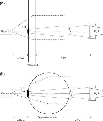
The experimental setup for testing the CIC method is shown in . To determine the particle size, we used a water tank in which polystyrene latex (PSL) spheres were suspended () for calibration. The water tank was made of acrylic plastic with a volume of 5 (depth) × 40 (width) × 30 (height) cm3. We used a round-bottom flask made of Pyrex glass with a 10-L volume as an expansion chamber (), in which cloud droplets formed under supersaturation of water vapor. A tungsten incandescent bulb was used as a white-light source for the formation of coronae by diffraction. The spectral distribution of the light source is smooth in the visible range, making it suitable for comparison with theoretical calculations. A commercial USB digital camera (CMS-V30SETBK_SV, Sanwa Supply Inc., Okayama, Japan) designed for white light was used for automated acquisition of corona images. A particle container (water tank or expansion chamber) was placed between the light source and the camera. There was a distance of 5 m between the light source and the container and 0.2 m between the container and the camera; therefore, the white light incident on the sample approximated a parallel beam.
FIG. 4 Schematic diagrams of the parameters for calculating scattering angles.
2.2. Scattering Angles
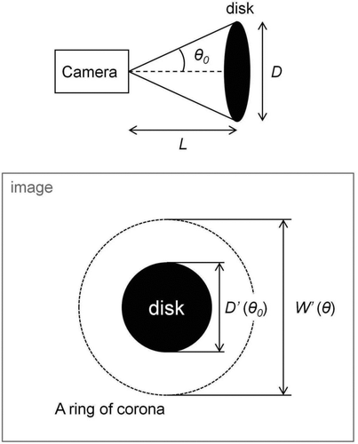
As shown in , a black circular disk of known diameter was attached to the container (water tank or expansion chamber) as an object of known viewing angle used for calculating the viewing angles of the corona rings from the two-dimensional image. In addition, this disk blocked the direct beam from the light source. The viewing half-angle of the corona ring corresponds to the scattering angle of the light. shows the parameters used in the present analysis. The scattering angle θ is calculated by
Typical values of θ and errors were estimated from typical values and measurement errors of the parameters listed above. Namely, using the values D = 2.5 ± 0.02 cm, D′ = 100 ± 1 pixels, W′ = 200 ± 1 pixels, and L = 20 ± 0.3 cm, the scattering angle and error are calculated to be θ ≅ 7.1 ± 0.1°.
2.3. Theoretical Model of a Corona
A theoretical model was developed to predict the angular color pattern of a corona from a given size and refractive index of the droplet relative to the suspension medium and spectral intensity of the incident light. The light scattering calculation was based on Mie theory, as was done by previous studies (Gedzelman and Lock Citation2003; Laven Citation2004). This model was used to determine the droplet size from an observed corona. For quantitative comparison of the model results with observed image data, the calculated spectral intensity must be converted to standard colorimetric parameters (xy-color coordinates) based on the theory of colorimetry.
The differential scattering cross section ΔC sca of a sphere can be predicted by Mie theory (Mie Citation1908; Debye Citation1909) from its size and relative refractive index and the wavelength of the incident light. We used the Mie scattering code BHMIE (Bohren and Huffman Citation2004) for this calculation. By assuming a monodisperse size distribution of droplets, the single scattering intensity I(θ, λ) from an ensemble of droplets is given by
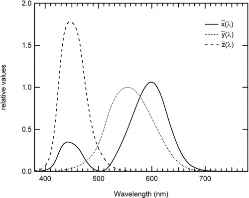
We converted the spectral intensity I(θ, λ) given by EquationEquation (2) to standard colorimetric parameters for quantitative comparisons of the experimental data with the model calculations in color space. Color can be defined by a set of three parameters called tristimulus values, because human eyes have three kinds of photoreceptors, each of which is sensitive to light in a different range of wavelengths (Berns Citation2000). In this study, we used the standard definition of tristimulus values defined in the CIE 1931 XYZ color space. In this color space, any color is expressed by a set of tristimulus values X, Y, and Z. The tristimulus values are calculated from the spectral intensity of observed light and color-matching functions (CIE Citation2004), a relationship that is a practical proxy of the spectral sensitivity curves of the eye's three photoreceptors. shows the CIE 1931 color-matching functions used in our study. The CIE XYZ tristimulus values of a corona are calculated from the scattering intensity and the color-matching functions as
FIG. 5 CIE 1931 color-matching functions.
The coefficient K, proportional to the absolute intensity, can be eliminated by the following normalization
The (x, y) values are normalization parameters called color coordinates. Any color can be represented numerically as a point on the xy plane, independent of absolute intensity. Thus, the angular color distribution of a corona can be represented in the form of (x(θ), y(θ)). The experimental and model-calculated values of this parameter were compared, as described below.
2.4. Transformation of RGB Values to XYZ Values
The RGB data obtained by the camera are also tristimulus values, but the values depend on the type of camera. For quantitative analysis, we converted the RGB values obtained by the camera to the CIE 1931 XYZ tristimulus values. We developed a scheme to transform the RGB values to XYZ values.
We assumed that the XYZ values could be expanded as a Taylor series of RGB parameters. By truncating higher order terms, the expansion can be written as
The expansion coefficients aij determine the transformation from RGB to XYZ. EquationEquation (6) can be rewritten in matrix form as
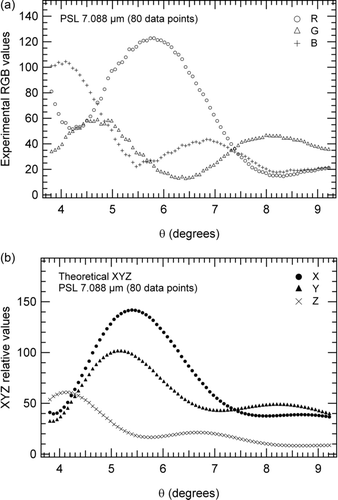
FIG. 6 Examples of RGB and XYZ values used for determining the transformation matrix. The RGB values were obtained for PSL particles with a diameter of 7.088 μm.
Here, the subscript m is the number of the data set, and T, D, and e refer to the corresponding matrices in EquationEquation (8). The matrix A can be determined by the least-squares method, symbolically given by
Transposed matrices (D t) are multiplied because D is not a square matrix. We measured the coronae of PSL particles of known size suspended in the water tank to obtain sets of measured RGB values and known XYZ values. We calculated the XYZ values according to the model described in Section 2.3. For each PSL particle size, we used 80 data sets of measured RGB and calculated XYZ values obtained at different scattering angles to determine A (i.e., m = 80 in EquationEquation (8)). shows an example of the 80 data sets of RGB values obtained for PSL particles (diameter 7.088 μm) and the corresponding theoretical XYZ values.
2.5. Comparing Angular Profiles of Color Coordinates
For quantitative comparison of the measured and calculated values of the angular color coordinates of a corona (x(θ), y(θ)), we defined a fitting parameter (FP), which represents the degree of agreement. The FP includes two distinct factors: the distance between the experimental and theoretical (x(θ), y(θ)) data points in the xy plane and the difference in shapes between the experimental and theoretical x(θ) (or y(θ)) curves as a function of θ. The former factor Dsum , defined as the sum of the squares of the distances over all θ, is expressed as
FIG. 7 Examples of originally defined parameter drvxe (θ) × drvxt (θ).
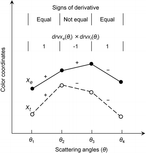
Following this definition, drvxe (θ) × drvxt (θ) (drvye (θ) × drvyt (θ)) is a parameter indicating the agreement between the signs of the derivatives of the experimental and theoretical x(θ) (y(θ)) curves. shows examples of this parameter. If the signs of the derivatives are the same, drvxe (θ) × drvxt (θ) is 1; otherwise, it is −1. The parameter Dx (Dy ) is defined as the average of drvxe (θ) × drvxt (θ) (drvye (θ) × drvyt (θ)) over all θ as
Using the two parameters D sum and Dx,y , we define the fitting parameter FP as
The droplet diameter is estimated from the color image of a corona by finding the global minimum of the FP as a function of the theoretical droplet diameter. When searching for the global minimum, theoretical values of (x(θ), y(θ)) and the corresponding FP were calculated as a function of diameter in 0.1-μm steps.
TABLE 1 Estimated PSL particle diameters
3. VALIDATION
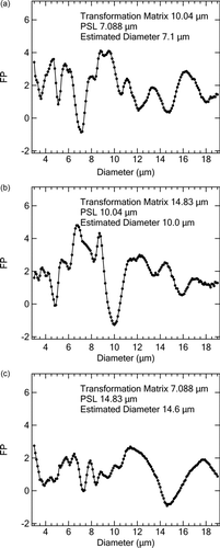
We assessed the accuracy of the CIC method by using PSL particles with known sizes: nominal diameters of 7.088, 10.04, and 14.83 μm (Dynospheres, JSR, Japan). We derived transformation matrices A 7μm, A 10μm, and A 15μm, determined by the coronae of the PSL particles of 7.088-, 10.04-, and 14.83-μm diameters, respectively. shows the PSL particle diameters determined by the CIC method using these transformation matrices and the nominal diameters. The scattering angles for the CIC method were chosen from the smallest scattering angles in which the color of the corona was apparently identified in this experiment. The data for clear coronae were obtained in these scattering angle ranges. Color variation of the coronae was apparent near the forward scattering peak, and the intensity of scattered light was higher for smaller scattering angles. The comparison demonstrates that the particles diameters were estimated with an accuracy of about 2% or better by the CIC method when compared with the nominal diameters.
FIG. 8 Fitting parameters (FP) as a function of assumed diameter for the model calculations. Assumed diameters were given every 0.1 μm. RGB data taken with PSL particles with diameters of 7.088, 10.04, and 14.84 μm were used. The diameters of PSL particles used to derive transformation matrices are also given in individual panels. The most probable particles diameter value was estimated by the value giving the minimum FP.
shows images of coronae obtained from both the experiment and theoretical calculations. The left half of shows a color picture of a corona produced by PSL particles (7.088 μm). A background image of the water tank filled with water without suspended PSL particles was subtracted from the raw picture, and the resultant RGB values were averaged circularly. Only data that were used in the CIC method in the scattering angle range of 4.25°–9.64° () are shown here. The right half of shows a theoretically calculated color pattern. The angular XYZ values of the corona were calculated using the nominal diameter of PSL (7.088 μm) and the refractive index (1.195) in the way described in Section 2.3. The corresponding RGB values were obtained by a universally defined XYZ-to-RGB transformation matrix (Pascale Citation2003). In a strict sense, the resultant RGB values are not appropriate for quantitative comparison with the experimentally obtained RGB values, but the rough agreement between the color patterns demonstrates that our definition of the fitting process in colorimetric parameters (x(θ), y(θ)) reflects real color matching. For the purpose of roughly comparing colors, the coefficient K, which determines the magnitude of the theoretical XYZ values (brightness of color), is not specified, and any value will do so as long as the RGB values transformed from the XYZ values are in the range of 0–255.
Plots of the relevant parameters in the CIC method are shown in and . shows FP as a function of the assumed particle diameter for the three PSL particle diameters in the study. In each plot, the particle diameter associated with the minimum of FP corresponds to the estimated particle diameter. shows the experimental and theoretical x(θ) and y(θ) values as functions of θ after the minimization of FP for each PSL particle size.
FIG. 9 Angular profiles of color coordinates (normalized parameters: x and y). Open circles indicate experimentally obtained color coordinates. Solid lines indicate theoretically calculated color coordinates minimizing FP.
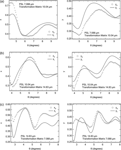
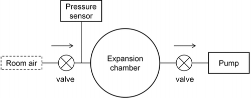
FIG. 10 Schematic diagram of the expansion chamber experiment.
4. MEASUREMENT OF DROPLET GROWTH BY THE CIC METHOD
We applied the CIC method to the time-resolved measurement of cloud droplets in the laboratory. shows a schematic diagram of this experiment. Room air containing aerosol particles was admitted into the same flask as shown in . The air was moderately humidified by passage through a wet filter before introduction into the flask. Then the pressure of the flask was reduced by a pump, and the change of pressure in the flask was measured by a barometric pressure sensor (Setra276; Setra Systems Inc., MA, USA).
A corona was observed associated with the formation of water droplets under water vapor supersaturation caused by adiabatic expansion of the air. A picture of the corona was taken by the camera every 0.15 s. The RGB values were obtained from each image of the corona at 80 different scattering angles in a scattering angle range of about 3.52°–8.59°. For calculating the scattering angles, the effect of the curved surface of the chamber was estimated to be much smaller than the experimental error, discussed in Section 2.2. Every 0.15 s, the droplet diameter was estimated using transformation matrices (A 7μm, A 10μm, and A 15μm), which were determined from the measurements obtained by using the PSL particles. Three droplet diameters, D (7 μm), D (10 μm), and D (15 μm), were derived every 0.15 s by using the three transformation matrices A 7μm, A 10μm, and A 15μm. shows the temporal variation of the mean values of D (7 μm), D (10 μm), and D (15 μm) (solid circles) and the minimum and the maximum values (bars) derived by the CIC method. The derived droplet diameters showed a monotonic increase with decreasing pressure in the flask (open diamonds) except for a few data points, qualitatively consistent with the induced process of the condensational growth of droplets.
FIG. 11 Diameters of growing cloud droplets versus time in the expansion chamber measured by the CIC method. The starting time (0 s) was set to the time when a corona was initially detected by the camera. Closed circles indicate mean values of the three estimated diameters obtained by the use of different transformation matrices. The vertical bars indicate the minimum and the maximum values of the three diameters. Diamonds are the pressures in the chamber.
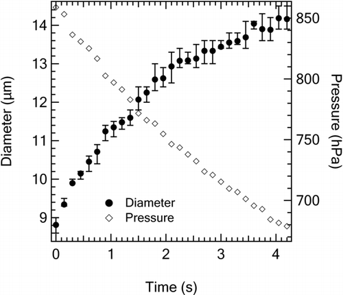
The differences of the three diameters were about 1%–5%, which is slightly larger than the differences of less than about 2% estimated by using the PSL particles (). In other words, the dependence of the estimated diameter on the transformation matrix A was larger than that of the PSL particles. A possible cause of this is that cloud droplets may be less monodisperse than the PSL particles. The precision of the mean value would be improved by increasing the number of transformation matrices obtained for PSL particles of different sizes.
is an example of a color picture of a corona produced by cloud droplets in the expansion chamber. The left side is the experimentally observed corona at a pressure of about 790 hPa. The right side shows the corresponding theoretical values determined by the CIC method.
The particle diameter range shown in extends to a large size parameter range (50–100). It would be difficult to apply the conventional CAMS method in this range, because the data analysis would be complicated by the very large number of complex maxima and minima of the time series data of Mie scattering intensity.
5. APPLICABILITY AND INFLUENCE OF POLYDISPERSITY
The CIC method can be applied when the rings of a corona are clearly observable. Under the illumination of white light, cloud droplets in the diameter range between 6.5 and 25 μm produces multiple-ring corona (Lock and Yang Citation1991). These can be regarded as the upper and lower size limits of the CIC method. The applicability of this method also depends on the concentration of the number of droplets in the chamber. At very low number concentrations, the intensity of the scattered light by the droplets will be too weak for accurate image detection by the digital camera. At very high number concentrations, the single-scattering approximation assumed in the CIC method will not be valid.
We assumed a monodispersed droplet size in applying the CIC method to the cloud chamber experiment. The actual size distributions of droplets in the chamber will not be monodisperse in a strict sense, although the appearance of clear coronae suggests that the droplets are nearly monodisperse. We have estimated theoretically this effect on the determination of the mean droplet size by the CIC method. For this analysis, we assumed “virtual droplets,” which have Gaussian size distributions with the same mean diameter as derived by the measurements of coronae but with a standard deviation (σ). First we calculated x(θ), y(θ) values for corona produced by these “virtual droplets.” Then we estimated the mean diameter by the CIC method by using these x(θ), y(θ) values following the same fitting procedure (). shows the results calculated for different σ values. Generally, the estimated diameters for the “virtual droplets” were equal to or larger than the actual mean diameters (σ = 0 μm). The estimated diameter for the “virtual droplets” with nonzero σ is very close to the actual mean diameter (σ = 0 μm) for σ = 0.5 μm and increases with increasing σ. The overestimation of the mean diameter for polydisperse cloud droplets may be caused by the greater contribution of particles with diameters larger than the mean diameters to the observed intensity of scattered light. A more quantitative estimate of the effect of polydispersity would be possible by estimating theoretically the σ values of the droplets formed in the chamber.
TABLE 2 The influence of polydispersity
6. CONCLUSION
We have developed the CIC method, a new technique for estimating the size of cloud droplets in an expansion chamber by the use of color pictures of coronae, without the need of a laser or specially designed optical system. We confirmed the accuracy of the CIC method by measuring PSL particles of known size comparable with that of cloud droplets (∼10 μm). For each PSL particle size, the estimated diameter agreed well with the nominal size to within 2%, confirming the validity of the overall procedures of the CIC method. The method was successfully applied to measure time-dependent cloud droplet growth in an expansion chamber. The diameter using three transformation matrices agreed to within 1%–5%. The CIC method is powerful for measuring droplets of a size range between 6.5 and 25 μm and can estimate the diameter independently of the growth history of the droplets. This means the CIC method is suitable for measuring droplet sizes comparable with atmospheric cloud droplets. Because of these characteristics, the CIC method complements the conventional CAMS method, which is adequate for a smaller size parameter range.
Acknowledgments
This work was supported by the Ministry of Education, Culture, Sports, Science, and Technology (MEXT), the Strategic International Cooperative Program of the Japan Science and Technology Agency (JST), the Global Environment Research Fund of the Japanese Ministry of the Environment (A-0803 and A-1101), and the GRENE Arctic Climate Change Research Project. The authors would like to thank S. Ohata for his support of the laboratory experiments.
REFERENCES
- Berns , R. S. 2000 . Billmeyer and Saltzman's Principles of Color Technology. , 3rd ed. , New York : John Wiley & Sons .
- Bohren , C. F. and Huffman , D. R. 2004 . Absorption and Scattering of Light by Small Particles , KGaA , Weinheim : Wiley-VCH Verlag GmbH & Co .
- CIE 15. 2004 . Technical Report Colorimetry. , 3rd edn , Central Bureau , Vienna : Commission International de l’Eclairage .
- Chuang , P. Y. 2002 . Analysis of the Influence of Film-Forming Compounds on Droplet Growth: Implications for Cloud Microphysical Processes and Climate . J. Atmos. Sci., , 59 : 2006 – 2018 .
- Davies , J. F. , Haddrell , A. E. and Reid , J. P. 2012 . Time-Resolved Measurements of the Evaporation of Volatile Components from Single Aerosol Droplets . Aerosol Sci. Technol., , 46 : 666 – 677 .
- Debye , P. 1909 . Der Lichtdruck anf Kugeln von beliebigem Material . Ann. Phys., , 30 : 57 – 136 .
- Fairman , H. S. , Brill , M. H. and Hemmendinger , H. 1997 . How the CIE1931 Color-Matching Functions Were Derived from the Wright–Guild Data . Color Res. Appl., , 22 : 11 – 23 .
- Gedzelman , S. D. and Lock , J. A. 2003 . Simulating Corona in Color . Appl. Opt., , 42 : 497 – 504 .
- Grassmann , A. and Peters , F. 2004 . Size Measurement of Very Small Spherical Particles by Mie Scattering Imaging (MSI) . Part. Part. Syst. Char., , 21 : 379 – 389 .
- Han , X. , Ren , K. F. , Wu , Z. , Corbin , F. , Gouesbet , G. and Gréhan , G. 1998 . Characterization of Initial Disturbances in Liquid Jet by Rainbow Sizing . Appl. Opt., , 37 : 8482 – 8503 .
- Kerker , M. 1997 . Light Scattering Instrumentation for Aerosol Studies: An Historical Overview . Aerosol Sci. Technol., , 27 : 522 – 540 .
- König , G. , Anders , K. and Frohn , A. 1986 . A New Light-Scattering Technique to Measure the Diameter of Periodically Generated Moving Droplets . J. Aerosol Sci., , 17 : 157 – 167 .
- Laven , P. 2004 . Simulation of Rainbows, Coronas and Glories using Mie Theory and the Debye Series . J. Quant. Spectrosc. Radiat. Transfer., , 89 : 257 – 269 .
- Lock , J. A. and Yang , L. 1991 . Mie Theory of the Corona . Appl. Opt., , 30 : 3408 – 3414 .
- Mie , G. 1908 . Beiträge zur Optic trüber Medien speziell kolloidaler Metallösungen . Ann. Phys., , 25 : 377 – 445 .
- Pascale , D. 2003 . A Review of RGB Color Spaces… from xyY to R’G’B’ , Montreal : The Babel Color Company .
- Sassen , K. 2003 . Cirrus Cloud Iridescence: A Rare Case Study . Appl. Opt., , 42 : 486 – 491 .
- Semyanov , K. A. , Tarasov , P. A. , Zharinov , A. E. , Chernyshev , A. V. , Hoekstra , A. G. and Maltsev , V. P. 2004 . Single-Particle Sizing from Light Scattering by Spectral Decomposition . Appl. Opt., , 43 : 5110 – 5115 .
- Shaw , J. A. and Neiman , P. J. 2003 . Coronas and Iridescence in Mountain Wave Clouds . Appl. Opt., , 42 : 476 – 485 .
- Stratmann , F. , Kiselev , A. , Wurzler , S. , Wendisch , M. and Heintzenberg , J. 2004 . Laboratory Studies and Numerical Simulations of Cloud Droplet Formation Under Realistic Supersaturation Conditions . J. Atmos. Ocean. Technol., , 21 : 876 – 887 .
- Wagner , P. E. 1985 . A Constant-Angle Mie Scattering Method (CAMS) for Investigation of Particle Formation Processes . J. Colloid Interface Sci., , 105 : 456 – 467 .
- Ward , A. D. , Zhang , M. and Hunt , O. 2008 . Broadband Mie Scattering from Optically Levitated Aerosol Droplets Using a White LED . Opt. Express, , 16 : 16390 – 16403 .
- Winkler , P. M. , Vrtala , A. , Rudolf , R. , Wagner , P. E. , Riipinen , I. Vesala , T. 2006 . Condensation of Water Vapor: Experimental Determination of Mass and Thermal Accommodation Coefficients . J. Geophys. Res., , 111 : D19202 doi: 10.1029/2006JD007194