Abstract
Copyright 2014 American Association for Aerosol Research
INTRODUCTION
Ice nucleating particles (INP) are particles that play a critical role in the formation of mixed phase clouds. The INP initiate glaciation which disturbs the colloidal equilibrium of the suspended supercooled drops and, in turn, results in precipitation (Pruppacher and Klett Citation1996). Ice crystals are frequently observed at temperatures warmer than −15°C (Murray et al. Citation2012) and biologically-derived particles are a plausible source of INP in this temperature regime (Möhler et al. Citation2007; Prenni et al. Citation2009). Biological particles have been identified in cloud ice-crystal residues (Pratt et al. Citation2009), rain and snow (Christner et al. Citation2008), and ground-level air surrounding rain (Huffman et al. Citation2013; Prenni et al. Citation2013). Nonetheless, the role of biological ice nuclei in cloud glaciation remains elusive because direct links between emission and cloud ingestion processes have not been identified. Here we present observations from case studies that suggest that the active release of biological INP is causally coordinated with the arrival of a cold-frontal boundary, which lofts the nuclei to seed the frontal cloud band.
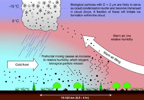
Our proposed mechanism is summarized in , which shows a conceptualization of how pre-frontal mixing at the edge of a cold front will decrease surface temperatures, leading to an increase in relative humidity (RH). Biota responds to this increase in RH by actively releasing biological aerosols. Mixing along the frontal boundary allows for the biological material to be entrained into the warm air that is being lifted over the cold front. The biologically-derived aerosols, along with any other particles present, will then be able to rise and form cloud droplets by acting as cloud condensation nuclei (Möhler et al. Citation2007). A fraction of the immersed biological particles are able to serve as immersion mode INP while droplets cool during ascent. This letter provides observational evidence for the proposed mechanism. Specifically, we will show that the INP release is triggered by high RH, that the INP are efficient immersion mode nuclei at T ∼ −12 to −15°C, that the source of the INP are likely biological particles with diameters ranging from 2–10 μm, and that the INP are also present in precipitation collected from the frontal rainbands.
METHODS
The presented observational evidence centers around two case studies consisting of 24-h Eulerian sampling of aerosol and rainwater in early August in Raleigh, NC, USA. Supporting material is provided in the form of online supplementary information, hereafter referenced as supplementary information (SI) Sn, where Sn denotes Section n. Ambient aerosol was sampled with an impinger sampler (Willeke et al. Citation1998) at ∼23 m a.g.l. in an elevated and open area of the roof of a building. The impinger sampler was operated at 12.5 L/min using 3-h sampling intervals. Ice nucleating particle concentrations were measured using the drop freezing method (Vali Citation1971; Hader et al. Citation2014), resulting in 3-h averaged concentrations of INP active at T = −15, −20, and −25°C (see the SI Section 1). Drop-freezing experiments were performed using a cooling rate of 1 K/min. Rainwater was collected in pyrex dishes during the passage of rainbands and the same method was used to measure INP within the collected rainwater. Analysis of impinger and rainwater solutions was performed immediately after collection. Experimental procedures were similar to those used previously (Hader et al. Citation2014). Further details are provided in the SI Section 1.
A collocated Wideband Integrated Bioaerosol Sensor (WIBS-4A, DMT, Inc.; Huffman et al. Citation2013; Robinson et al. Citation2013) measured particle size and UV fluorescence for individual particles having optical diameter 0.5 < D < 15 μm. The instrument illuminates each particle with UV light at 280 and 370 nm. Fluorescence is measured in the 310 to 400 nm and 420 to 650 nm spectral bands, resulting in three channels. Channels F1 (excitation 280 nm/emission 310–400 nm), F2 (excitation 370 nm/emission 310–400 nm), and F3 (excitation 370 nm/emission 420–650 nm) are indicative of the possible presence of tryptophan, riboflavin, and NAD(P)H, respectively (Després et al. Citation2012). Excitation and emission in the above wavelengths only indicates a likelihood that the listed fluorphores are present (Huffman et al. Citation2010) as other substances can generate fluorescence signals in various combinations of the channels (Pöhlker et al. Citation2012). Particles were grouped together by characteristic excitation–emission profiles (EEP). Seven EEPs are designated: fluorescing particles (FP), fluorescing biological aerosol particles (FBAP; Toprak and Schnaiter Citation2013), and five subgroups of fluorescing particles (FP1–FP5). The FP group includes particles that fluoresce in at least one channel. The FBAP group includes particles that fluoresce in two of the channels, but not a third. FBAP fluorescence is generally attributed to the presence of biofluorophores like tryptophan and NAD(P)H (Després et al. Citation2012). The FP1–FP5 subgroups are subsets of the entire 3-D state space created by the three fluorescing channels and do not overlap. Signal thresholds that delineate these groups are defined in Table S1. This study identifies a group of fluorescing particles that strongly correlated with the 3-h averaged INP concentrations: particles that when excited by λ = 280 nm light strongly emitted in the 310 to 400 nm spectral band, but at the same time either did not or only weakly fluoresced in the other two detection channels. We denote this group of particles as FP3. Further details regarding the fluorescence signal data reduction are provided in the SI Section 2. Potential interference from other weakly fluorescing aerosol types (Pöhlker et al. Citation2012) such as dust and polycyclic aromatic hydrocarbons unlikely played a role here (SI Section 2).
RESULTS AND DISCUSSION
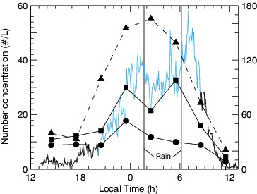
The main case study spanned the passage of a cold front across central North Carolina that brought with it moderate precipitation (detailed meteorological conditions are provided in the SI Section 3). From the start of sampling, at 13:00 LT, until after sunset the temperature decreased and the RH increased. shows that during this time, the INP concentrations at T = −15, −20, and −25°C and the FP3 particle concentrations remained nearly constant. At ∼21:00 LT, the RH increased above ∼75% and from this point until the arrival of the frontal boundary (∼1:30 LT) the INP at −15°C roughly doubled and INP at −25°C roughly tripled. This coincided with a fourfold increase in the FP3 particle concentration. The arrival of the cold front brought with it a band of moderate rain. After the rain had ended, the RH increased to ∼95% and remained high throughout the rest of the night. During this period, the concentration of INP and FP3 remained relatively constant. A second rainband passed near 6:00 LT on 2 August. Subsequently, the temperature increased and the RH decreased substantially. The concentration of FP3 particles peaked at ∼8:30 LT and then fell sharply toward noon. Simultaneously the INP concentrations decreased, approaching pre-frontal concentrations. The RH deviated from the typical diurnal cycle ∼4–6 h ahead of the precipitation suggesting that the spatial scale of the emission footprint corresponds to a 120–180 km wide emission band. The nocturnal boundary layer was sufficiently turbulent to permit shear-induced mixing throughout the stable layer within ∼30 min. To first approximation, the increase in concentration observed at 23 m a.g.l. is due to a distributed source with a footprint on the order of several 10s of km (see the SI Section 3 for further discussion on the interplay between boundary layer turbulence and concentration observations). The August 7–8 case study had a very similar diel cycle, but without an airmass change due to a frontal passage (SI Section 3). This second case study is provided to demonstrate conditions without frontal motion or rainfall so as to provide a control experiment to compare against. These observations excite interest in a causal link between the increase in RH and the emission of biological ice nuclei.
FIG. 2. Particle number concentrations over 24 h, starting on 1 August, 13:00 local time. Measured 3-h ambient INP concentration at T = −15ºC (circles), −20ºC (squares), −25ºC (triangles), and FP3 fluorescent particle concentration (solid line). Solid and dashed lines correspond to the left and right ordinal axes, respectively. Grey shading indicates times of rainfall. The lighter portion (blue) of the FP3 concentration corresponds to when RH > 75%.

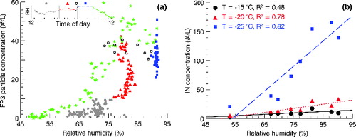
suggests that there may be a release mechanism that is active when RH exceeds a threshold value. Prior to the front, when the RH was less than ∼75% (a, filled circles), there was minimal impact on the concentration of FP3 particles with changes in RH. As the front approached and the RH increased, a burst of FP3 particles was detected (a, triangles). After the rain, but before sunrise, the particle concentration remained high (a, squares). The FP3 particles decreased only after sunrise when the RH started decreasing (a, stars). Very similar increases in the FP3 particle concentration with increasing RH were observed during the 7–8 August case study (SI Section 3). We note that correlation between FP and RH was much weaker than the correlation with FP3 particles. The size distribution of the FP3 group was broad and dominated by particles larger than 2 μm (SI Section 4). Based on particle sizes and timing of the release, a plausible candidate for FP3 particles are fungal spores. Most fungal spores are larger than 2 μm (Després et al. Citation2012) and many fungi actively eject spores at high RH (Rockett and Kramer Citation1974; Huffman et al. Citation2013; Schumacher et al. Citation2013; Toprak and Schnaiter Citation2013; Healy et al. Citation2014) via Buller's drop mechanism (Hirst and Stedman Citation1963; Webster et al. Citation1984; Webster et al. Citation1989; Pringle et al. Citation2005; Joly et al. Citation2014). Furthermore, some studies have demonstrated that selected fungal species (e.g., Fusarium avenaceum) can nucleate ice at temperatures warmer than −15°C (Huffman et al. Citation2013), although most species require T ≤ −20°C to serve as efficient INP (Iannone et al. Citation2011; Haga et al. Citation2014). b demonstrates that the concentration of immersion mode INP also correlate with RH (R2 = 0.48, 0.78, and 0.82 for T = −15, −20, and −25°C, respectively). This correlation points to a link between the RH-controlled biological particle release and the INP population and is consistent with the observation that INP and FP3 concentrations fluctuate in tandem (). The corresponding R2 = 0.7 for T = −20 and −25°C. In contrast, the R2 values for the correlation between FP and INP and FBAP and INP range between 0.25 and 0.4 (SI Section 4). These correlations suggest that FP, FP3, or FBAP may serve as reasonable predictors for INP concentration, with the FP3 group correlating best. We caution, however, that correlation studies cannot establish causation. Actual flux measurements are needed to determine the intensity and scale of emissions in the context of the evolution of the turbulence structure of the atmospheric boundary layer (SI Section 3.3).
FIG. 3. Correlation of RH and FP3 particle concentration (a) and RH and INP concentration (b) for the 1–2 August frontal passage. In the left panel, each of the five markers corresponds to a continuous time period indicated by the inset RH plot. For expanded temporal RH data see the SI Section 3.

Assuming that the INP are biological in origin, the fraction of FP that serve as INP active at T = −20°C is ∼10%. In contrast, almost all of the FP3 particles acted as INP, which aligns with the observation that FP3 particles were between ∼5 and 10% of FP (Table S2). These values are comparable to results from the Amazon rainforest (Prenni et al. Citation2009), where INP concentrations could be modeled assuming that 21% of all fluorescent particles measured by a UV aerodynamic particle sizer (excitation 355 nm/emission 420–575 nm) serve as INP. The FP concentration corresponds to a lower estimate of the total biological aerosol concentration (Hummel et al. Citation2014) and thus actual fractions of biological particles serving as INP are likely smaller, which is more consistent with laboratory studies (Morris et al. Citation2013; Haga et al. Citation2014). A subset of the INP concentration correlated with the FP3 particle concentration, but this does not necessarily imply that the INP and FP3 particles are the same. It appears that either FP or FP3 may serve as a rough proxy for biological INP in this environment. Nevertheless, we point out that parameterizations that scale INP with the number concentration of FBAPs that are larger than 0.5 μm (Tobo et al. Citation2013) or number concentration of all particles larger than 0.5 μm (DeMott et al. Citation2010) significantly underestimate the INP concentration for both measurement days (SI Section 5). This discrepancy likely arises from multiple sources, with the first being climatological differences between this and previous studies. In addition, the fraction of total INP contributed by particles that are smaller than 0.5 μm is unknown and may have been different here compared to the cases underlying the parameterizations. Finally, the measurements used to construct the parameterizations required the removal of particles with an aerodynamic diameter larger than 2.4 μm (Tobo et al. Citation2013). The presumably biologically-derived and fluorescing INP reported here (SI Section 4) and elsewhere (Huffman et al. Citation2013; Prenni et al. Citation2013) are dominated by particles with optical or aerodynamic diameters of D > 2 μm. Therefore, the processes identified in this work would likely have gone unnoticed in previous experimental studies and would be missed in model simulations that must rely on such parameterizations.
CONCLUSIONS
The main process we propose here is the prefrontal release of INP due to increasing RH, which in turn may seed the frontal cloud band. The freezing spectra obtained from rainwater collected during the rain storm shows the presence of a population of efficient INP starting at T ∼ −12°C (SI Section 1). Particles within the rainwater were presumably derived from two principal sources. The first source is particles that were incorporated into the cloud water via droplet activation and in-cloud scavenging processes. The second source is sweepout from the air column below the frontal cloud system. For the given conditions, we estimate that less than 10% of the INP present in the rainwater were scavenged from the column via sweepout (SI Section 1). Under the assumption that sweepout did not significantly add to the rainwater INP, we estimate that cloud-level concentrations of the T ∼ −12 to 15°C population exceeded 100 m−3 air (SI Section 1). The presence of warm INP provides some indication that the prefrontal particle release may have seeded the clouds with INP. This is significant since the frontal rainband is contingent on the presence of seeder ice crystals (Matejka et al. Citation1980). If active nuclei are present they may initiate freezing and release latent heat. Subsequent buoyancy production may generate updrafts under conditions where static stability would have otherwise limited the formation of embedded convective cells that are thought to be required for generating the seeder ice crystals. The ability of a propagating frontal boundary to trigger the release of INP that then seed the frontal cloud systems is in marked contrast with the process involved in strong convective systems which have some difficulty in entraining lofted dust particles produced by their cold pools (Seigel and van den Heever Citation2012). Modelling studies and carefully designed observations, including micrometeorological, microbiological, and molecular biological analyses, are needed to quantify the specific sources, the INP emission fluxes, cloud ingestion rates for the frontal sweep, and impacts on the frontal precipitation.
SUPPLEMENTAL MATERIAL
Supplemental data for this article can be accessed on the publisher's website.
Supplementary_Information_-_Release.zip
Download Zip (561.7 KB)ACKNOWLEDGMENTS
We thank Chris Osburn for providing us with ultrapure water. We thank Droplet Measurement Technologies, Inc., for the use of the WIBS-4A.
FUNDING
This research was funded by the National Science Foundation (NSF) award NSF-AGS 1010851.
REFERENCES
- Christner, B. C., Morris, C. E., Foreman, C. M., Cai, R., and Sands, D. C. (2008). Ubiquity of Biological Ice Nucleators in Snowfall. Science, 319(5867).
- DeMott, P. J., Prenni, A. J., Liu, X., Kreidenweis, S. M., Petters, M. D., Twohy, C. H., Richardson, M. S., Eidhammer, T., and Rogers, D. C. (2010). Predicting Global Atmospheric Ice Nuclei Distributions and Their Impacts on Climate. P. Natl. Acad. Sci. USA, 107(25):11217–11222.
- Després, V. R., Huffman, J. A., Burrows, S. M., Hoose, C., Safatov, A. S., Buryak, G., Frölich-Nowoisky, J., Elbert, W., Andreae, M. O., Pöschl, U., and Jaenicke, R. (2012). Primary Biological Aerosol Particles in the Atmosphere: A Review. Tellus B, 64, 15598.
- Hader, J. D., Wright, T. P., and Petters, M. D. (2014). Contribution of Pollen to Atmospheric Ice Nuclei Concentrations. Atmos. Chem. Phys., 14(11):5433–5449.
- Haga, D. I., Burrows, S. M., Iannone, R., Wheeler, M. J., Mason, R. H., Chen, J., Polishchuk, E. A., Pöschl, U., and Bertram, A. K. (2014). Ice Nucleation and Its Effect on the Atmospheric Transport of Fungal Spores from the Classes Agaricomycetes, Ustilaginomycetes, and Eurotiomycetes. Atmos. Chem. Phys. Discuss., 14(4):5013–5059.
- Healy, D. A., Huffman, J. A., O’Connor, D. J., Pöhlker, C., Pöschl, U., and Sodeau, J. R. (2014). Ambient Measurements of Biological Aerosol Particles Near Killarney, Ireland: A Comparison Between Real-Time Fluorescence and Microscopy Techniques. Atmos. Chem. Phys. Discuss, 14(3):3875–3915.
- Hirst, J. M., and Stedman, O. J. (1963). Dry Liberation of Fungus Spores by Raindrops. J. Gen. Microbiol., 33(2):335–344.
- Huffman, J. A., Treutlein, B., and Pöschl, U. (2010). Fluorescent Biological Aerosol Particle Concentrations and Size Distributions Measured with an Ultraviolet Aerodynamic Particle Sizer (UV-APS) in Central Europe. Atmos. Chem. Phys., 10(7):3215–3233.
- Huffman, J. A., Prenni, A. J., DeMott, P. J., Pöhlker, C., Mason, R. H., Robinson, N. H., Fröhlich-Nowoisky, J., Tobo, Y., Després, V. R., Garcia, E., Gochis, D. J., Harris, E., Müller-Germann, I., Ruzene, C., Schmid, B., Sinha, B., Day, D. A., Andreae, M. O., Jimenez, J. L., Gallagher, M., Kreidenweis, S. M., Bertram, A. K., and Pöschl, U. (2013). High Concentrations of Biological Aerosol Particles and Ice Nuclei During and After Rain. Atmos. Chem. Phys., 13(13):6151–6164.
- Hummel, M., Hoose, C., Gallagher, M., Healy, D. A., Huffman, J. A., O’Connor, D., Pöschl, U., Pöhlker, C., Robinson, N. H., Schnaiter, M., Sodeau, J. R., Toprak, E., and Vogel, H. (2014). Regional-Scale Simulations of Fungal Spore Aerosols using an Emission Parameterization Adapted to Local Measurements of Fluorescent Biological Aerosol Particles. Atmos. Chem. Phys. Discuss, 14(7):9903–9950.
- Iannone, R., Chernoff, D. I., Pringle, A., Martin, S. T., and Bertram, A. K. (2011). The Ice Nucleation Ability of One of the Most Abundant Types of Fungal Spores Found in the Atmosphere. Atmos. Chem. Phys., 11(3):1191–1201.
- Joly, M., Amato, P., Deguillaume, L., Monier, M., Hoose, C., and Delort, A. (2014). Direct Quantification of Total and Biological Ice Nuclei in Cloud Water. Atmos. Chem. Phys. Discuss, 14(3):3707–3731.
- Matejka, T. J., Houze, R. A., and Hobbs, P. V. (1980). Microphysics and Dynamics of Clouds Associated with Mesoscale Rainbands in Extratropical Cyclones. Q. J. Roy. Meteorol. Soc., 106(447):29–56.
- Möhler, O., Demott, P. J., Vali, G., and Levine, J. (2007). Microbiology and Atmospheric Processes: The Role of Biological Particles in Cloud Physics. Biogeosciences, 4(6):1059–1071.
- Morris, C. E., Sands, D. C., Glaux, C., Samsatly, J., Asaad, S., Moukahel, A. R., Gonçalves, F. L. T., and Bigg, E. K. (2013), Urediospores of Rust Fungi are Ice Nucleation Active at > −10°C and Harbor Ice Nucleation Active Bacteria. Atmos. Chem. Phys., 13(8):4223–4233.
- Murray, B. J., O’Sullivan, D., Atkinson, J. D., and Webb, M. E. (2012). Ice Nucleation by Particles Immersed in Supercooled Cloud Droplets. Chem. Soc. Rev., 41:6519–6554.
- Pöhlker, C., Huffman, J. A., and Pöschl, U. (2012). Autofluorescence of Atmospheric Bioaerosols—Fluorescent Biomolecules and Potential Interferences. Atmos. Meas. Tech., 5(1): 37–71.
- Pratt, K. A., DeMott, P. J., French, J. R., Wang, Z., Westphal, D. L., Heymsfield, A. J., Twohy, C. H., Prenni, A. J., and Prather, K. A. (2009). In Situ Detection of Biological Particles in Cloud Ice-Crystals. Nat. Geosci., 2(6): 398–401.
- Prenni, A. J., Petters, M. D., Kreidenweis, S. M., Heald, C. L., Martin, S. T., Artaxo, P., Garland, R. M., Wollny, A. G., and Pöschl, U. (2009). Relative Roles of Biogenic Emissions and Saharan Dust as Ice Nuclei in the Amazon Basin. Nat. Geosci., 2(6):402–405.
- Prenni, A. J., Tobo, Y., Garcia, E., DeMott, P. J., Huffman, J. A., McCluskey, C. S., Kreidenweis, S. M., Prenni, J. E., Pöhlker, C., and Pöschl, U. (2013). The Impact of Rain on Ice Nuclei Populations at a Forested Site in Colorado. Geophys. Res. Let., 40(1):227–231.
- Pringle, A., Patek, S. N., Fischer, M., Stolze, J., and Money, N. P. (2005). The Captured Launch of a Ballistospore. Mycologia, 97(4):866–71.
- Pruppacher, H. R., and Klett, J. D. (1996). Microphysics of Clouds and Precipitation, 2nd ed. Springer, New York.
- Robinson, N. H., Allan, J. D., Huffman, J. A., Kaye, P. H., Foot, V. E., and Gallagher, M. (2013). Cluster Analysis of WIBS Single-Particle Bioaerosol Data. Atmos. Meas. Tech., 6(2):337–347.
- Rockett, T. R., and Kramer, C. L. (1974). Periodicity and Total Spore Production by Lignicolous Basidiomycetes, Mycologia, 66(5):817–829.
- Schumacher, C. J., Pöhlker, C., Aalto, P., Hiltunen, V., Petäjä, T., Kulmala, M., Pöschl, U., and Huffman, J. A. (2013). Seasonal Cycles of Fluorescent Biological Aerosol Particles in Boreal and Semi-Arid Forests of Finland and Colorado. Atmos. Chem. Phys., 13(23):11987–12001.
- Seigel, R. B., and van den Heever, S. C. (2012). Dust Lofting and Ingestion by Supercell Storms. J. Atmos. Sci., 69(5):1453–1473.
- Tobo, Y., Prenni, A. J., DeMott, P. J., Huffman, J. A., McCluskey, C. S., Tian, G., Pöhlker, C., Pöschl, U., and Kreidenweis, S. M. (2013). Biological Aerosol Particles as a Key Determinant of Ice Nuclei Populations in a Forest Ecosystem. J. Geophys. Res.-Atmos., 118(17): 10, 100–10, 110.
- Toprak, E., and Schnaiter, M. (2013). Fluorescent Biological Aerosol Particles Measured with the Waveband Integrated Bioaerosol Sensor WIBS-4: Laboratory Tests Combined with a One Year Field Study. Atmos. Chem. Phys., 13(1):225–243.
- Vali, G. (1971). Quantitative Evaluation of Experimental Results on the Heterogeneous Freezing Nucleation of Supercooled Liquids. J. Atmos. Sci., 28:402–409.
- Webster, J., Davey, R. A., and Turner J. C. R. (1989). Vapour as the Source of Water in Buller's Drop. Mycol. Res., 93(3):297–302.
- Webster, J., Davey, R., and Ingold, C. T. (1984). Origin of the Liquid in Buller's Drop, T Brit. Mycol. Soc., 83(3):524–527.
- Willeke, K., Lin, X., and Grinshpun, S. A. (1998). Improved Aerosol Collection by Combined Impaction and Centrifugal Motion, Aerosol. Sci. Tech., 28(5):439–456.

