
Abstract
This article presents analytical and experimental results for the velocity distribution and transport of expiratory particles from an artificial cough. The stream-wise penetration distance and velocity field of the cough jet were determined through a combination of dimensionless analysis and experimental techniques. The experiments were conducted in a well-controlled environmental chamber with simplified thermal manikins to simulate human coughs and buoyant thermal plumes, and involved flow visualization, velocity measurements employing high and low velocity hot-wire anemometers, and particle size and concentration measurement. The study analyzed three particle sizes—0.77, 2.5, and 7 μm—to examine the impact of particle size on particle transport in the cough jet region and in the vicinity of a receiver occupant positioned in close proximity to the coughing source. The results indicate that the cough jet has a lower axial velocity but higher span-wise expansion rate than a steady jet with an identical discharge velocity. The particles of three sizes have a similar trajectory when considering the transport in the cough jet region. However, particle concentration distributions of the three size particles show that size is an important factor for particle transport in the vicinity of the receiver occupant where airflow velocity decays to the room background air velocity. Furthermore, the results suggest that a cough jet is able to overcome the buoyant human thermal plume and travel further ahead in the region behind the receiver occupant.
Copyright 2014 American Association for Aerosol Research
INTRODUCTION
Epidemic diseases are believed to be related to respiratory airborne transmission (Roy and Milton Citation2004), and human coughing promotes the spread of human released particles. By discharging a large quantity of airborne particles with a high discharge velocity, coughing releases particles with a higher concentration than breathing or talking. Lindsley et al. (Citation2012) found that the average number of particles expelled by each cough ranges from 900 to 302,200. For an influenza-infected subject, it is very likely for a cough jet to contain pathogens and spread airborne diseases that can be inhaled into the respiratory tracts of other individuals. Furthermore, coughed particles have a wide range of sizes, with different transport characteristics. Lindsley et al. (Citation2012) also observed that coughed particles have a size range of 0.35 to 10 μm and Yang et al. (Citation2007) pointed out that the entire average size distribution of the coughed droplets was 0.62–15.9 μm with nuclei sizes of 0.58–5.42 μm. An extensive review conducted recently by Gralton et al. (Citation2011) summaries the size range of coughed particles from a large quantity of studies and concludes that the size scale of particles ranges from 0.1 to 100 μm for individuals prior to and after 1979. Without conducting another review about particle size, this article focuses on the airborne phase particles as they stay longer before deposition and have a higher potential for a long-term transmission. World Health Organization (Citation2007) employs a 5 μm cut-off to delineate between droplet (>5 μm) and airborne (≤5 μm) transmission. Due to the large-size range of and possibly long-term transmission of airborne-coughed particles, it is of importance to comprehend the dynamics of coughed particles with different sizes for understanding interpersonal exposure mechanisms.
A cough jet dominates the transport and dispersion of released coughed particles. The velocity field of human coughs has been extensively examined using various experimental methods, including particle image velocimetry (PIV) (Zhu et al. Citation2006; Chao et al. Citation2009; VanSciver et al. Citation2011; Nishimura et al. Citation2013) and Schlieren imaging (Tang et al. Citation2009). A typical cough has a peak velocity in a range of 6–22 m/s with a duration ranging from 0.25 s to 0.8 s (Zhu et al. Citation2006; Gupta et al. Citation2009). A theoretical analysis of coughed droplets analyzed the trajectories of different size droplets driven by a cough jet assuming the cough jet was steady (Xie et al. Citation2007). Although this analysis is very helpful to understand the dynamics of different size droplets in a jet, the simplification related to the steady-state jet limits the findings from being generalized to real cough conditions. Kouros et al. (Citation1993) found that an unsteady-state jet, such as puff, has significantly different properties from a steady-state jet with the same inlet velocity. Another study has shown that the momentum of a puff is constant in time and flow, and self-persevering (self-similarity) can be achieved downstream from the puff source (Kovasznay et al. Citation1975). The short duration of a cough jet classifies it as a puff and using a puff theory helps the theoretical analysis of the velocity field of a cough, and consequently the trajectories of various size particles within a cough jet.
There are numerous numerical and experimental studies that consider the dispersal of airborne particles expelled from a cough in different indoor environments. Yin et al. (Citation2011) presented the spatial distribution of particles coughed and breathed out by a recumbent patient in a medical ward with two ventilation modes. They concluded that the difference in particle distribution between two cases of coughing and breathing was insignificant only for mixing ventilation. A numerical simulation of an airline cabin showed that the expelled particles from a subject are predominately affected by the bulk airflow (Gupta et al. Citation2011). Modeling of coughs in railway cabins showed that the transmission of particles expelled from a 0.4 s cough contains two sets of events: particles generated in the first 0.2 s escaping from the body plume, and particles generated in the next 0.2 s following the upward body plume (Zhang and Li Citation2012).
While the previously mentioned studies concerned the dispersion of coughed particles in indoor environments, Lai et al. focused on the interpersonal transport of expiratory particles in scaled and full-scale chambers (Berrouk et al. Citation2010; Lai and Wong Citation2010; Seepana and Lai Citation2012). In these studies, coughs were simulated by using a spray gun with the results providing fundamental data regarding interpersonal transmission of coughed particles. However, further improvement of experimental studies regarding interpersonal transport is possible, especially when considering the size of the discharge opening (“human mouth”) and more details on discharge concentration. The discharge concentration of a cough allows an analysis of to what extent expiratory particle concentration decays during particle transport. Furthermore, much less is known about the effect of particle size on coughed particles transporting in the jet and vicinity of a receiver occupant. Most particle sampling instruments, e.g., optical particle counters (OPC) and aerodynamic particle sizes (APS), have a minimum response time not less than 1 s. This prevents the measurement of particle concentration in the cough jet region where coughed particles pass through a position quickly, especially when the cough velocity is high. This may explain why previous studies measured only long-duration and low-discharge-velocity coughs (Lai and Wong Citation2010; Seepana and Lai Citation2012; Chen et al. Citation2013). Detailed studies about the transport of particles expelled from a typical cough are very scarce.
To address the challenges and knowledge gaps identified by previous research on human coughs, this article provides an empirical analysis of an unobstructed coughed jet regarding penetration distance and velocity field. The unobstructed cough refers to a cough that is not covered by hand, tissue, or other covering cases. Even though most people attempt to cover their mouth while coughing, a survey conducted at a train station, a shopping mall, and a hospital shows that approximately one in four people uncover their mouths when coughing or sneezing (Barry et al. 2009). This study focuses on the unobstructed cough jet, since it has a larger penetration distance and particle dispersion than an obstructed jet. The general objective of the present work is to explore the transport of coughed particles in the cough jet region and the vicinity of a receiver occupant. Specific objectives are the following: (1) analyze the velocity field of a cough jet; (2) evaluate the transport of particles expelled from both a cough jet and steady jet with an identical discharge velocity; (3) examine the transport of coughed particles affected by an occupant's thermal plume; (4) and explore the impact of particle size. To fulfill these objectives, the article discusses the relation of cough penetration distance and time using dimensionless analysis and flow visualization. They characterize the cough jet's velocity field as measured further by anemometers. Moreover, this work measures and analyzes the interpersonal transport of coughed particles while considering different particle sizes.
The following sections of the article provide the theoretical background in which a similarity analysis is used to analyze flow dynamics of a cough jet and the governing equations for the trajectories of coughed particles. Since flow dynamics becomes much more complicated when the cough jet reaches the region of human thermal plume, it is difficult to predict particle transport in such region by employing an analytical method. Therefore, this work applies experimental methods to investigate particle distribution in this region and to validate the results for the analytical analysis. The methodology section describes experimental facilities and setup. And the results and discussion section presents the cough jet velocity findings, and the transport of coughed particles as obtained by theoretical analysis and measurements.
THEORETICAL BACKGROUND
This section provides a mathematical description of the penetration distance and velocity field of a cough jet, and the trajectories of particles driven by the jet. As the short duration of a cough prevents detailed measurements of particle dynamics due to the limited capabilities of particle counters, theoretical or numerical analysis are the alternatives. In this study, analytical methods were applied to analyze the flow and particle dynamics in the cough jet region where the airflow was dominated by the jet. In this jet-dominant region, particles especially fine ones (PM 2.5), strictly follow jet streamlines because of the high discharge velocity. The dispersion of particles is mainly driven by drag force for fine particles and by gravity for coarse ones. When the cough jet decays and travels to another occupant, the body's thermal plume may affect dispersion. Other factors, such as receiver occupant's movement and respiratory activities, might also impact the transport and dispersion of coughed particles. The influence level of these factors depends on the characteristics of a cough jet.
Most coughs last 1 s or less with a velocity of 6 m/s or higher (Gupta et al. Citation2009; Nishimura et al. Citation2013). For a commonly used particle sampler, e.g., OPC or APS, it is improbable that the sampler will have enough time to measure particle dispersion accurately in the cough jet region. Fortunately, momentum conservation allows a parameter analysis in the jet region before the jet decays to room background (Kovasznay et al. Citation1975). In the vicinity of a receiver occupant, including the breathing zone, the momentum conservation of the cough jet is broken by additional buoyancy force due to the body thermal plume. In such region, flow and particle dynamics become increasingly complex and limit the theoretical analysis.
The characteristics of an unsteady cough jet are strongly related to: injected volume flow rate and duration of the jet besides the parameters defining a steady jet such as discharge velocity and inlet geometry. Due to the self-preserving (or momentum conservation) of a cough jet flow in the downstream region, the penetration distance Sp referring to the distance from injection source to jet frontal tip, can be written as EquationEquation (1)[1] :
[1] where U0 is discharge velocity, τ is time, and D is the diameter of jet source. The volume flow rate of the jet is
. A dimensionless analysis results in a relationship for the jet at a constant discharge velocity U0,
[2] where X0 is the virtual origin. The power n (n = 1/2) reflects the relationship between penetration distance with time, which varies in different studies (Lee and Chen Citation1998; Sangras et al. Citation2002; Song and Abraham Citation2003). It is worthy noted that EquationEquation (2)
[2] is valid for the case of a jet with a constant discharge velocity.
After taking the derivative of the penetration distance, Sp, with respect to time, τ, we arrive at EquationEquation (3)[3] ,
[3] where Uom is the maximum centerline velocity at the jet tip with a penetration of Sp. Uom can also refer to the peak average velocity at the cough jet center following the tip. The cough jet center is defined as a location of maximum velocity. EquationEquation (3)
[3] shows that the tip velocity of a cough jet has a similar relationship with the dimensionless distance, Sp /D, as a steady jet (Chen and Rodi Citation1980). The coefficient K1 is a constant value that can be determined via experiments.
Ghaem-Maghami and Johari (Citation2010) reported that the axial velocity profile through a puff center follows a Gaussian function similar to a steady jet as described in EquationEquation (4)[4] ,
[4] where r* = r/Sp, r0* = r0/Sp, and r is the vertical distance from the cough jet center. r0* is used because the cough jet center may be slightly off-center.
Furthermore, the velocity profiles of a steady jet with an identical Reynolds number as the cough jet in the literature were used for comparison with the cough jet (Wygnanski and Fiedler Citation1969; Bocksell Citation1998),[5]
[6] where B is velocity-decay constant, 5.5, and
is 10.4 through curve-fitting.
Once the velocity field is determined, the trajectory of a particle injected with a cough jet can be obtained according to Newton's second law of momentum conservation. Because the particle sizes concerned in this study were in the range of 0.35–10 μm and the density of coughed particles is approximately equal to water liquid (Havel et al. Citation1955), only drag, buoyant, and gravity forces dominate particle momentum. For the unobstructed cough and particle sizes considered in this study, these forces are several orders of magnitude greater than Brownian force, thermophoresis force, and lift force (Talbot et al. Citation1980; Li and Ahmadi Citation1992). It is worth noting that this study focuses on an isothermal cough jet. This phenomenon can be reflected by r0* in EquationEquation (4)[4] . Furthermore, the particle momentum and displacement equations can be written as follows:
[7]
[8] where
is velocity, ρ is density, rp is particle size, and
refers to gravitational acceleration. The subscript, p and a, represents particle phase and air phase, respectively. Cd is the drag coefficient represented by a general function of particle Reynolds number, Re, as shown in EquationEquation (7)
[7] ,
[9] where a1, a2, and a3 are constants that apply to spherical particles over a large range of Re (Morsi and Alexander Citation1972).
In terms of EquationEquations (2)[2] –(Equation9
[9] ), the trajectory of a single particle in a cough jet or steady jet can be determined. In this study, the fourth-order Runge-Kutta method was employed to numerically solve the differential equations. The calculation applied the same initial condition as experiments described in next section.
METHODOLOGY
The impact of particle sizes (0.77 μm, 2.5 μm, and 7 μm) was examined by comparing their trajectories in the high velocity jet region. In addition, the transport of coughed particles of the three sizes, released by a source occupant, in the vicinity of a receiver occupant was examined and compared to the results in the cough jet region regarding the influence of particle sizes. The receiver occupant was positioned in a further region of the source where velocity decayed to a low level. Experimental setup and instrumentation are described in the following three methodology subsections that provide specifics about a cough generator, velocity measurement at various locations in the cough jet region and stream-wise penetration distance, and the concentration of coughed particles of three sizes surrounding the receiver occupant. The velocity field in the jet region and particle concentration distribution at the locations further from the jet source will be experimentally validated in the result section.
Cough Box/Generator
A cough box/generator depicted in , was constructed to model a human cough. The cough box has dimensions of 0.25 m × 0.25 m × 0.25 m (15.6 L), and a cough jet was released by injecting pressurized air into the box. The coughed flow was controlled by a fast-response solenoid valve actuated in an “on-off” (square wave) manner. An airflow straightener was built inside to create a piston-like unidirectional flow. In ideal conditions, the unidirectional flow in the box carries particles at a low deposition rate. When the particle generator was turned on, a pump connected to the box drawed air to maintain a slightly negative pressure inside the box. This prevented particles from leaving the box before coughing. In addition, a small computer fan was placed at the center bottom to create a well-mixed condition. The cough box used a 6 cm long stainless steel tube with an inner diameter of 2.4 cm as the discharge opening to simulate a human mouth, 4 cm2 (Gupta et al. Citation2009). In this study, the cough jet was adjusted to have a discharge velocity of 6.08 m/s (Reynolds number 9700), and turbulence intensity 4.25% at the center of the tube opening. The duration of the cough jet was 1 s.
FIG. 1. Schematic of the cough box/generator and the particle generators.
Particle sizes of 0.77 μm, 2.5 μm, and 7 μm, were employed in this study. For 0.77 and 2.5 μm particles, a Collison nebulizer sprayed particle solution into the box. The solution contained latex spherical mono-dispersed particles (coefficient of size variation 1%–3%) with a density of 1.05 g/cm3. Careful attention was paid to ensure that all solution except particle nuclei was evaporated in the box before coughing. Such mono-dispersed particles can be monitored without size categorization issues if the background concentration is negligibly low. However, the Collison nebulizer had difficulties generating enough mono-dispersed 7 μm particles. Therefore, in this study we developed a large-particle disperser that can generate a large quantity of Arizona test dust (ATD) (nominal 5–10 μm) particles ().
Axial Velocity Field in the Cough Jet Region
A one-dimensional hot-wire anemometer and a hot-sphere anemometer measured axial velocity in the center-section plane (76 positions) downstream the cough jet. The hot-wire anemometer measured high velocities (>0.1 m/s), while the hot-sphere anemometer measured velocities between 0.05 m/s to 0.1 m/s. The hot-wire anemometer was calibrated using a Pitot-static system with a pressure transducer (0.1 Pa resolution). The measurement at each position was repeated three times. Furthermore, flow visualization offered an illustration of a cough jet penetrating into an environment. The penetration distance of a cough jet as a function of time and the spreading rate can be easily obtained from visualization. The visualization was created by using a water based fog machine (Model EF-1000, Eliminator Lighting, Los Angeles, CA, USA) that atomizing unscented water based fog juice (density of 1.043 × 10³ kg/m3 at temperature 23.0°C) into particles with 60% in number smaller than 1.0 μm and 99% smaller than 2.5 μm. It is believed that the cough jet profile was captured by such smokes. The smoke temperature was only slightly higher than background yielding a nearly isothermal turbulent cough jet. The visualization video was recorded by a camera at 60 fps (frames per second).
Particle Dispersion in the Vicinity of a Receiver Occupant
To examine how different size particles from coughing dispersed around a receiver occupant, this study carried out experiments of interpersonal transport of coughed particles in a stainless-steel chamber with two thermal manikins insides as shown in . The chamber had a ventilation rate of approximately 3.5/h and supply air was filtered with a high efficiency filter. The temperatures of supply air and exhausted air were 21.6–21.9°C and 24.2–24.6°C during the experiments, respectively. Thermal plumes were created by two thermal manikins, referred to as source and receiver, which were coated with electric heater sheets generating 45 W convective heat each. We compared the velocity 0.25 m above the cylindrical manikin with that in our previous study using a full-scale manikin. It was observed that 45 W heat can generate a thermal plume with a velocity of 0.19 m/s at the region of 0.25 m above the head which was consistent to that for the full scale thermal manikin (Rim and Novoselac Citation2009). In order to decrease the radiant heat transfer between the manikins and interior surfaces of the chamber, aluminum foil with very low emissivity was used to cover the exterior surfaces of the manikins.
FIG. 2. (a) Schematics of the experimental setup and (b) measurement locations of particle concentration.
Two particle sensors sampled particles in the chamber and cough box, respectively. An APS is capable of measuring the concentration of particles from 0.5 to 20 μm using a sophisticated time-of-flight technique which enables high resolution sizing over the entire particle size range. Due to its “high” sampling frequency (up to 1 Hz), the APS was used to monitor the dispersed particles in the surrounding of the receiver occupant as depicted in , whereas an OPC sampled particles in the cough box to measure the injection concentration of a cough. The performance of two particle sensors was calibrated side-by-side before experiments. Before tracking the particle dispersion of an individual cough, the chamber was stabilized and cleaned by ventilation to minimize background concentration. In the data interpretation, the background concentration in the chamber was subtracted in the “Results and Discussion” section (Liu and Novoselac Citation2014). Table S1 shows the coordinates of sample locations of coughed particles. Although the concentration at other locations in the vicinity of the receiver occupant was also measured, the readings were too low to achieve statistically reliable results. The measurements at each location were repeated at least three times; for example, measurements at P1 were repeated 13 times for 0.77 μm particles. To faciliate the comparison of number concentrations of different size particles, all experimental results were normalized by the injection concentration in the cough box.
RESULTS AND DISCUSSION
This section presents the experimental results of penetration distance as a function of time, and velocity field of a cough jet including visualization of the cough jet and correlation of measured axial velocity with the self-preserving scaling law from EquationEquations (2)[2] –(Equation4
[4] ), which verifies the theoretical analysis. Next, the trajectories of three single particles of size 0.77, 2.5, and 7 μm were calculated in terms of cough flow velocity. Finally, the variation of normalized concentrations of three size particles at different locations is discussed.
Penetration Distance and Velocity Field of a Cough Jet
The hot-wire anemometer measured the velocity profile of the cough jet with a discharge velocity of 6.08 m/s at the center of the source opening. The velocity distribution across the source opening follows the velocity profile of a fully developed turbulent pipe flow. compares the area-weighted average velocities of the cough jet varying with time in this article with those reported in three previous studies by Khan et al. (Citation2004), Gupta et al. (Citation2009), and Nishimura et al. (Citation2013), respectively. The velocity profile in the paper of Gupta et al. (Citation2009) is calculated according to recommended regressed models by assuming a standard male occupant with a height of 1.7 m and weight of 70 kg. Real cough jets have a large uncertainty in velocity profile, and the peak velocity ranges from approximately 5.5 m/s to 21 m/s. The artificial cough jet in this article has a simple square wave manner profile that is much different from a real one as shown in . The total air volume of a cough presented in shows that a cough can generate 0.15–2.2 L of air.
FIG. 3. Velocity profiles and total air volume of different cough jets: (a) velocity profile; (b) volume of coughed air.
Snapshot images of a cough jet with a discharge velocity of 6.08 m/s at the center and duration of 1 s at various times after initiation of the flow are provided in . The snapshots show that the cough jet is broken into a vortex ring at the tip, referred to leading tip, followed by the main flow in the first 0.1 s. However, the vortex ring decays shortly in 0.15 s after triggering the cough jet. Therefore, the penetration distance is measured from the main flow tip instead of vortex ring to the source opening. The turbulent region starts from the transition condition to a turbulent jet. The cough jet transports further than 0.2 m during the first 0.1 s. Since the jet spreads linearly, the spreading rate of the jet can be obtained via the jet shape of the visible edges. The averaged spreading rate (drp/dxp), is in a range of 0.21–0.26.
FIG. 4. Flow visualization of a cough jet (D = 0.024 m, U0 = 6.08 m/s, and Re = 9700).
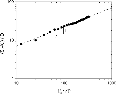
shows the normalized stream-wise penetration distances of the cough jet as a function of normalized time according to the self-preserving scaling of EquationEquation (2)[2] ,
. The nondimensional times for 0.1 s, 0.4 s, 0.7 s, and 1.0 s in are 25.3 and 101.3, 177.1 and 253, respectively. The curve fit shows that the power n in EquationEquation (2)
[2] is 1/2 that is consistent to the study by Sangras et al (Citation2002). The virtual origin X0 is approximately 5.1 times jet exit diameter. The penetration distance in follows the correlation of EquationEquation (2)
[2] well in the whole range of dimensionless time investigated in experiments.
FIG. 5. Penetration distance of a cough jet as a function of time.
Self-preserving in EquationEquation (3)[3] shows that the cough jet also has a linear decay of centerline velocity similar to a steady jet. The average centerline velocities of the cough jet and steady jet with an identical discharge velocity are plotted in . When the axial distance X > 3D, self-similarity is achieved for the cough jet, and the velocity in the jet center reasonably follows the scaling law of EquationEquation (3)
[3] , with a constant coefficient K1 = 0.29. In the region of X < 3D where potential core and mixing layer exist, the center velocity is equal to the discharge velocity in the initialization. It is observed that the centerline velocity in the cough jet, maximum velocity, is lower than that of the steady jet at the same location. This indicates that the calculation of particle trajectories of coughed particles employing a steady jet velocity field is not accurate.
FIG. 6. The comparison of velocity fields of a cough jet and steady jet. (a) The centerline velocity of a cough jet and steady jet as a linear function of dimensionless distance; (b) axial velocity profile through a cough jet center at various X/D: 4.2; 6.3; 8.3; 16.7; 25.0; 33.3, and 41.7. Experiments for a steady jet at an identical Reynolds number were conducted by Wygnanski and Fiedler (Citation1969) as described by EquationEquations (5)[5] and Equation(6)
[6] .
![FIG. 6. The comparison of velocity fields of a cough jet and steady jet. (a) The centerline velocity of a cough jet and steady jet as a linear function of dimensionless distance; (b) axial velocity profile through a cough jet center at various X/D: 4.2; 6.3; 8.3; 16.7; 25.0; 33.3, and 41.7. Experiments for a steady jet at an identical Reynolds number were conducted by Wygnanski and Fiedler (Citation1969) as described by EquationEquations (5)[5] and Equation(6)[6] .](/cms/asset/79f27e74-ff62-4b1b-85de-a5754bbc7e5c/uast_a_968655_f0006_b.gif)
Ghaem-Maghami and Johari (Citation2010) showed that the axial velocity profile through the cough jet center follows a Gaussian function regardless of dimensionless distance, X/D. The mean velocity profile for the cough jet is normalized by the velocity at the cough jet center (close to the centerline), U0m and plotted versus radical coordinates, r/Sp in . The axial velocity profiles through the cough jet center collapse onto a Gaussian function EquationEquation (4)[4] ,
, which is wider than the function for a steady jet with an identical Reynolds number reported by Wygnanski and Fiedler (Citation1969). In other words, the cough jet has a wider radical penetration distance than the steady jet.
Particle Transport in the Cough Jet Region
This subsection describes the transport/trajectories of individual particles injected by both the cough jet and steady jet. Three particle sizes, 0.77, 2.5, and 7 μm, were considered, consistent with the sizes used in the transport around the receiver occupant (). It should be pointed out that the particles were injected simultaneously with the cough jet injection. In the potential core and mixing layer, additionally, the time of particles residing is negligibly small compared to the time spent in the main region (Bocksell Citation1998). Therefore, the axial velocity in this region was assumed identical to the discharge velocity.
shows the trajectories of three size particles following the steady jet and cough jet. It is observed that larger particles have an increased deposition distances than smaller ones. However, the distance even for 7 μm particles is only few millimeters, negligible comparing to horizontal penetration. As described in , the curves of horizontal penetration for three size particles collapse into one. This suggests that the transport characteristics of coughed particles in the range of 0.58–5.42 μm are very similar in the cough jet region. The range of 0.58–5.42 μm is the average size of droplet nuclei coughed from healthy subjects (Yang et al. Citation2007).
FIG. 7. Comparison of trajectories of different size particles following a steady jet and a cough jet with the same discharge velocity. (a) Particle trajectories; (b) the variation of horizontal positions with time.
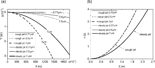
Furthermore, the particles—regardless of size—show a substantial deviation when injected from a steady jet comparing to a cough jet. Overall, the deposition distance of particles in the cough jet is larger than the steady jet because of relatively low velocity magnitude. Therefore, particles in the cough jets take longer time to reach a prescribed position. For instance, coughed particles need approximately 1.2 s to transport 1.1 m further away from the jet exit comparing to 0.8 s for particles in the steady jet.
Particle Transport in the Vicinity of the Receiver Occupant
The concentration distribution for particles of three sizes (0.77 μm, 2.5 μm, and 7 μm) expelled from a cough jet was measured in the vicinity of the receiver occupant and at the exhaust of the chamber (). shows the variation in the normalized distribution of 0.77, 2.5, and 7 μm particles emitted by a cough jet. The uncertainties of the concentration (vertical bars) and the concurrence time (horizontal bars) are represented by standard deviations among repeats. Overall, the concentration decays with the increase of particle size in all measured locations around the receiver occupant after the impingement of the cough jet. It illustrates that coughed particles with various sizes in the vicinity of the receiver occupant perform different transport characteristics as in the cough jet region shown in . The measured peak concentration of 0.77, 2.5, and 7 μm particles in the breathing zone of the receiver occupant, P1, decays to 4.93%, 3.68%, and 1.74% of the initial level in the cough box, respectively. It is observed that particle clouds take approximately 1–2 s to reach the breathing zone (P1) and rise up to the maximum in 3 s for the three size particles. Right above the receiver occupant, P2, the concentration is approximately one order of magnitude lower than that in the breathing zone, P1. This suggests that even 7 μm particles can be transported upwards by human thermal plume.
FIG. 8. The variation of coughed particle concentration in the vicinity of receiver occupant.
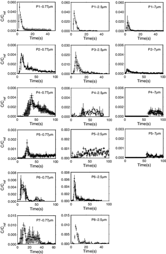
Two side-locations (P6 and P7) that are 0.3 m away from the receiver occupant on the side have slightly higher concentrations for 0.77 μm particles than the position above (P2). However, the concentrations of 2.5 and 7 μm particles were too low to be detected at the two side locations. The reason is that large particles disperse a shorter distance in the span-wise direction due to increased inertia. The study also measured the concentration of three size particles behind the receiver occupant, P4. The result shows a concentration equivalent to that above the receiver occupant (P2) for both 0.77 and 2.5 μm particles. The results of the particle concentration indicate that the cough jet is still strong enough in the zone behind the occupant to overcome the thermal plume. The level of exhaust concentration (P5) demonstrates particle removal by both ventilation and deposition that increases with particle size. shows that peak normalized exhaust concentrations of three size particles are approximately 0.16%, 0.10%, and 0.042%, respectively. The concentrations of 0.77 μm particles achieve the maximum at around 25 s. However, 7 μm particles cannot be sampled until 55 s at the exhaust. Since the concentration of 2.5 μm particles is not significantly higher than the background, does not show a substantial peak at the exhaust, P5.
The Transport Performance of Latex Particles and Arizona Test Dust (ATD)
The mono-dispersed latex particles, 0.77 and 2.5 μm, enable the particle sensors to analyze particle concentration without the uncertainty of particle sizing. However, ATD contains particles in a large range of sizes. The accuracy of 7 μm particle sampling depends on the capacity of size categorization of the particle sensors. In addition, the latex particle and Arizona test dust show different characteristics, such as density and refractive index. Therefore, the results using the two types of particles (latex and ATD) may lead to increasing uncertainties. To examine the extent to which the measured concentration varied using latex and ATD, this study compared the concentration of 2.5 μm particles at location P1 obtained from using mono-dispersed particles and ATD. The comparison plotted in Figure S1 shows that the two curves agree well with each other. The uncertainty of the results obtained from ATD is not shown here. The deviation of the peak concentrations using the two particle types is approximately 8%. The comparison indicates that the experiments using latex particles and ATD can generate repeatable results. In these experiments, however, care must be taken to ensure that the size distributions in the cough box and at each location do not vary significantly when ATD is used.
Limitations
The article employs a simply artificial cough jet that has a square wave manner of velocity profile to investigate the transport of coughed particles. The cough jet with such profile might create a different velocity field from real ones in the jet region even though the total air volume of the artificial cough is reasonable in the range of real coughs. Future studies should examine how different velocity profiles affect flow entrainment, velocity field, and consequently particle transport in the cough jet and vicinity of the recipient. In addition, the study concerns the transmission of airborne particles smaller than or equal to 7 μm. Nevertheless, further exploration is required to include a larger size range and the shrink of large droplets to nuclei by evaporation, especially for low relative humidity environments. The effects of buoyancy force because warm cough jets and turbulent mixing of particles during transmission should be also addressed in the future studies.
CONCLUSION
The transport characteristics of coughed particles in the jet region and the vicinity of a receiver occupant were examined analytically and experimentally. The stream-wise penetration distance and the velocity field of a cough jet were determined through a combination of dimensional analysis and flow visualization. In addition, the transport of coughed particles in the vicinity of a receiver occupant 1.1 m away from the coughing source was determined by measuring the concentration variation of particles with three sizes, 0.77, 2.5, and 7 μm. The findings of this study show that:
A cough jet has a lower axial velocity but higher expansion rate than a steady jet with an identical discharge velocity. Therefore, coughed particles take longer time to transport to a specified location in the stream-wise direction. The transport of coughed particles based on steady jet theory is not valid for coughed particles.
The particle trajectory of coughed particles in the jet region states that the size of coughed particle nuclei (0.35 to 10 μm) has a negligible effect on transmission. Large particles, 7 μm, only settle few millimeters due to gravity in the jet region. However, different size particles exhibit different performance in the vicinity of the receiver occupant. Overall, large particles have a decreased concentration in this zone because the airflow velocity is relatively low.
The normalized concentrations based on the source value in the breathing zone of the receiver occupant at a distance of 1.1 m from the source occupant are 4.93%, 3.68%, and 1.74% for 0.77, 2.5, and 7 μm particles, respectively.
The peak concentrations show decreased levels at the position directly above the occupant when compared to the receiver occupant's surroundings. This indicates that a cough jet presents a greater impact on dispersing particles than thermal plume, especially for a short distance between the two occupants. Furthermore, the concentration level in the position behind the receiver occupant suggests that a cough jet is able to overcome human thermal plume and travel past the receiver occupant.
SUPPLEMENTAL MATERIAL
Supplemental data for this article can be accessed on the publisher's website.
Supplementary_Materials_968655.zip
Download Zip (64.4 KB)REFERENCES
- Barry, T., Manning, S., Lee, M. S., Eggleton, R., Hampton, S., Kaur, J., et al. (2011). Respiratory Hygiene Practices by the Public During the 2009 Influenza Pandemic: An Observational Study. Influenza Respir. Viruses 5(5):317–320.
- Berrouk, A. S., Lai, A. C. K., Cheung, A. C. T., and Wong, S. L. (2010). Experimental Measurements and Large Eddy Simulation of Expiratory Droplet Dispersion in a Mechanically Ventilated Enclosure with Thermal Effects. Build. Environ., 45(2):371–379.
- Bocksell, T. L. (1998). An Enhanced DRW Model for Turbulent Particle Diffusion, Master's thesis, University of Illinois at Urbana—Champaign, Champaign, Illinois, USA.
- Chao, C. Y. H., Wan, M. P., Morawska, L., Johnson, G. R., Ristovski, Z. D., Hargreaves, M., et al. (2009). Characterization of Expiration Air Jets and Droplet Size Distributions Immediately at the Mouth Opening. J. Aerosol Sci., 40(2):122–133.
- Chen, C., Liu, W., Li, F., Lin, C.-H., Liu, J., Pei, J., et al. (2013). A Hybrid Model for Investigating Transient Particle Transport in Enclosed Environments. Build. Environ., 62(0):45–54.
- Chen, C. J., and Rodi, W. (1980). Vertical Turbulent Buoyant Jets: A Review of Experimental Data. NASA STI/Recon Technical Report A, 80, 23073.
- Ghaem-Maghami, E., and Johari, H. (2010). Velocity Field of Isolated Turbulent Puffs. Phys. Fluids (1994-present) 22(11):115105.
- Gralton, J., Tovey, E., McLaws, M., and Rawlinson W. D. (2011). The Role of Particle Size in Aerosolized Pathogen Transmission: A Review. J. Infect., 62(1):1–13.
- Gupta, J. K., Lin, C. H., and Chen, Q. (2009). Flow Dynamics and Characterization of a Cough. Indoor Air, 19(6):517–525.
- Gupta, J. K., Lin, C. H., and Chen, Q. Y. (2011). Transport of Expiratory Droplets in an Aircraft Cabin. Indoor Air, 21(1):3–11.
- Havel, R. J., Eder, H. A., and Bragdon, J. H. (1955). The Distribution and Chemical Composition of Ultracentrifugally Separated Lipoproteins in Human Serum. J. Clin. Invest., 34(9):1345.
- Khan, T. A., Higuchi, H., Marr, D. R., and Glauser, M. N. (2004). Unsteady Flow Measurements of Human Micro Environment Using Time-Resolved Particle Image Velocimetry. Proceedings of RoomVent 2004, the 9th International Conference at the University of Coimbra, Portugal, 5–8 September, 2004, p. 6.
- Kouros, H., Medina R., and Johari, H. (1993). Spreading Rate of an Unsteady Turbulent Jet. AIAA J., 31(8):1523–1526.
- Kovasznay, L. S., Fujita, H., and Lee, R. L. (1975). Unsteady Turbulent Puffs. Adv. Geophys. B, 18:253.
- Lai, A. C. K., and Wong, S. L. (2010). Experimental Investigation of Exhaled Aerosol Transport Under Two Ventilation Systems. Aerosol Sci. Technol., 44(6):444–452.
- Lee, J. H. W., and Chen, G. Q. (1998). A Numerical Study of Turbulent Line Puffs via the Renormalization Group (RNG) k-Epsilon Model. Int.l J. Numer. Methods Fluids, 26(2):217–234.
- Li, A., and Ahmadi, G. (1992). Dispersion and Deposition of Spherical Particles from Point Sources in a Turbulent Channel Flow. Aerosol Sci. Technol., 16(4):209–226.
- Lindsley, W. G., Pearce, T.A., Hudnall, J.B., Davis, K.A., Davis, S.M., Fisher, M.A., et al. (2012). Quantity and Size Distribution of Cough-Generated Aerosol Particles Produced by Influenza Patients During and After Illness. J. Occupat. Environ. Hyg., 9(7):443–449.
- Liu, S., and Novoselac, A (2014). Lagrangian Particle Modeling in the Indoor Environment: A Comparison of RANS and LES Turbulence Methods (RP-1512). HVAC&R Res., 20(4):480–495.
- Morsi, S. A., and Alexander, A. J. (1972). An Investigation of Particle Trajectories in Two-Phase Flow Systems. J. Fluid Mech., 55(02):193–208.
- Nishimura, H., Sakata, S., and Kaga, A. (2013). A New Methodology for Studying Dynamics of Aerosol Particles in Sneeze and Cough Using a Digital High-Vision, High-Speed Video System and Vector Analyses. PloS one, 8(11):e80244.
- Rim, D., and Novoselac, A. (2009). Transport of Particulate and Gaseous Pollutants in the Vicinity of a Human Body. Build. Environ., 44(9):1840–1849.
- Roy, M., and Courtay, C. (1991). Daily Activities and Breathing Parameters for Use in Respiratory-Tract Dosimetry. Radiat. Prot. Dosim., 35(3):179–186.
- Roy, C. J., and Milton, D. K. (2004). Airborne Transmission of Communicable Infection—The Elusive Pathway. New Eng. J. Med., 350(17):1710–1712.
- Sangras, R., Kwon, O. C., and Faeth, G. M. (2002). Self-Preserving Properties of Unsteady Round Nonbuoyant Turbulent Starting Jets and Puffs in Still Fluids. J. Heat Trans., 124(3):460–469.
- Seepana, S., and Lai, A. C. K. (2012). Experimental and Numerical Investigation of Interpersonal Exposure of Sneezing in a Full-Scale Chamber. Aerosol Sci. Technol., 46(5):485–493.
- Song, L., and Abraham, J. (2003). Entrainment Characteristics of Transient Turbulent Round, Radial and Wall-Impinging Jets: Theoretical Deductions. J. Fluid Eng., 125(4):605–612.
- Talbot, L. R., Cheng, R. K., Schefer, R. W., and Willis, D. R. (1980). Thermophoresis of Particles in a Heated Boundary Layer. J. Fluid Mech., 101(04):737–758.
- Tang, J. W., Liebner, T. J., Craven, B. A., and Settles, G. S. (2009). A Schlieren Optical Study of the Human Cough with and Without Wearing Masks for Aerosol Infection Control. J. R Soc., Inter, 6:s727–s736.
- VanSciver, M., Miller, S., and Hertzberg, J. (2011). Particle Image Velocimetry of Human Cough. Aerosol Sci. Technol., 45(3):415–422.
- World Health Organization. (2014). Infection Prevention and Control of Epidemic- and Pandemic-Prone Acute Respiratory Diseases in Health Care: WHO Guidelines.
- Wygnanski, I., and Fiedler, H. (1969). Some Measurements in the Self-Preserving Jet. J. Fluid Mech., 38(03):577–612.
- Xie, X., Li, Y., Chwang, A. T. Y., Ho, P. L., and Seto, W. H. (2007). How Far Droplets Can Move in Indoor Environments–Revisiting the Wells Evaporation–Falling Curve. Indoor Air, 17(3):211–225.
- Yang, S., Lee, G. W., Chen, C. M., Wu, C. C., and Yu, K. P. (2007). The Size and Concentration of Droplets Generated by Coughing in Human Subjects. J. Aerosol Med., 20(4):484–494.
- Yin, Y., Gupta, J. K., Zhang, X., Liu, J., and Chen, Q. (2011). Distributions of Respiratory Contaminants from a Patient with Different Postures and Exhaling Modes in a Single-Bed Inpatient Room. Build. Environ., 46(1):75–81.
- Zhang, L., and Li, Y. G. (2012). Dispersion of Coughed Droplets in a Fully-Occupied High-Speed Rail Cabin. Build. Environ., 47:58–66.
- Zhu, S., Kato, S., and Yang, J. H. (2006). Study on Transport Characteristics of Saliva Droplets Produced by Coughing in a Calm Indoor Environment. Build. Environ., 41(12):1691–1702.