
Abstract
Bacteria, viruses, fungus, and other biological components (toxins, membranes, spores) can spread in the air through various aerosolization processes (breathing, bubbling, explosion, evaporation) and travel until they reach a surface or a host. Nosocomial diseases are an example of illnesses caused by a human contact with such pathogen vectors in hospital settings. Very little is known about the aerosolization processes of viruses and bacteria and their potential to infect people after their passage in the airborne state and about the microbial burden carried by individual aerosol particles. Here we propose a novel approach to study the aerosolization mechanisms of bacteria in single particles using fluorescence spectroscopy and a homemade system allowing the control of the aerosolization and the impaction of bacteria on a black filter. We validated the concept using P. fluorescence and E. coli. The results show that independently of the amount of P. fluorescens and E. coli aerosolized the average distribution of cells impacted on a black filter is described by a Poisson fit with λ ∼ 0.6 ± 0.2. This means that using this aerosolization process, an aerosol will present no bacterium, but when it does, the number of bacteria per particle in the distributions will more probably be one. We also observed that the aerosolization processes of these two bacterial species allow P. fluorescens to be preferentially aerosolized against E. coli. These results demonstrate that fluorescence spectroscopy is a powerful tool to study bioaerosols in single particles. This technique can be used to study several phenomena like preferential aerosolization.
Copyright 2015 American Association for Aerosol Research
INTRODUCTION
The study of the aerosolization mechanisms of microorganisms is an underexploited field of research from which few is being published from a fundamental point of view. Sparse literature is available on single particle bioaerosols and their behavior during their airborne lifetime, impaction on a surface or even the number of microorganisms clustered within individual aerosol particles, hence the difficulty to understand and predict inhalation and exposure doses. It is already known that microorganisms can be ejected in the air through liquid–air aerosolization mechanisms and potentially be inhaled (Blanchard and Syzdek Citation1970). The potential of microorganisms to be aerosolized depends on their shape, size, or hydrophobicity. These factors can be significantly different amongst species (Moletta-Denat et al. Citation2010; Hinds Citation1999). These phenomena must be studied and understood in order to prevent the spreading of microbes over long distances or from infected or colonized sites.
Studying aerosols in single particles is a mandatory step to understand the aerosol behaviour and the adaptation to airborne state of different species and microbe types. In other research fields, understanding single particle behavior leads to crucial information on the pathway of diseases (membrane proteins gating in electrophysiology), conception of drugs against membrane proteins malfunction (pharmacology) understanding and conception of biopesticides (Groulx et al. Citation2010; Laedermann et al. Citation2014; Wagner et al. Citation2014) and powerful tools are now available. The morphology of single particles has a direct impact on the aerosol behavior. As an example, aerosols with smaller aerodynamic diameter will reach deeper in the respiratory track and will remain airborne for a longer period. Several individual bioaerosol particles containing one respiratory pathogenic microbe resistant to aerosolization stress could be able to cause more damages to lungs than bigger aerosols that can be trapped by respiratory tract natural filtration systems.
Knowing the distribution of microorganisms within single aerosol particles will help understanding the potential exposure routes and transport and help evaluate the respiratory doses and the lung zones exposed to those microorganisms sheltered in single particles. Moreover, phenomena involved in the spreading of airborne diseases such as preferential aerosolization can be more easily recorded and analyzed. Preferential aerosolization is a phenomenon which occurs when aerosolization conditions favors one microorganisms against others. Parker et al. were the first to suggest that water to air pathway (bubbling, jet droplet mechanisms) favors M. intracellulare against M. scrofulaceum. Legionella pneumophilia, a pathogenic bacterium, was also found to be easily aerosolized from water surfaces, supporting high environmental exposure (Parker et al. Citation1983). Only one other research on this misunderstood topic has been undertaken few years ago. Moletta et al. showed that different bacteria species (Archaea, Synergistes, Staphylococcus spp., and Propionibacterium acnes) were more efficiently aerosolized in biogas than others (Moletta-Denat et al. Citation2010). The molecular biology approach used in this study wasn't precise enough to explain the aerosolization mechanisms responsible for this phenomenon.
This work aims to develop a simple method for studying bacterial aerosols in single particles in order to analyze fundamental processes such the clustering of bacteria inside droplets or preferential aerosolization.
MATERIAL AND METHODS
Purification, Labeling, and Aerosolization
Eschericha coli (strain ATCC 25422) and Pseudomonas fluorescens expressing green fluorescent protein (GFP; strain A506, Unge A. Citation1997) were grown separately in 400 ml of tryptic soy broth (TSB) medium overnight. Cells were washed with milli-Q® water by centrifugation at 6,500 rpm for 20 minutes (Sorvall, RC5B plus). Cells were resuspended in milli-Q® water at a concentration factor of 1/20. E. coli was subsequently incubated with 1 mM 4′,6-diamidino-2-phenylindole (DAPI [Sigma], E-0835) for 5 min and the bacteria were washed three times by centrifugation at 6,000 rpm for 15–20 min (Eppendorf, 5430) and concentrated 2.5× in milli-Q® water. DAPI will stain E. coli in blue, while P. fluorescens will express GFP and therefore will be stained in green. E. coli and P. fluorescens were diluted and mixed in proportions 1:1; 1:10, and 10:1. Those preparations were adjusted with the optical densities of both preparations, which were shown by microscopy to be directly proportional (data not shown). However, the exact number of each species inside the atomizer was counted and validated under fluorescence microscopy for all experiments.
Bioaerosols were produced by an aerosol generator (atomizer 9302, TSI). The nebulization solution was composed of 65 ml milli-Q® water supplemented with 5 ml of bacterial mix described earlier (1:1, 1:10, 10:1). The aerosol were dried through a diffusion dryer (TSI 3062) and size classified with an electrostatic classifier (3081L, TSI). The electrostatic classifier will mono-disperse as much as possible the aerosol and is also an important asset in the dilution process of the aerosol before it is sent to a 3.36 L aerosol chamber (SCL GenaMini) and then directed to an impaction system. The aerosol chamber was placed inside a biosafety level 2 cabinet. Airborne microorgansims were impacted on polycarbonate black filter of 400 nm porosity (Isopore, HTB04700) using an impactor inlet (TSI 3306) mounted on an aerodynamic particle sizer (APS) (TSI 3321) that was used to size and count particles. (). The sampling was performed during a short period included between 3–5 min at a rate of 21 L per minute. Preferential aerosolization was observed through six different aerosolization experiments in this work, and at least 215 isolated spots were analyzed for every processed aerosolization. Through those six experimentations, three different proportions of the species were studied in the same time (for a total of at least 633 independent spots per condition). During the experiments, the relative humidity and the temperature inside the aerosol chamber reached 10.6 ± 3.6%, and 24.9 ± 1.1°C, respectively. Black filters were then mounted between slide and coverslip and scanned with a motorized epifluorescence microscope. Pictures were taken with an inverted microscope (Axio Observer Z1; Zeiss) with a plan-apochromatic 63X objective. GFP excitation was produced at 470/40 nm, while the emission was read at 525/50 nm. In the case of DAPI, the latest were 365 nm for the excitation and 445/50 nm for the emission. Between 50–100 mosaics containing between 9 and 16 fields of views each were used for each aerosolization. In order to avoid bleaching, the experiments were performed in a light-free environment and the images were taken the same day they were acquired. As DAPI is a DNA marker, one can ask if the aerosolization process could provoke a DNA leak resulting in empty shelves undetectable in fluorescence spectroscopy. The stability of the DAPI labeling during the aerosolization was confirmed by comparing the distribution of bacteria inside the aerosols whilst coated with paraformaldehyde (in order to seal bacteria) against non-f bacteria. As the distributions were similar (data not shown), it was concluded that the labeling of E. coli with DAPI is stable through aerosolization and sampling processes.
FIG. 1. Bioaerosol were produced by an aerosol generator (atomiser, TSI); the aerosol was dried and size classified (electrostatic classifier, TSI); the size selected aerosol was sent to an aerosol chamber and then directed to an impactor system. Airborne microorganisms were impacted on a black filter (impactor inlet, TSI) and numbered by an aerodynamic particle sizer (APS, TSI).
Images were analyzed using a Matlab (Mathworks) routine written in-house. As most of the fields of view were empty on the filter, all the surface area containing bacteria were analyzed.
Counting statistics were first fitted to a Poisson distribution by minimizing the difference between the data and the fit as a function of the average (λ):[1] P(k) represents the Poisson distribution probability as a function of k, the number of bacteria in the particles, and λ is the average number bacteria in a single particle. The preferential aerosolization analyses were made by establishing the distribution of each species within all aerosol particles counted on the filters and comparing it to the distribution of those two species contained inside the aerosolization media.
RESULTS
Single Particles of Bioaerosols can be Spatially Distinguishable on a Black Filter
The two species of bacteria in milli-Q water could be aerosolized with this system. Both species were chosen because they are physically similar. In our pictures, both E. coli's and P. fluorescens’ length ranged between 1 and 2.5 μm while their width ranged between 0.3 and 0.5 μm (see the online supplemental information, Figure S1). The medium mass aerodynamic diameter (MMAD) of the two species was different (0.793 μm for P. fluorescens and 0.909 μm for E. coli, ). One of this study's objectives was to determine the number of total bacteria distributed within each single particle, hence, histograms were established for all dilutions proposed earlier using fluorescence spectroscopy.
FIG. 2. The aerodynamic size distribution of E. coli and P. florescens as measured by an APS.
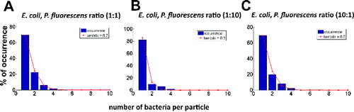
The preliminary results of the aerosolization of E. coli and P. fluorescens revealed that isolated spots in different fields of view were composed of never more than five bacteria. Most of them contained only 1 bacterium. Based on these first observations, it was decided, evaluating that the length of five bacteria (roughly 1 μm long) standing side by side will occupy a maximum area defined by a disk of radius of 2.5 μm (19.6 μm2). One could ask if the observation of bacteria under the microscope could be the result of multiple impactions of several aerosol particles at the same point. We calculated that the filter, using a 100× objective, contains about 3.0×10⁵ fields of view. Two aerosols impacting at the same point will need to follow the same line of flux, which is less likely to happen, as the aerosols are strongly diluted and that the sampling time is very short. Therefore, in each field of view, every bacterium localized within an area of 19.6 μm2 was considered to be part of a single particle.
They all could be fitted by a Poisson distribution describing a system with λ = 0.6 ± 0.2. This means that among droplets formed inside the atomizer, most of them didn't have any bacteria clustered inside. However, when they did, the value of λ was driven by the number of droplet containing only 1 bacterium (Figures a–c). The average χ2 was 89 ± 5%, which gives a good thrust interval for those experiments and validate the use of a Poisson distribution to describe the experimental data.
FIG. 3. Total bacterial distribution in single particles when bacteria species (E. coli, P. fluorescens) were aerosolized, respectively, in proportions: (a) 1:1, (b) 1:10, and (c) 10:1.
As both species were present in the aerosols, it was asked if specific mechanisms could favor one species against the other during the aerosolization process. In order to answer this question, the specific distributions of the species in single particle were established ().
P. fluorescens is More Easily Aerosolized than E. coli in Our Setup
describes how each bacteria species can be distributed over all possibilities of aerosols composed of 1, 2, 3, 4, or 5 bacteria. Between 633 and 1001 single particles were analyzed for each condition applied upon two distinct aerosolizations. Not more than five bacteria were found within individual aerosol particle numbered on the filters.
shows how many bacteria of each species were aerosolized in total compared with the number of bacteria present in the atomizer. The occurrences (in %) of E. coli and P. fluorescens were plotted against the proportions found inside the atomizer's content. The targeted bacterial proportions in the atomizer were 1:1, 1:10, 10:1. However, the exact bacterial proportions were evaluated using fluorescence microscopy, as displayed on .
FIG. 4. Bacterial distributions in single particles were analyzed in three conditions of co-aerosolization of E. coli and P. fluorescens. At least 633 particles were counted over two different aerosolizations. The proportion of each bacterium in the atomizer was measured by fluorescence microscopy.
Whilst both types were present in the same proportions in the atomizer (about 51.5% of E. coli and 48.5% of P. fluorescens), a maximum of 44.7% of the total bacteria aerosolized were E. coli against a maximum of 71.6% of P. fluorescens (). When the proportion of E. coli cells in the atomizer was increased about 10 times (), the amount of E. coli found on the filter was significantly lower (between 51.4 and 77.3%) than the 90% expected. The number of P. fluorescens was comprised between 22.7% and 48.6%, which is significantly higher than the 10% expected. When the concentration of P. fluorescens was nearly 10 times higher than that of E. coli in the atomizer liquid, the same observation was made, even if the total bacteria aerosolized was closer to the amount found inside the atomizer's media ().
DISCUSSION
A controlled aerosolization setup in a confined environment was built in order to study bioaerosols in single particles. To the author's knowledge, this is the first study to propose a simple method to investigate the content of individual particles in fundamental phenomena related to the aerosolization mechanisms.
Furthermore, this approach allowed performing an analysis of the total bacteria clustered within single particles aerosolized and an analysis of the specific species introduced inside the aerosolization media. Upon aerosolization, bacteria follow a stochastic law, which describes a system of aerosols composed of one bacterium in average (). This was true for all tested dilutions and shows that these two species follow similar aerosolization mechanisms. As an excess of E. coli was expected whilst it had higher concentration in the aerosolization media, we observed a significant higher amount of P. fluorescens on the filter (Figures and ). This phenomenon is also clearly observable if the same quantity of each type of bacteria is used in the nebulizer. Even in the case where P. fluorescens is in excess, the phenomenon is still observable, although it is less obvious. The results revealed that P. fluorescens is more easily aerosolized than E. coli in this set up and is thus enriched in the aerosol state.
Previous authors suggested that P. fluorescens cells vary their water content with regards to moisture level, making them well adapted to aerosolization and desiccation on different types of surfaces (Cox Citation1989; Robine et al. Citation2000). In other words, cells will experience fast shrinking upon aerosolization and survive for days on dehydrated surfaces, keeping their metabolic functions unaltered (Cox Citation1989; Robine et al. Citation2000). This result is supported by our finding as it was already shown earlier that upon aerosolization, P. fluorescens has a peak MMAD shifted of about 100 nm compared to E. coli's one (). If upon dehydration, the aerodynamic size and the density of P. fluorescens decrease significantly, it may travel faster across the tubing and experience less loss inside the system caused by inertial forces (Hinds 1999). Other lethal species of bacteria, such as M. tuberculosis, have been shown to have similar survival capabilities upon big mechanical stresses during aerosolization and could preserve its viability capacity for several minutes in airborne state (Clark et al. Citation2011). E. coli can be less resistant than P. fluorescens to those mechanical stresses. As a second peak was observed in all size distributions of E. coli (), one could argue that it corresponds to fragments coming from E. coli's membrane which didn't resist to the aerosolization process. Indeed, the humidity inside the aerosol chamber is low (about 10%, see introduction) and if E. coli presents a resistance to desiccation lower than P. fluorescens, then it can explain at least partially why it is less concentrated on the filters. In that case, producing the experiments at 50% of humidity should decrease the gap between the two species on the filters, but this condition can't be tested inside the aerosol chamber. Another study also observed preferential aerosolization of P. fluorescens compare to Bacillus subtilis bacterial endospores and Penicillium melinii fungal spores from metal working fluids. The author suggested that preferential aerosolization of P. fluorescens could be because of its hydrophilic characteristics inside metalworking fluids (Wang et al. Citation2004). It is interesting to note that we observed similar phenomenon using a different nebulization fluid and a different aerosolization mechanism.
Study of single bioaerosol particles is a new approach to determine the aerosolization mechanisms of airborne pathogens. It allows analysis of the individual particles contain instead of the average of the particles collected like qPCR. This tool could be very useful in the study of airborne disease spread.
CONCLUSION
A simple method was developed in order to study bioaerosols in single particle. Having two species of bacteria, E. coli labeled with DAPI and P. fluorescens expressing GFP, it was possible to use fluorescence spectroscopy in order to study the distribution of those microorganisms within single aerosols on a black filter in several conditions. These experiments revealed that using this system these two bacterial species can be aerosolized and they will most likely travel alone during their airborne lifetime. Results may differ if a physiological process like bubbling occurs and will need further investigations. The co-aerosolization of these two species allowed studying preferential aerosolization, a phenomenon which could contribute significantly to the knowledge about the spreading mechanisms of airborne diseases. Interestingly, E. coli is less aerosolized than P. fluorescens in all conditions tested. This method will be adapted in order to study viral aerosols in single particles as well.
SUPPLEMENTAL MATERIAL
Supplemental data for this article can be accessed on the publisher's website.
UAST_1019606_Supplemental_Information.zip
Download Zip (6 MB)ACKNOWLEDGMENTS
We are grateful to Dr. Kerstin Bellmann for her support with the microscope and to Dr. Hugo McGuire for useful discussions on the interpretations of the results and the statistical analysis.
Funding
N. Groulx is the recipient of studentships from the biophotonic (CREATE) program. C. Duchaine is a FRQS senior scholar, and a member of the FRQ-S Respiratory Health Network.
Related Research Data
REFERENCES
- Blanchard, D. C. and Syzdek, L. (1970). Mechanism for the Water-to-Air Transfer and Concentration of Bacteria. Science 170:626–628.
- Clark, S. O., Hall, Y., Kelly, D. L., Hatch, G. J., Williams, A. (2011). Survival of Mycobacterium Tuberculosis During Experimental Aerosolization and Implications for Aerosol Challenge models. Journal of applied microbiology 111:350–359.
- Cox, C. S. (1989). Airborne Bacteria and Viruses. Science progress 73:469–499.
- Groulx, N., Juteau, M., Blunck, R. (2010). Rapid Topology Probing Using Fluorescence Spectroscopy in Planar Lipid Bilayer: The Pore-Forming Mechanism of the Toxin Cry1Aa of Bacillus Thuringiensis. The Journal of general physiology 136:497–513.
- Hinds, W. C. (1999). Aerosol Technology: Properties, Behavior, and Measurement of Airborne Particles. Wiley-Interscience, New York.
- Laedermann, C. J., Decosterd, I., Abriel, H. (2014). Ubiquitylation of Voltage-Gated Sodium Channels. Handbook of experimental pharmacology 221:231–250.
- Moletta-Denat, M., Bru-Adan, V., Delgenes, J. P., Hamelin, J., Wery, N., Godon, J. J. (2010). Selective Microbial Aerosolization in Biogas Demonstrated by Quantitative PCR. Bioresource technology 101:7252–7257.
- Parker, B. C., Ford, M. A., Gruft, H., Falkinham, J. O., 3rd (1983). Epidemiology of Infection by Nontuberculous Mycobacteria. IV. Preferential Aerosolization of Mycobacterium Intracellulare from Natural Waters. The American review of respiratory disease 128:652–656.
- Robine, E., Derangere, D., Robin, D. (2000). Survival of a Pseudomonas Fluorescens and Enterococcus Faecalis Aerosol on Inert Surfaces. International journal of food microbiology 55:229–234.
- Unge A. T. R., Möller, A., Jansson, J. K. (1997). Optimization of GFP as a Marker for Detection of Bacteria in Environmental Samples. In Bioluminescence and Chemiluminescence: Molecular Reporting with Photons., L. J. K. J.W. Hastings, P.E. Stanley, ed., John Wiley & Sons, Sussex, UK., 391–394.
- Wagner, L. E., 2nd, Groom, L. A., Dirksen, R. T., Yule, D. I. (2014). Characterization of Ryanodine Receptor Type 1 Single Channel Activity Using “On-Nucleus” Patch Clamp. Cell calcium 56:96–107.
- Wang, H. X., Reponen, T., Adhikari, A., Willeke, K., Grinshpun, S. A. (2004). Effect of Fluid Type and Microbial Properties on the Aerosolization of Microorganisms from Metalworking Fluids. Aerosol Sci. Technol 38:1139–1148.