Abstract
Reviews of the effects of solar UV radiation on the survival rate of aerosolized biological material found that the current understanding of environmental viability degradation in response scenarios is insufficient to inform appropriate emergency response measures. We evaluated the effects of UV degradation, in terms of the number of viable, culture forming units as a function of spore cluster size on the downwind hazard presented by a release of a biological organism such as Bacillus anthracis into the environment. We used experimentally derived survival rates for B. atrophaeus var. globigii (BG) spores and BG spore clusters (as a surrogate for Bacillus anthracis) of various sizes exposed to UVC fluences to derive predicted survival rates for single spores and spore clusters of up to 10 µm. For the range of weather conditions encountered in hazard estimates, as characterized by Pasquill-Gifford-Turner classes, we calculated and compared the downwind inhalation and deposition hazards for single spores versus spore clusters up to 10 μm diameter based on standard plume dispersion and particle deposition models. These models can be used to predict survival rates for solar exposure taking into account differences in plume depletion due to deposition, and differences in dose–response due to particle size. The combined effects of solar degradation and size-dependent deposition resulted in clusters presenting from a few to up to 10 orders of magnitude greater hazards than single spores depending on meteorological conditions and downwind distance.
Copyright 2015 American Association for Aerosol Research
INTRODUCTION
In the release of Bacillus spores into the environment, whether for nefarious purposes such as Bacillus anthracis (Ba) in the form of a biological warfare agent (BWA), or Bacillus atrophaeus var globigii (BG) or Bacillus thuringiensis (Bt) for pest control, environmental conditions impact the viability of the organism and resulting dissemination and dispersion effectiveness. One of the likely dominant factors in survivability of spores in a daytime outdoor release within a plume is mutagenesis of DNA through exposure to ultraviolet (UV) light (Harm Citation1969; Mitchell and Karentz Citation1993). There are a number of environmental factors that play a role in germicidal efficiency of sunlight including solar composition, solar angle, solar intensity, and transmittance through the atmosphere. All of these environmental conditions, as well as others including wind speed, direction, topography, and turbulence, can be used to predict resulting clouds and areas of deposition of a released agent. When spraying Bacillus thuringiensis for pest control, releases of the insecticide typically occur at night with near zero solar UV, minimal wind speed, and large droplets; all in an attempt to minimize unintended dispersion of the released material (Van Cuyk et al. Citation2011, Citation2012). Current threat assessment and hazard prediction models do not take into consideration particle size with respect to potential UV shielding effects, or self-encapsulation, which provide protection to spores within the middle of the cluster from UV exposure. Ignoring the effect particle size has on the viability of the particle could lead to a miscalculation of hazards associated with an outdoor release of a biological warfare agent intentional or accidental, such as the 1979 accidental release of Bacillus anthracis in Sverdlovsk, Russia (Meselson et al. Citation1994). The importance of particle size is not only considered for transport and dispersion, but also in its ability to be an inhalational hazard to humans. Several publications report that there is a range in particle size between 1 and 10 µm in which Bacillus spore clusters are likely to be able to be an inhalational threat and the number of particulates needed for an infection is size dependent (Druett et al. 1953; Bartrand et al. Citation2008). Larger particulates typically do not reach the deep lung while smaller particulates traverse through the respiratory system much easier. The inhalational threat of aerosolized material is size dependent for both distance traveled and likelihood for infection.
Reviews of the effects of solar UV radiation on the survival rate of dispersed biological hazards have found that “the current level of understanding and representation of environmental viability degradation in response models is inadequate to inform appropriate emergency response measures (Stuart and Wilkening Citation2005).” Relation of measurements and models of UV degradation of single Bacillus spores to environmental releases in air due to bioterrorism or biowarfare is complicated by the fact that measurements have generally been performed on single spores, whereas bioterror or biowarfare releases are expected to involve spore clusters or spores encapsulated with weaponizing material (Lighthart et al. Citation1991; Matsumoto Citation2003). Clustering and encapsulation of spores can serve to protect the inner spores from the effects of the UV radiation and enhance survivability of the spores and thus, impact degradation rates used in cloud dispersion models (Kesavan et al. Citation2014). Experimental results have recently been reported evaluating the effect of UVC, from low pressure lamps producing a wavelength of 254.3 nm, on inactivating BG spores in different size clusters as well as single spores on both surfaces and as an aerosol (Kesavan et al. Citation2014). Kesavan et al. demonstrated that there is, in fact, a size-dependent degradation rate variance between different sized BG particles (Kesavan et al. Citation2014). However, the effects of those differences in dispersion models were not demonstrated.
We evaluated the effects of UV degradation, in terms of the number of viable, culture forming spores, or clusters of spores after exposure, and as a function of spore cluster size on the hazard presented by a release of a biological agent such as B. anthracis spores. Experimentally derived survival rates for BG spores as reported in Kesavan et al. are used to demonstrate the effects particle size induced shielding has on inhalational and deposition hazards resulting from a dispersed biological agent (Kesavan et al. Citation2014). We evaluated the experimental survival rates of single spores (approximately 1 µm in diameter) and spore clusters up to 4.4 µm in diameter, and developed predictive models to estimate the degradation of spores, under UV exposure, due to increased cluster size and compare CFUs between 10 µm clusters and single spores. Further, we used standard plume dispersion models to assess the effect of survival rate differences due to particle size on downwind inhalation and deposited hazards.
METHODS
Modeling Cluster Decay
Approaches to modeling the decay of single spores under UV irradiation include the classical exponential decay model with modifications to account for low fluence threshold effects and the effects of multiple subpopulations of differing sensitivity to decay, and the multihit model with a single or multiple subpopulations (Kowalski et al. Citation2000). We used a single population multihit model to describe the decay of single spores[1] where S is the surviving fraction of single spores, k is the rate constant, F is the fluence of UV on the spore surface, and n is a constant representing the number of sites within the spore that are required to photo-react to render the spore non-culturable. Fitting the multihit model to the single spore survival data given in Kesavan et al. gives k = 0.12 m2/J and n = 2.8. gives the results of fitting the multihit model to the single spores, the 2.8 μm clusters, and the 4.4 μm clusters. The multihit model performed worse, as measured by the mean square error (mse) over the variance and the confidence intervals for the parameters, in fitting the data as the cluster size increases. In addition, the reduction in sensitivity of the spores to incident UV, as apparent in the reduced values of k, as well as the reduction in n, which is an indicator of the number of sites per spore required to photo-react to result in inactivation, had no clear physical interpretation. Since there was no apparent physical interpretation in terms of the cluster size dependence of the multihit parameters, there was no physical rationale for extending the results to larger (or simply different sized) clusters without additional experimental data.
TABLE 1 Multihit model fits to the inactivation data of Kesavan et al. (Citation2013)
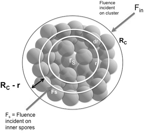
A physical model for the dependence of survival rate on cluster size can be obtained by considering the attenuation, by spores in the outer layers of the cluster, of the UV fluence reaching spores inside the cluster. To extend the multihit model to spore clusters, we considered the survival of spores throughout the cluster (). The fluence, Fs, incident on an inner spore at radius r, within a radial shell dr, within the spore cluster attenuated by transmission through general encapsulating material is
The double exponential integral is an example of a Gompertz distribution developed for application to human mortality (Gompertz Citation1825; Skiadas and Skiadas Citation2010). It has no known analytical integral but can be computed numerically. The values of k and n in the expression are those derived from the single spore data and the unknown parameters are rs, the outer boundary of the innermost spore or spores relevant for UV incidence, and τ, the UV attenuation of the spore material. We expected rs to be on the order of the spore diameter, between 0.65 μm and 1.22 μm from Carrera et al. Citation(2007), and the value of τε to be about 1.5 μm−1 (Hill et al. Citation2013).
Cluster Decay Model Parameters Derived from Experimental Data
A nonlinear regression of Equation (6) was performed on the combined data for survival fractions of the 2.8 μm and 4.4 μm diameter spore clusters and the results are given in . Plots of the fitted curves versus experimental data are given in . The value obtained for rs is 0.95 μm, about midway between the length and the width of the spores. The value for τε is 1.56, which, for ε = 0.78, gives a value of approximately 2.0 μm−1 for τ. This value can be compared to the extinction coefficient for Bacillus spores based on first principles calculations of the complex index of refraction, mi, as 0.048 at 266 nm, which gives (Hill et al. Citation2013). This extinction coefficient resulted in reducing the intensity of incident flux across one spore diameter by about exp(−2.0 × 0.65) to roughly 30% of the incident flux, which agreed well with calculations of extinction in spheres of Bacillus material with the index of refraction derived in Hill et al. Citation(2013).
FIG. 2. Gompertz model fits and experimental data. Data for single spores are shown as triangles, 2.8 μm clusters as squares, and 4.4 μm clusters as circles. The dotted lines show the regression fits to the data, the multihit model for single spores, and the combined Gompertz model for the 2.8 and 4.4 μm cases. The solid line shows the Gompertz fit prediction for 10 μm clusters.
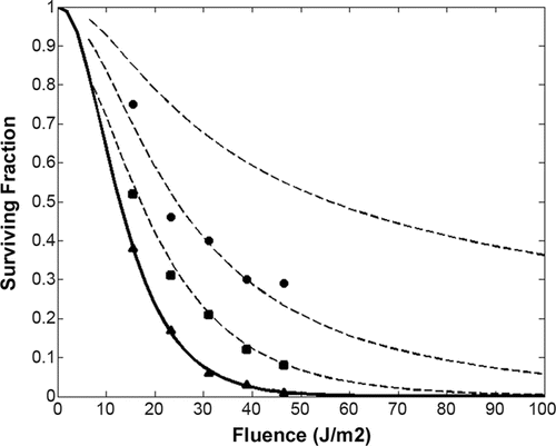
TABLE 2 Regression results for spore cluster survival
Predictions for Larger Clusters
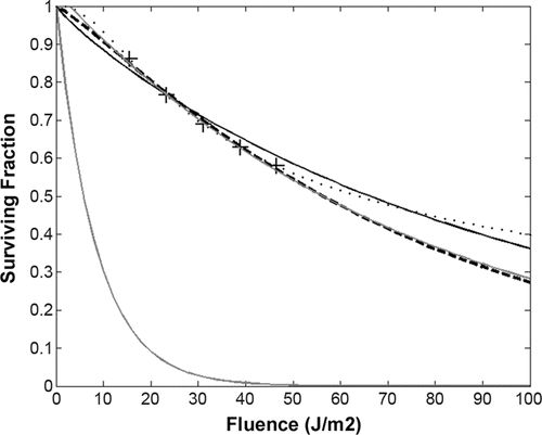
The values of τ and rs derived from the data in Kesavan et al. Citation(2014) can be used to predict the survivability of larger clusters using the Gompertz model multihit extension. Prediction for 10 μm clusters showed orders of magnitude difference in survival rates at larger fluences compared to the rates for single spores. Even when fit to experimental data, the simple multihit model diverged from the Gompertz model extension significantly. plots survival rates for 10 μm clusters at 51 values of fluence evenly distributed from 0 to 100 J/m2 calculated from the Gompertz model extension using the experimentally derived values for τ and rs. The dashed and solid lines show two multihit fits on the calculated Gompertz extended values for the survival rate, the dashed line using just five data points at the fluences used in the experiments of Kesavan et al. Citation(2014) and the solid line using all 51 calculated points in the regression (Skiadas and Skiadas Citation2010). The multihit fits approximate the survival rates for fluences close to the values used for regression, but diverge significantly as fluence increases. Without cluster decay data, both the single spore multihit model and a simple single spore exponential model failed to capture the cluster behavior by orders of magnitude. With cluster decay data as in Kesavan et al. Citation(2014), simple exponential models diverged significantly from measured behavior at the fit cluster size and produce unphysical results when extrapolated straightforwardly to larger cluster sizes (see figures in the online supplementary information [SI]). The Gompertz extension we show here provides a basis for extension of single spore decay data to clusters without cluster decay data using information on the UV attenuation of the spore material, the size of the spores, and the packing fraction of spores within clusters. We describe results here using a multihit single spore model but we have also applied the technique to the phenotypic persistence and external shielding (PPES) model fit to the Kesavan et al. Citation(2014) data.
FIG. 3. Gompertz model prediction for 10 μm cluster compared to multihit and simple exponential models. Black circles plot the Gompertz model prediction for 10 μm clusters using parameters obtained from the fit to data for 2.8 and 4.4 μm clusters for 51 evenly spaced fluence values from 0 to 100 J/m2. The solid black line is a fit to all 51 data points of a multihit model. The dashed black line is a fit of just five data points at the fluences used in the experiments of reference 10. The gray lines show a simple exponential fit to the 10 μm points at the experimental fluences (upper gray line) and a simple exponential fit of the single spore experimental data (lowest gray line).
Modeling Plume Dispersion and Particle Size Effects on Inhalation and Deposited Hazards
The degradation of biological aerosols by solar radiation is a function of the time, the aerosols are suspended in the process of plume dispersion downwind to the point of deposition. We used standard engineering models for plume dispersion since our aim was not to evaluate hazards from specific release scenarios but to compare the relative hazards presented by single spores versus spore clusters under generic dispersion phenomena. We consider the standard meteorological wind conditions described by the various Pasquill-Gifford-Turner stability classes A, B, C, D, E, F (Turner Citation1971), where A is most unstable, D is neutral, and E and F are stable. We considered several wind speeds: 0.2, 0.5, 1, 2, 3, 5, and 6 m/s to span a range of relevant conditions. Each combination of wind speed, insolation conditions, and season corresponded to a particular PGT class. Since we were interested in the differences in dispersion due to cluster size, we used plume calculations that follow standard engineering methods, and are given in the SI. Here, we briefly sketch the calculation and defer details to the SI.
Each PGT class has associated an evolution of the dispersions in the x, y, and z directions, which has a resulting Gaussian dispersion pattern characterized by σx, σy, σz, empirically fit for each of the PGT classes, which are functions of downwind distance the plume travels. The puff evolves in three dimensions as a Gaussian distribution, given by[7] where x0 = vwindt0 = the x position reached by the center of the puff after t0 seconds, σx = σy and σz evolution is approximated by the empirical plume relations, evaluated at t0. This approximates the puff as distributed at t0, moving with velocity vw past x0 without evolving the distribution in time as it passes. The simple form of Equations (7) allows analytical expressions to be obtained for the concentration and deposition as a function of downwind distance for the various wind speeds and PGT classes.
The concentration is given by Equation (7) with x(t) = x0 and we incorporated the effect of UV degradation due to solar exposure simply by multiplying the concentration by the surviving fraction as a function of times of exposure at distances downwind as[8]
We used the times calculated by t = x0/vw and standard plume dispersion solar elevation angles and sky cover categories (described in detail in the SI) for estimating the suspension time and resulting total UV insolation for particles deposited at varying distances downwind.
The UV fluence is reduced as a function of solar elevation angle by the increase in attenuation due to increasing optical path length through the atmosphere, given by the commonly used Beer–Lambert law and adjusted for optical air mass (Kasten and Young Citation1989; Kirchoff et al. Citation2001) appropriate to various seasonal and cloud cover conditions to obtain germicidal effective doses (see details in the SI).
The deposited hazard on the centerline is given by the plume concentration times the particle deposition velocity, vD, integrated over the time the plume passes the downwind distance x0,[9] where we have integrated the Gaussian plume passing the point x0 as characterized by its distribution at x0 and constant for the time the plume passes. The deposition velocity, vD, is calculated with standard empirically calibrated models (Sykes et al. Citation2011) as
, where vg is the gravitational settling velocity and vd is the dry deposition velocity as a function of the wind speed ur, friction velocity u*, and a deposition efficiency E,
. The deposition efficiency is a function of aerodynamic quantities and the particle size (see details in the SI). Finally, we estimated the depletion of the plume concentration resulting from the amount deposited by integrating Equation (9) from plume initiation at xl to downwind distance x0 for
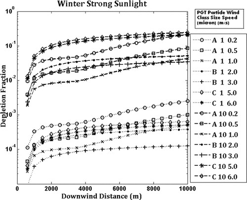
[10] where we used a simplified power law for σz to allow closed-form expression. The results for the various cases are shown in the SI, illustrating the depletion fraction due to deposition for strong and moderate sunlight, respectively. The differences are due to the effect of sunlight on the PGT classes for each wind speed and the resulting differences in the empirical plume spread as a function of downwind distance (see details in the SI). Generally, single spores deposit less than about 0.4% even out to 10 km downwind distance whereas 10 μm clusters can deposit as much as 70% at 19 km downwind distance and well over 10% in as little as 1 km downwind.
Relative Effects of Decay of Single Spores and Clusters During Seasonal Solar Insolation Scenarios
The inactivation rates we used here for a specific strain of B. atrophaeus single spores and clusters for controlled exposure to UVC fluences cannot be used directly to estimate absolute inactivation rates under solar exposure for spores and clusters of the same strain of B. atrophaeus, for other strains, or for other spore forming species such as B. anthracis. This is due to in part to variations (between inactivation under UVC versus solar irradiation) within and among (single spore) species due to cellular protective mechanisms such as (1) absorbing pigments in spore outer layers (Nicholson et al. Citation2005), (2) saturation of the DNA with small acid-soluble proteins (SASP) that inhibit production of UV-induced photoproducts (Desnous et al. 2010), or (3) the amount of dipicolinic acid (DPA) that can increase the formation of UV photoproducts (Setlow and Setlow Citation1993). In addition, the photochemistry differs between single spore exposure to UVC versus full solar irradiance in that the relative contributions to lethality differ in each case due to formation of cyclobutane pyrimidine dimers, spore photoproduct (SP, identified as 5-thyminyl-5,6-dihydrothymine), other DNA photoproducts, and both single strand and double strand breaks in the DNA as well as the repair pathways associated with each damage mechanism (Setlow Citation2001; Desnous et al. 2010). Further there are wide seasonal and geographic variations in total solar incident UV irradiation and relative spectral flux composition due to diurnal variations, dependence on latitude, solar zenith angle, elevation, clouds, aerosols, and ozone levels (Weatherhead Citation2005).
Therefore, we parameterized these variations, as described below, based on results from spore dosimetry experiments to illustrate the relative effect of clustering compared to single spores rather than absolute inactivation rates. The parameterization is chosen to reflect either (1) variation in the relation of UVC decay rates to total solar decay rates or (2) variation in the solar irradiance with diurnal, geographical, and other factors. Coohill and Sagripanti Citation(2009) reviewed measurements of various workers of the decay of B. subtilis spores exposed to both UVC and to solar irradiation at various locations, dates, and times, with most exposures bracketing noon. The values were reported as the ratio of solar/UVC sensitivity, i.e., the time in minutes of solar exposure to obtain F−1log decay divided by the UVC fluence in Jm−2 for F−1log decay. The ratios reported range from 1.5 min/Jm−2 to about 8 solar min/Jm−2. The ratios were reported to be about a factor of 2 larger for winter versus summer, reflecting differences in solar zenith angle and atmospheric composition found in surveys of solar UV (Weatherhead Citation2005). To illustrate the relative effects of clustering, we evaluated decays by adjusting the experimental sensitivities determined by Kesavan et al. Citation(2014) to solar minutes by the factor 1.5 to reflect the most sensitive case and strong sunlight and also by twice that, a factor 3, to reflect either less sensitivity to solar wavelengths in strong sunlight or a reduction of solar irradiance in the highly sensitive case. Examining the UVC data for single spore B. atrophaeus from Kesavan et al. Citation(2014), we found that a one log10 decrement is reached at a UVC fluence of about 27.5 J/m2, so using the factor of 1.5 adjustment that implies that in solar exposure (at high noon and mid latitudes) one log10 decrement in survival should occur at about 41 min. For reduced sensitivity or for high sensitivity/reduced solar irradiance, the factor 3 yielded one log10 decrement at about 82 min. These parametrizations place the decay results in the context of solar irradiation, but should not be interpreted as estimates of absolute decay rates. Instead, we illustrated the relative effects of cluster versus single spore decay by calculating the ratio of the surviving fraction of clusters to the surviving fraction of single spores.
Empirically Derived Decay Curves for Model Fitting
Spore clusters of known sizes, single spores, 2.8 μm, and 4.4 μm, were subjected to a known flux of a wavelength of 254 nm for pre-determined periods of time to obtain the total fluence. The total fluence was then plotted against the surviving fraction of spores comprising the clusters to produce a decay curve. A detailed description of the methodology for generating the empirical data used to produce the decay curves for the model fit can be found in Kesavan et al. Citation(2014).
RESULTS
Effects of Particle Size on Projected Hazards
To assess the differences between hazards presented by clusters and single spores, we estimated and compared the ratios of surviving concentrations and depositions for 4.4 μm clusters and 10 μm clusters to surviving concentrations and depositions for single spores, adjusting for (1) UV degradation of the dispersing aerosolized particles, (2) depletion of the plume due to deposition, and (3) differences in infective dose–response due to particles size.
For the range of weather conditions encountered in hazard estimates, as characterized by the PGT classes, we compared the downwind surviving inhalation hazards presented and the downwind surviving deposition hazards based on the standard plume dispersion models and particle deposition rates as described above, and the survival rates for solar exposure derived from the experimental data. We used exposure times, at downwind distance x0 with wind speeds vw, calculated by t = x0/vw for estimating the suspension time and fluence received for particles dispersed and deposited at varying distances downwind. We found that the combined effects of solar degradation and size-dependent deposition, plume depletion by deposition, and particle size differences in dose–response resulted in both 4.4 μm and 10 μm clusters presenting from a few to up to 10 orders of magnitude greater deposition and inhalation hazards than single spores, depending on meteorological conditions and downwind distance. The differences in surviving deposited hazards presented by the two sizes are increased, relative to the inhalation hazards, due to the difference in deposition rates as a function of particle size.
As noted above, deposition depletion is greater for the 4.4 μm and 10 μm particles still left more than 75% of the concentration intact. Deposition depletion was relatively negligible (compared to orders of magnitude differences) with respect to UV degradation when comparing single spores to 4.4 μm but were significant for 10 μm cluster hazards.
Particle size influences infective dose–response significantly. One contributing factor is that retention in both the trachea and the lungs depends strongly on particle size, increasing from about 1 μm particles up to a peak around 5 μm and then decreasing for larger particle sizes (Hofmann Citation2011). Experimental determination of LD1, LD10, and LD50 for various species found an increase of about a factor of 20 in the respective doses required for equivalent effect for particle sizes above about 5 μm (Druett et al. 1953; Bartrand et al. Citation2008).
The ratios of surviving inhalation hazards for clusters to that for single spores, adjusted for deposition depletion as calculated and dose–response differences by reduction of a factor 1/20 are shown in for the strong sunlight case and high solar sensitivity (solar minutes/UVC = 1.5 min/Jm−2) for various wind speeds from 0.2 m/s to 3 m/s. Also presented are results for a solar sensitivity factor of 3 solar minutes/UVC min/Jm−2, which represents either a reduced solar sensitivity in strong sunlight or high sensitivity in reduced sunlight. The plots essentially show the log difference in surviving hazard presented by the plume concentrations. For 4.4 μm clusters, UV degradation dominated deposition depletion and dose–response effects in inhalation hazard differences at both high and low sensitivity (or reduced sunlight), more at low wind speeds and less at higher wind speeds, due to differences in exposure times to disperse to a given distance. Low sensitivity (or lower sunlight), the solar minutes/UVC fluence = 3 Jm−2 case, reduced the log difference between single spores and 4.4 μm clusters by about a half. In contrast, the left side of shows that 10 μm clusters present greater inhalation hazards (ratio greater than 1) only for the lowest wind speeds in which the time to reach a given distance is sufficient to degrade the single spores enough to compensate for the large deposition and the reduced dose–response of the 10 μm clusters.
FIG. 4. Calculated inhalation hazard ratios (cluster hazard/single spore hazard) including dose–response (1/20 decrement for 10 μm clusters) and deposition depletion. The deposition at downwind distance x0 is given by Equation (9), with the number concentration depleted by the depletion fraction given by Equation (11). The hazard for 10 μm clusters is further multiplied times a factor 1/20 to account for the relative difference between single spore and 10 μm cluster dose–response. The solar/UVC ratio value of 3 represents either reduced sensitivity in strong sunlight or higher sensitivity at reduced solar irradiance.
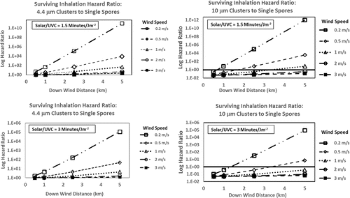
The ratios of surviving deposited hazards for 4.4 μm and 10 μm clusters to single spores, also adjusted for plume depletion and dose–response, are shown in for the strong sunlight, high sensitivity (solar/UVC = 1.5) case and the low sensitivity (or high sensitivity low sunlight) case. In these cases, both 4.4 μm and 10 μm clusters produced greater deposited hazards at all wind speeds since both reduced UV degradation and increased deposition serve to increase the relative deposited hazards of clusters. The combined effects are sufficient to overcome the reduced dose–response for 10 μm clusters.
FIG. 5. Calculated deposition hazard ratios (cluster hazard/single spore hazard) including dose–response (1/20 decrement for 10 μm clusters) and deposition depletion. The depletion fraction is given by Equation (10) and the hazard is obtained as the product of Equations (10) and (8). The hazard for 10 μm clusters is further multiplied times a factor 1/20 to account for the relative difference between single spore and 10 μm cluster dose–response. Hazard ratios are given at the time of deposition. The solar/UVC ratio value of 3 represents either reduced sensitivity in strong sunlight or higher sensitivity at reduced solar irradiance.
DISCUSSION
Enhanced UV survivability of micro-organisms through clumping, clustering, or encapsulation of organisms, has been considered in the context of biowarfare and disease propagation (where enhanced survivability is harmful) as well as for improved microbial pesticide effectiveness (where enhanced survivability is beneficial). To our knowledge, Kesavan et al. Citation(2014) is the first study to provide data with sufficiently well-characterized cluster sizes, dosimetry, and methodology to support reliable determination of surviving fraction of aerosolized clusters as a function of UV fluence and cluster size. Severin proposed and applied the multihit and series-event kinetic models for batch process water disinfection and suggested, but did not demonstrate, that they could be applied to clusters (Severin et al. Citation1983). Current literature addressing ultraviolet germicidal effectiveness provides extensive quantitative information on the decay rates of various single organisms but does not include information characterizing cluster decay. Clustering, or clumping, of organisms is suggested as being a possible effect of increasing RH, a potential source of rate constant error, or an outcome to be avoided for biological warfare agents or a possible explanation for threshold effects (Kowalski et al. Citation2000; Nicholson and Galeano Citation2003). Previous experiments report clumping only in the context of an undesirable artifact of the aerosol generation process to be minimized as much as possible in order to obtain single organisms (King et al. Citation2011; Peccia et al. Citation2001). One reason cluster decay kinetics have not been treated previously is that as Nicholson reports, only when a variety of variables, such as spore purification, irradiation, dosimetry, and survival determination are controlled and assayed in parallel can consistent UV inactivation kinetics be identified and compared (Nicholson and Galeano Citation2003).
The plume dispersion and deposition models we have used are standard engineering models adjusted to agree with a large body of experimental data (Sykes et al. Citation2011) and reasonably well confirmed by recent deposition experiments and applied to deposition onto relatively smooth (non-vegetative) surfaces such as concrete or soil (Roupsard et al. Citation2003; Pryor et al. Citation2008; Sykes et al. Citation2011). While the plume dispersions calculated here are sufficient for comparison of effects between different particle sizes, detailed scenario calculations require more detailed representations of actual meteorological conditions of interest and application to surfaces (such as vegetation) and topographies (such as urban canyons). In addition, application to a specific scenario will require detailed characterization of the specific germicidal effectiveness of the solar spectrum on the specific species concerned in the time and place of interest.
To extend our results to specific pathogens of interest, simulants, and configurations, such as degradation on surfaces and survival during UVGI, requires, as Nicholson notes, experiments in which a variety of variables, such as spore purification, cluster generation composition and size statistics, irradiation, dosimetry, and survival determination were controlled and assayed in parallel (Nicholson and Galeano Citation2003). Cluster decay modeled via a Gompertz function as we have demonstrated also requires controlled measurements of the index of refraction of the spore material and any other material included in the cluster packing. Realistic dispersions of biological agents include not only dry clusters of spores, but may also include clusters of vegetative bacteria and viruses, as well as liquid suspensions or agglomerations involving other materials such as starches, silica derivatives, or polymers. More accurate estimates of UV degradation of clusters involving such characteristics will require addressing three key issues. First, the UV degradation of the single organism needs to be determined under the conditions encountered in the cluster. For example, for dry clusters one needs the aerosolized single spore degradation behavior but for water suspensions one needs the degradation of the organism immersed in water. Second, the packing fraction within the cluster or suspension needs to be assessed. This is required to account accurately for the shielding provided to inner spores as well as the probabilistic accounting for number of spores degraded. Third, the complex indices of refraction for both the bioagent material and the suspending material are needed. Water is relatively transparent to UV on the scales relevant here, but other materials are not. Our derived results for these values for BG are consistent with measurements and calculations for similar spores, but detailed assessments require data specific to the pathogen or simulant in question. For decay on surfaces, the physical configuration of the cluster on the surface needs to be assessed to incorporate actual geometry into an integral of the form of Equation (6). For aerosolized clusters, we assumed spherically symmetric incident fluence; for clusters on a surface, the incident flux would likely be plane or nearly plane incident and the geometry of the cluster may be modified by deposition and surface interaction. Finally, our results indicate that while 4 μm and 10 μm clusters survive UV exposure much more than single spores, the larger clusters deplete the plume by deposition more and require about a factor of 20 greater to provide the same dose–response as single spores. This suggests that a particle size around 5 μm might present the greatest hazards by optimizing UV survival, dispersion, and dose–response.
We have shown that better representations of UV decay of clusters are essential to avoid large errors in estimates of hazards of realistically dispersed biological agents. Even for single spores, simple exponential decay models significantly underestimate experimentally derived behavior at small fluences and overestimate the decay at larger fluences. Several alternative models exist, and we have illustrated the application of one such, the multi target model for a single population. Other models linked to the biochemistry of degradation exist, including multiple population multitarget, and series event type models and the phenotypic persistence and external shielding (PPES) model (Severin et al. Citation1983; Pennell et al. Citation2008). When derived from sufficient experimental data, such models allow estimation of UV fluence decay rates for single spores over a large range of fluences with reasonable accuracy for modest computational burden.
Realistic releases of biological agents typically involve clusters of organisms and we have shown that the models used for single spores inadequately represent the experimentally determined behavior of clusters of spores. Further, there is no formal mechanism within the single spore models to extend them to clusters of spores. We have shown here that an application of the Gompertz model can account for the shielding of inner spores in the cluster from incident fluences due to the UV attenuation of the outer spores, and that such a model can be used to relate single spore experimental UV degradation data to clusters of dry spores of any realistic size using a multitarget model for single spores as a starting point. We further demonstrated that the model interpretation of experimental data is consistent with spore properties derived from first principles, such as spore sizes, UV attenuation, and packing fractions. Elsewhere we show that the approach (modified Gompertz) can be similarly applied using other models, such as series event type models (in progress).
In summary, models of UV degradation of realistic dispersed biological agents are possible based on experimental data and models with physical and biochemical rationales for extension to clusters. Such models can reduce hazard assessment errors in predictions of operational scenarios potentially by many orders of magnitude.
SUPPLEMENTAL MATERIAL
Supplemental data for this article can be accessed on the publisher's website.
UAST_1102857_Supplementary_File.zip
Download Zip (532.7 KB)REFERENCES
- Bartrand, T. A., Weir, M. H., and Haas, C. N. (2008). Dose-Response Models for Inhalation of Bacillus Anthracis Spores: Interspecies Comparisons. Risk Anal., 28:1115–1124.
- Carrera, M., Zandomeni, R. O., Fitzgibbon, J., and Sagripanti, J.-L. (2007). Difference Between the Spore Sizes of Bacillus Anthracis and Other Bacillus Species. J. Appl. Microbiol., 102:303–312.
- Coohill, T. P., and Sagripanti, J.-L. (2009). Review: Bacterial Inactivation by Solar Ultraviolet Radiation Compared with Sensitivity to 254 nm Radiation. Photochem. Photobiol., 85:1043–1052.
- Davidson, G. A. (1990). A Modified Power Law Representation of the Pasquill-Giffod Dispersion Coefficients. J. Air Waste Manag. Assoc., 40(8):1146–1147.
- Druett, H. A., Henderson, D. W., Packman, L., Peacock, S. (1953). Studies on respiratory infection. I. The influence of particle size on respiratory infection with anthrax spores. J Hyg (Lond), 51:359–371.
- Gompertz, B. (1825). On the Nature of the Function Expressing of the Law of Human Mortality. Philosoph. Trans. R. Soc., 36:513–585.
- Harm, W. (1969). Biological Determination of Germicidal Activity of Sunlight. Radiat. Res., 40:63–69.
- Hill, S. C., Pan, Y. L., Williamson, C., Santarpia, J. L., and Hill, H. (2013). Fluorescence of Bioaerosols: Mathematical Model Including Primary Fluorescing and Absorbing Molecules in Bacteria. Opt. Exp., 21:22285–22313.
- Hofmann, W. (2011). Modelling Inhaled Particle Deposition in the Human Lung-A Review. J. Aerosol Sci., 42:693–724.
- Kasten, F., and Young, A. T. (1989). Revised Optical Air-Mass Tables and Approximation Formula. Appl. Opt., 28:4735–4738.
- Kesavan, J., Schepers, D., Bottiger, J., and Edmonds, J. (2014). UV-C Decontamination of Aerosolized and Surface Bound Single Spores and Bioclusters. Aerosol Sci. Technol., 48:450–457.
- King, B., Kesavan, J., and Sagripanti, J.-L. (2011). Germicidal UV Sensitivity of Bacteria in Aerosols and on Contaminated Surfaces. Aerosol Sci. Technol., 45:645–653.
- Kirchoff, V. W. J. H., Silva, A. A., Costa, C. A., Paes Leme, N., Pavao, H. G., and Zaratti, F. (2001). UV-B Optical Thickness Observations of the Atmosphere. J. Geophys. Res.-Atmos., 106:2963–2973.
- Kowalski, W. J., Bahnfleth, W. P., Witham, D. L., Severin, B. F., and Whittam, T. S. (2000). Mathematical Modeling of Ultraviolet Germicidal Irradiation for Air Disinfection. Quant. Microbiol., 2:249–270.
- Lighthart, B., Shaffer, B. T., Balkumar, M., and Ganio, L. (1991). Trajectory of Aerosol Droplets from a Sprayed Bacterial Suspension. Appl. Environ. Microbiol., 57:1006–1012.
- Matsumoto, G. (2003). Bioterrorism: Anthrax Powder: State of the Art? Science, 302:1492–1497.
- Meselson, M., Guillemin, J., and Hugh-Jones, M. (1994). The Sverdlovsk Anthrax Outbreak of 1979. Science, 266:1202–1208.
- Mitchell, D. L., and Karentz, D. (1993). The Induction and Repair of DNA Photodamage in the Environment. Plenum Press, London.
- Nicholson, W. L., and Galeano, B. (2003). UV Resistance of Bacillus Anthracis Spores Revisited: Validation of Bacillus Subtilis Spores as UV Surrogates for Spores of B-Anthracis Sterne. Appl. Environ. Microbiol., 69:1327–1330.
- Nicholson, W..L, Shcuerger, A. C., and Setlow, P. (2005). The Solar UV Environment and Bacterial Spore UV Resistance: Considerations for Earth-to-Mars Transport by Natural Processes and Human Spaceflight. Mutat. Res., 571:249–264.
- Peccia, J., Werth, H. M., Miller, S., and Hernandez, M. (2001). Effects of Relative Humidity on the Ultraviolet Induced Inactivation of Airborne Bacteria. Aerosol Sci. Technol., 35:728–740.
- Pennell, K. G., Aronson, A. I., and Blatchley, E. R. (2008). Phenotypic Persistence and External Shielding Ultraviolet Radiation Inactivation Kinetic Model. J. Appl. Microbiol., 104:1192–1202.
- Pryor, S. C., Gallagher, M., Sievering, H., Laresen, S. E., Barthelmie, R. J., Birsan, F., Nemitz, E., Rinne, J., Kulmala, M., Gronholm, T., Taipale, R., and Vesala, T. (2008). A Review of Measurement and Modelling Results of Particle Atmosphere–Surface Exchange. Tellus, B:42–75.
- Roupsard, P., Amielh, M., Maro, D., Coppalle, A., Branger, H., Connan, O., Laguionie, P., Hebert, D., and Talbaut, M. (2003). Measurement in a Wind Tunnel of Dry Deposition Velocities of Submicron Aerosol with Associated Turbulence Onto Rough and Smooth Urban Surfaces. J. Aerosol Sci., 55:12–24.
- Severin, B. F., Suidan, M. T., and Engelbrecht, R. S. (1983). Kinetic Modeling of UV Disinfection of Water. Water Res., 17:1669–1678.
- Setlow, B., and Setlow, P. (1993). Dipicolinic Acid Greatly Enhances Production of Spore Photoproduct in Bacterial Spores upon UV Irradiation. Appl. Environ. Microbiol., 59:640–643.
- Setlow, P. (2001). Resistance of Spores of Bacillus Species to Ultraviolet Light. Environ. Mol. Mutagen., 38:97–104.
- Skiadas, C. H., and Skiadas, C. (2010). Comparing the Gompertz-Type Models with a First Passage Time Density Model. Statistics for Industry and Technology, Springer, pp. 203–209.
- Stuart, A. L., and Wilkening, D. A. (2005). Degradation of Biological Weapons Agents in the Environment: Implications for Terrorism Response. Environ. Sci. Technol. 39(8):2736–2743.
- Sykes, I. R., Parker, S. F., Henn, D., and Chowdhury, B. (2011). SCIPUFF Version 2.7 Technical Documentation. Sage Management, Princeton, NJ, pp. 85–89.
- Turner, B. D. (1971). Workbook of Atmospheric Dispersion Estimates. United States Environmental Protection Agency, Research Triangle Park, NC, 6.
- Van Cuyk, S., Deshpande, A., Hollander, A., Duval, N., Ticknor, L., Layshock, J., Gallegos-Fraves, L., and Omberg, K. (2011). Persistence of Bacillus Thuringiensis Subsp Kurstaki in Urban Environments Following Spraying. Appl. Environ. Microbiol., 77:7954–7961.
- Van Cuyk, S., Deshpande, A., Hollander, A., Franco, D., Teclemariam, N., Layshock, J., Ticknor, L., Brown, M., and Omberg, M. (2012). Transport of Bacillus Thuringiensis var. Kurstaki from an Outdoor Release into Buildings: Pathways of Infiltration and a Rapid Method to Identify Contaminated Buildings. Biosecurity Bioterror, 10:215–227.
- Weatherhead, E. (2005). Report on Geographic and Seasonal Variability of Solar UV Radiation Affecting Human and Ecological Health. Task (c) Report, Contract 4D-5888-WTSA. Environmental Protection Agency, Research Triangle Park, NC.

