ABSTRACT
A combination of high-repetition rate imaging, laser extinction measurements, two-color soot pyrometry imaging, and high-resolution transmission electron microscopy of thermophoretically sampled soot is used to investigate the long-term and permanent effects of rapid heating of in-flame soot during laser-induced incandescence (LII). Experiments are carried out on a laminar non-premixed co-annular ethylene/air flame with various laser fluences. The high-repetition rate images clearly show that the heated and the neighboring laser-border zones undergo a permanent transformation after the laser pulse, and advect vertically with the flow while the permanent marking is preserved. The soot volume fraction at the heated zone reduces due to the sublimation of soot and the subsequent enhanced oxidation. At the laser-border zones, however, optical thickness increases that may be due to thermophoretic forces drawing hot particles towards relatively cooler zones and the rapid compression of the bath gas induced by the pressure waves created by the expansion of the desorbed carbon clusters. Additionally sublimed carbon clusters can condense onto existing particles and contribute to increase of the optical thickness. Time-resolved two-color pyrometry imaging show that the increased temperature of soot both in the heated and neighboring laser-border zones persists for several milliseconds. This can be associated to the increase in the bath-gas temperature, and a change in the wavelength-dependent emissivity of soot particles induced by the thermal annealing of soot. Ex-situ analysis show that the lattice structure of the soot sampled at the laser-border zones tend to change and soot becomes more graphitic. This may be attributed to thermal annealing induced by elevated temperature.
Copyright © 2017 American Association for Aerosol Research
EDITOR:
Introduction
Particulate matter (PM) emitted during the combustion of hydrocarbons is a major issue due to the adverse impact it has on human health and the environment. A comprehensive understanding of soot formation processes is necessary to develop new combustion concepts and energy conversion systems that help reduce these emissions. Powerful diagnostic tools are required that measure the properties of soot during the combustion event to support and assess studies aimed at reducing soot emissions. Laser-induced incandescence (LII) is an optical in situ technique for measuring the soot volume fraction and primary particle size. In this technique, soot particles are heated via absorption of light from either a pulsed or a continuous-wave (CW) laser to temperatures high enough to emit detectable blackbody-like radiation. Over the course of laser heating and immediately after, the soot particles continue to exchange energy and mass until they reach thermal equilibrium with their surroundings, i.e. bath gas.
For soot volume fraction measurements, it is convenient to detect the LII signal when the soot particles are at their peak temperature, where the LII signal is also at its maximum. Assuming a uniform temperature and particle size distribution among the laser-heated soot particles, the magnitude of the LII signal is linearly proportional to the soot volume fraction. This technique, standalone, provides qualitative measurements of the soot volume fraction, and for quantitative analysis it requires either a true-light calibration of the LII system (Snelling et al. Citation2005), or a calibration with a secondary technique such as light extinction method (Musculus and Pickett Citation2005). Particle sizing with LII, on the other hand, is based on the fact that smaller particles cool down faster than large ones due to their larger surface-to-volume ratio (Melton Citation1984), and a time-resolved LII signal is required for such analysis. In a quantitative approach, particle-size information can be obtained from a best-fit comparison of the temporal LII signal during the heating and cooling phases, and simulations based on the particle's energy- and mass-balance equations (Michelsen et al. Citation2007).
One challenge in particle sizing with LII is that the energy absorbed by the particles and resulting high temperatures may distort the particles and the surrounding bath gas permanently from their initial state. Such changes affect the particle's energy- and mass-balance equations, and must be considered in simulations to describe the LII process accurately. Soot particles may undergo a thermal annealing process which leads to a permanent change of the optical and thermodynamic properties of soot. These laser-induced changes on the soot structure are identified with transmission electron microscopy (TEM) for soot types with various age or coating conditions (Vander Wal and Choi Citation1999; De Iuliis et al. Citation2011; Michelsen et al. Citation2007; Bambha et al. Citation2013). Optical diagnostics of such changes are also performed by using two pulse LII experiments where in-flame soot is preheated with the first pulse, and the LII signal from the consecutive second pulse is measured (Cenker and Roberts Citation2017; Vander Wal and Jensen Citation1998; Vander et al. Citation1998). In these measurements, however, the time delay between the heating and the probing were limited to only a few microseconds, and changes possibly affecting longer time scales could not be observed. At high laser fluences, particles are also subjected to sublimation leading to mass loss. These effects were assessed with LII coupled time-resolved light scattering and extinction (Saffaripour et al. Citation2015; Witze et al. Citation2005; Witze et al. Citation2001; Yoder et al. Citation2005), and it is reported that the time scales for subliming the soot at high laser fluences are on the order of nanoseconds. The possible effects of soot sublimation and effects on the bath-gas flow in longer time scales and larger spatial domains are not analyzed.
In the present work, potential long-term and permanent effects of laser heating on in-flame soot particles and bath gas are investigated by using a conventional pulsed LII setup and extended signal acquisition time durations. It is aimed to extract both qualitative and quantitative information about soot and bath-gas properties that can later be used in optimization of particle sizing with LII modelling. Additionally this study aims to expand the usual probe volume size to a much larger scale and detect signal not only from the heated soot, but also the neighboring zones and report if there is any valuable information that can be used for LII diagnostics. It must be remembered that the in-flame soot measurements have substantial differences compared to cold soot measurements. Therefore, the outcome of this work may not be directly useful for all kind of applications. However this first attempt to investigate the long-lasting effects of laser heating on particles may help develop a better understanding for modelling.
For a comprehensive analysis, four different diagnostic techniques are used in this work: high-repetition rate imaging of incandescence signal after the laser pulse, point-wise laser extinction measurements at the downstream of the laser heating zone, two-color soot pyrometry imaging, and high-resolution transmission electron microscopy (HRTEM) of thermophoretically sampled soot. All of these techniques are well-known and used in soot diagnostics for a large range of applications from the analysis of atmospheric aerosols to in-cylinder engine combustion. In this study, though, minor novel approaches are introduced in the implementation of these techniques such as, volumetric heating of all soot at a fixed height above the burner, coupling high-speed imaging with the single-pulse LII, a probe volume at the downstream of heating zone for extinction measurements, and thermophoretic soot sampling with a single direction pneumatic piston with extremely short sampling times (down to ∼1 ms). Measurements are performed with pulses at different fluences and the effects of increased heating are investigated. In the next section, each of these diagnostics and acquired data are introduced in their respective subsections. Advantages and disadvantages of applied techniques and related challenges are also discussed. The discussion section contains consolidated information about the long-term effects of LII, and potentials for implementation of this information are mentioned.
Experimental setup
Measurements were performed in a non-premixed laminar ethylene/air flame from a Santoro burner (Santoro et al. Citation1983). The burner was operated under standard conditions (C2H4: 0.232 standard liters per minute (slm), air co-flow: 43 slm). The flame was stabilized by a wire screen chimney with a diameter slightly larger than the co-flow and a height of 30 cm. Windows were opened in the chimney to provide optical access to the flame. The visible flame height was measured as 98 mm. A schematic of the experiment is shown in .
Figure 1. Illustration of the experimental set-up. Soot was generated using a Santoro burner under atmospheric conditions. An Nd:YAG laser (1064 nm) was used to irradiate the soot. The laser was first passed through a horizontal slit and then imaged to the flame at 50 mm HAB. A He-Ne laser at 633 nm was used to perform laser extinction measurements. The signal was collected using a photodiode and measured with an oscilloscope. An ICCD camera was used to perform two color pyrometery and a high-speed camera was used to image the flame during TEM sampling (not shown) and to obtain signal intensity measurements.
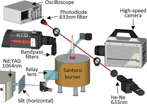
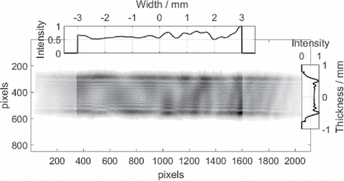
An Nd:YAG laser (SpectraPhysics Pro 250–10) was operated at its fundamental output wavelength of 1064 nm. The triggering of the laser and the synchronization of the laser pulse with photodetectors were realized by a delay generator. The laser energy was controlled with an automatic variable attenuator control system (Laseroptik AVACS) (not shown in the schematic). The laser beam was directly sent into a horizontal metal slit aperture, cropped into a sharp 1.5-mm thick and 10-mm wide horizontal sheet, and imaged onto the flame at 50 mm height above the burner (HAB) with a plano-convex C-coated cylindrical lens at a 2f distance. At this axial height, the flame diameter is smaller than the width of the laser sheet. Hence the soot particles at all radial locations, (i.e., particles along the line-of-sight of cameras and extinction setup) were heated. Such a volumetric heating strategy was preferred over the conventional small beam or thin vertical laser-sheet heating to avoid any complications and signal contribution from the unheated soot at the line-of-sight during the imaging of soot. It also provided setting an angle-independent laser extinction system and simplified the study into one-dimensional analysis along the flame axis. The energy distribution of the horizontal laser sheet pulse was measured with a beam profiler (Gentec Beamage-4M) at the flame region and shown in (the image is an average of ten single shot measurements). The line plots shown at the top and the right hand side were calculated by averaging all the pixels in each pixel column and pixel row, respectively. The resulting curves were then normalized to their respective peak values. At HAB 50 mm the flame diameter is less than 5 mm, thus only the middle section of the 10-mm wide laser sheet that is traversing the flame is of interest (the laser profile outside of the flame zone is faded out in the image, and ignored for the calculations of line profiles). Although displays substantial energy irregularities, these energy distribution measurements show that the laser sheet was sharp, and the laser imaging successfully avoided any unwanted expansion of the laser sheet due to the diffraction at the slit aperture. It can be concluded that the horizontal laser sheet had a quasi-top-hat profile. The energy variation with the variable attenuator control system has no effects on the spatial profile of the laser.
Figure 2. Energy distribution of the horizontal laser sheet. The intensity maps shown above and to the right of each laser profile are the normalized average of each row and column of the laser sheet respectively. The laser region of interest (location where the laser hits the flame) is at ± 3 mm in the horizontal direction. In the vertical direction, the laser sheet has a quasi-top hat laser profile approximately 2 mm thick.
High-repetition rate imaging
In a conventional pulsed LII setup, the lifetime of the detectable LII signal from soot particles is limited to a few hundred nanoseconds after the laser pulse at atmospheric pressure (at high pressure, the cooling process is much faster due to the enhanced conduction). To extract temporally-resolved LII information that is useful for particle-sizing with LII modelling, such time durations are too short for the state-of-the-art high-repetition cameras, and therefore this method has not received major attention so far in the diagnostic community. The aim of the current work is not to perform particle sizing, and the information is used to analyses the long-term effects of LII.
High-repetition rate images were acquired at frame rates up to 225,000 fps for a duration of 5 ms after the laser heating with a high-speed color camera (Photron SA4) coupled with a 60-mm lens (AF-S Micro Nikkor). In this work, images acquired at only 30,000 and 120,000 fps at 128 × 128 pixel resolution are used for the analysis. Each pixel shows a 56 µm distance in physical domain. A sample video acquired after a high fluence laser (1.26 J/cm2) pulse is available online and can be found in the Supplemental Materials section – Visualization 1 (A snapshot of the flame 500 µs after the laser heating is later shown in ). The camera was mounted at a 90° angle to the laser sheet as shown in and the image plane was focused onto the flame axis between HAB 48 and 55 mm. The 8-bit RGB information at each pixel was converted into the grayscale intensity by eliminating the hue and saturation information while retaining the luminance. The data acquisition starts 2 frames before the laser pulse.
One challenge for the analysis of in-flame soot with LII is that the initial temperature of the soot may be up to 2100 Kelvins. For a detectable LII signal, the heated soot must reach a substantially higher temperature than the initial state. The optical setup and the sensitivity settings of the photodetectors must be adjusted in a way that the detectors capture the LII signal without saturation by the peak luminosity while yielding a good signal to background ratio. This challenge is alleviated in soot volume fraction measurements by a short prompt (i.e., with the start of the laser pulse) detection, and choosing a high laser fluence (this also provides a uniform temperature distribution) (Michelsen et al. Citation2015). In particle sizing, on the other hand, the rapidly decreasing signal must be acquired for an extended period of time for an accurate comparison with the simulated curves. The signal to background ratio can be maximized by choosing a detection time period closer to the peak signal (such strategy ensures including decay information from all size classes in a poly-dispersed soot ensemble as well (Dankers and Leipertz Citation2004) and limiting the sensitivity of the optical setup. The limitation can be achieved by apertures, neutral density (N.D.) filters and low gain settings for the detectors. This limited sensitivity avoids saturation of the photodetectors with the strong LII signal, and the contribution of the relatively weaker background signal is minimized. The trade-off is that the photodetectors are not able to detect the signal at delayed times after the peak signal, and the total acquisition time is limited to a few hundred nanoseconds. Regardless of these constraints, in this work, a high sensitivity setting was selected for the high-speed camera with a large aperture. While this strategy causes a saturation of the initial and possibly a few consecutive images after the laser pulse, a good contrast can be achieved for the delayed images.
Laser extinction method
Laser extinction method (LEM) is a non-intrusive zero-dimensional line-of-sight technique based on the attenuation of light when a laser beam passes through a particle cloud. The attenuation of light is caused both by elastic scattering and absorption in the particles. Qualitative changes in the soot volume fraction were determined using LEM in this study to assess the long-term effects of rapid heating. The measurements were performed with a low power 632.8 nm CW helium-neon (HeNe) laser (ThorLabs Model HNL210L) 7 mm downstream from the pulsed laser zone. The heated volume arrived at the probe measurement zone after 4 milliseconds. The HeNe laser light was cropped with a 400 µm pinhole and one-to-one imaged onto the flame axis. The HeNe laser light was then collected and refocused to a photodiode (ThorLabs DET36A/M Si Biased detector) using a one-inch diameter convex lens. An ultra-narrow bandpass filter at 632.8 nm (SemRock Maxline Laser Line) was attached to the photodiode to minimize cross-talk from the broadband flame incandescence. The photodiode was connected to an oscilloscope (Keysight Technologies DSOS804A) and the signal was acquired every 1 µs. Details of the setup are illustrated in .
The intensity of the incident beam was measured several times without the presence of flame. Day-to-day variation of the signal was less than 4%. Time-resolved extinction measurements were performed for a duration of 8 ms after the LII pulse for 12 different laser fluences. For each measurement, 100 samples were collected and averaged. The standard deviation of these samples was calculated as less than 4% of the magnitude of the attenuated signal at steady-state. Background light and flame luminosity (without the presence of HeNe laser) were also measured and shown to be insignificant.
Two-color pyrometry imaging
The soot temperature at the LII affected zones was measured with two-color pyrometry imaging (Cenker et al. Citation2015; Zhao and Ladommatos Citation1998) at eight different instants after the laser pulse. In these measurements, the laser line was slightly modified and the horizontal laser sheet thickness was measured as 1 mm. Images were acquired with a 16-bit intensified CCD camera (PI-MAX) coupled with an f = 50 mm, f# = 1.2 prime lens. The camera was mounted at a 90° angle to the laser sheet as shown in , and the image plane was focused onto the flame axis between HAB 48 and 61 mm. The incandescence signals were selected with bandpass filters (Semrock, 435±20 and 655±20 nm) according to the recommendation of (Liu et al. Citation2009). At each measurement, one of these filters was mounted to the camera lens. For mapping of the images at two different colors, a strategy developed by (Tea et al. Citation2011) was used. The gate width of the camera for both color bands was set to 20 µs. The spectral calibration of the setup was performed with the steady flame of the Santoro burner as detailed in (Cenker and Roberts Citation2017). At each measurement, 100 images were acquired and averaged to reduce uncertainties related to camera shot noise, laser pulse-to-pulse variations and flame movements. The calibrated color ratio information was converted into temperature distribution images via a lookup table approach (Kuhn et al. Citation2011) based on Planck's theory. In these calculations, a wavelength-dependent soot absorption function, E(m), as expressed in (Snelling et al. Citation2004), was used. The accuracy of the temperature measurement depends on this E(m) which may change with the age and/or heating of soot. In this study, however, E(m) for a given spectral band was assumed to be constant for all locations.
Temperature measurements were performed at three different laser fluences, 0.18, 0.56 and 1.13 J/cm2. At 0.18 J/cm2, a long-term or permanent change in soot temperature was not observed and therefore those results are omitted. The temperature images for the mid- and high-range fluences are shown in for eight different delays after the laser pulse. Timestamps are shown at the lower left corner of each image. The upper row shows the results from 1.13 J/cm2 high-range fluence and the lower row shows the mid-range fluence at 0.54 J/cm2. Pixels that have intensities lower than 10% of the maximum signal in the images acquired with 655 ± 20 nm filter were equalized to zero. This spatial filtering provides contrast between the dark field information and the flame temperature. The resulting temperature shown here are calculated with the line-of-sight integrated incandescence signal. Thus, it is biased to the information at the flame edge (Musculus et al. Citation2008) where the soot temperature is maximum.
Soot sampling
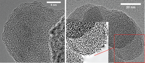
Thermophoretic sampling was achieved in this study for ex-situ characterization of the soot at the laser-border zones (not directly contacted by the LII laser). The analysis was aimed to determine whether the morphology and crystal structure of the soot at this neighboring zone changes during LII. The soot sampling setup consisted of a PLC (Master K series K7M-DR40S) programmed to trigger a relay device powering a solenoid valve. The solenoid valve was connected to lab air at 4 bar. When the solenoid valve is triggered, the valve opens and consequently actuates a Festo Double Action Pneumatic Roundline Cylinder, (DSNU-16-50-PPV-A). An adaptor was used to secure Dumont Style N5 tweezers to the cylinder. The tweezers directly gripped 300 mesh carbon coated copper grids during sampling. The sampling device was aligned to insert the probe directly through the flame and out the other side at approximately 60 mm HAB. The flame was extinguished immediately thereafter and the grid was retrieved. To trigger the PLC, a pulse generator (BNC Model 575) was used with a contact switch installed between the generator and the PLC.
Sampling was carried out while imaging the flame with a high-speed camera to verify that the probe passed through the LII affected region. shows time stamped (with respect to laser pulse) sampling of the soot at the laser-border zone upstream to the directly heated volume. The flame is shown in the center and is in orange color. The dark region to the right of the flame is due to shading issues with the camera. The probe enters the flame in the LII affected zone from the right hand side, traverses through the flame in ∼1 ms, and exits the flame. The probe can be seen to have passed through the flame in the last image where the fuel is shown being pushed out to the left of the flame. The synchronization of the LII pulse and the probe actuation was arranged in a way that the grid center enters the flame immediately after the heated volume passes the sampling zone. The probe, then, travels through the flame as quick as possible before the advection brings initially far upstream regions (that are loaded with unaffected fresh soot) into the sampling zone. Although the laser-border zone does not have a definite thickness, the high-speed images show that the grid center is exposed dominantly to the thin laser-border zone. The short sampling times and the piston moving in a single direction result in minimal flame perturbation. Samples were taken using an LII fluence of 1.13 J/cm2. To compare the possible permanent effects of LII, particles were also sampled without the presence of a laser pulse. At each condition, two samples were obtained. To prevent sample contamination, the tweezers were cleaned between samples using acetone. The cleaning method was tested and determined to be adequate by handling a TEM grid with cleaned tweezers and imaging the grid to search for particles.
Figure 3. Thermophoretic soot sampling was performed using a single action pneumatic piston to insert a TEM grid. High precision tweezers were used to hold the grid during sampling. A high speed camera was used to image the flame during sampling and the images at various time steps with respect to the initial laser prompt are displayed above. As the evaporated zone advects, the probe begins to enter the flame at approximately 6 ms, and exits at approximately 7.1 ms. Following the exit of the grid from the flame, the flame was extinguished and the sample collected.

Results
High-repetition rate imaging
shows the relative incandescence signal with respect to the flame luminosity at nine selected instants after the laser pulse (The first image before the laser pulse was subtracted from all the consecutive images). These profiles were extracted from the high-speed camera images along the flame axis (three neighboring pixel columns at the center were averaged horizontally). The original set of images were acquired at 120,000 fps and each image includes a time-integrated, i.e., gated, signal over a period of 8.3 µs. The prompt image at the time of laser pulse was saturated and is not shown in this analysis. The information is inherently line-of-sight integrated. Measurements were performed at different laser fluences and they are shown in different line colors. Regardless of fluence, the laser pulse causes a marking, i.e. a deviation of the incandescence signal at the heated and neighboring regions, which persists up to several hundreds of microseconds. At fluences above ∼0.4 J/cm2, these markings become permanent and can be tracked until the tip of the flame ( shows the signal only up to 55 mm HAB). The top left plot, 8.3 µs after the laser pulse, shows that the immediate marking is roughly 1.5 mm wide which is the same as the thickness of the horizontal laser sheet. As time marches, the marking travels towards the downstream due to advection. The permanent marking with the laser provides an opportunity for velocimetry in nanoparticle loaded laminar flows without additional tracers (Seitzman et al. Citation1999; Yang and Seitzman Citation2003; Yang et al. Citation2000). In the original works of the Seitzman group, the permanent marking was achieved by substantial vaporization of the soot. To ensure this vaporization, the marking laser fluence was recommended to be above 2—3 J/cm2, and in the actual studies much higher laser fluences (up to 20 J/cm2) were employed. In this current work, a trackable permanent marking was achieved in this non-premixed flame with much lower fluences via partial vaporization and annealing. To demonstrate this, a sample video acquired after heating with a 1 mm diameter laser beam (point-wise heating) at 0.4 J/cm2 laser fluence is added to online Supplemental Materials section – Visualization 2. The beam crosses the flame at 50 mm HAB and 1 mm off the flame axis. However, in an attempt to observe the flow velocity in a turbulent flame (Reynolds number 15,000) with the same technique it was not possible to extract useful information as the marking patterns dissipate too quickly and are not trackable.
Figure 4. Signal intensity profiles were obtained using a high speed camera. The profiles are an average of the three center pixels at 9 different times shown in the upper right hand of each image. The y axis shows the signal intensity difference between the flame with an LII pulse and the natural flame luminosity. The intensities were recorded at 10 different laser fluences shown on the right side of the image.
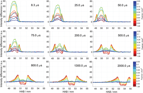
shows that the permanently affected volume is larger than the directly heated volume for laser fluences above ∼0.3 J/cm2. Immediately at the downstream and upstream of the heated volume, i.e., the laser-border zones, the incandescence signal increases systematically with increasing laser fluence. At delayed times, the signal profiles exhibit two obvious quasi-identical crests at these regions. Similarly, the relative signal at the directly heated volume differs more than the original luminosity with increasing fluences. The energy distribution irregularities within the beam profile (see ) causes an uneven heating of the soot in the direction of flame axis. This non-uniformity can be seen in signal profiles within the directly heated volume between 50 and 1300 µs time stamps.
Figure 5. Signal intensities from the heated soot were obtained using a high speed camera at 10 different laser fluences (shown on the right side). The relative intensities of the signal were tracked through time at 6 different locations in the flame. The tracked locations in the flame are shown in the upper left hand image. The images on the right show time in the x axis beginning at the laser prompt, and in the y axis the relative signal intensity between LII heated soot and the natural soot luminosity.
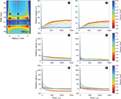
A negative value in indicates a loss of signal in the region with respect to the original flame luminosity before the laser pulse. Such reduction can be attributed to the soot mass loss, hence reduction of soot volume fraction, due to the sublimation of soot and/or oxidation. A positive value, on the other hand, indicates that the incandescence signal is higher with respect to the original flame luminosity. This increase can be attributed to an increase in the temperature, soot volume fraction, emissivity of the soot or a combination of these parameters. At high laser fluences, the laser heating of the in-flame particles is audible (in the form of a pop sound), indicating a sonic expansion of the sublimed clusters (Michelsen et al. Citation2003). In this study, such audible sound, hence expansion of sublimed soot, was observed at all fluences above 0.2 J/cm2. The increase in the soot volume fraction may be due to a possible compression of border zones induced by this expansion generated at the directly heated volume. The increased temperature in the affected volumes may cause accelerated annealing of particles, hence an increase in their emissivity. Furthermore, sublimed carbon clusters, which have high translational energy (Michelsen Citation2003), can escape the laser-heated region. When they reach the relatively lower temperature zones, they can either form new particles (Michelsen et al. Citation2007) or condense onto existing particles and increase their size. Such increase of the solid mass also causes an increase of the incandescence signal. However high-repetition rate imaging of incandescence signal, standalone, is not sufficient to show which of these parameters are dominant.
also shows that the midrange laser fluences cause a stronger long-lasting incandescence signal at the heated volume compared to low and high fluence experiments. In low fluence cases the incandescence signal is limited by the low residual temperatures, and at high fluence cases soot particles sublime rapidly and there is less material left to incandesce. An optimum fluence for the highest signal is around 0.4 J/cm2 for heating with 1064 nm.
Particle tracking
For a better understanding of the history of the heated volume and laser-border zones after the laser pulse, the incandescence signal from particles at six different locations were observed along their trajectory, and the results are shown in . The flame image on the left hand side was acquired 500 µs after the laser pulse and displays the location of six selected points. For this analysis the same image set introduced in the previous section was used. Location 1, 3 and 5 are at the flame edge where soot particles are more graphitic, and soot volume fraction and temperature are higher (López-Yglesias et al. Citation2014). Location 2, 4 and 6, on the other hand, represent soot at the flame center. It must be remembered that the signal coming from the axisymmetric flame is line-of-sight integrated and volumetric heating was applied. Therefore flame center locations also include information from the flame edge. However the contribution of relatively thinner edge layers on the overall signal is marginal.
Location 5 and 6 are within the directly heated volume. At the laser prompt image, both locations are saturated with low and midrange fluence heating, and the signal peaks can be seen in at time zero. At both locations the signal loss due to sublimation increases with increasing fluence, hence at high fluences the corresponding pixels at these two locations do not saturate. The persisting temperature increase and the continuous soot formation have competing effects on the long-lasting LII signal against the instantaneous sublimation. At midrange fluences the positive effects of temperature and soot formation overcome the negative effects of sublimation for up to 500 and 1200 µs at the edge and center locations, respectively. When soot is heated with fluences above ∼1 J/cm2, the absolute signal before subtracting the original flame luminosity at the flame edge (location 5) reaches absolute zero (level of background) within about 1 ms, whereas at location 6, a small residual signal persists.
Location 3 and 4 show the signal at the laser-border zones. At both locations, the absolute signal saturates for mid and high range fluences at the laser prompt image. Immediately after the laser prompt, a signal increase remains in these regions that is proportional to the laser fluence. The increase at the flame edge (location 3) is stronger than the center section (location 4). As mentioned in the previous section, the signal increase at the laser-border zone can be attributed to an increase in the temperature, soot volume fraction, emissivity of the soot or a combination of these parameters. This increased signal after the laser pulse gradually decreases in time. The rate of reduction varies for different laser fluences in the first few hundred microseconds, however it becomes identical after 400 µs in all cases.
Location 1 and 2 represent the far downstream of the directly heated volume. Repeating the particle tracking exercise in these locations is useful to understand the gradual signal decrease in Location 3 and 4. The added energy or material at the laser-border zones after the laser pulse slowly diffuse further downstream (or upstream for the lower part of the laser heated volume) and the incandescence signal gradually increases in these far downstream or upstream locations. Depending on the laser fluence, the slow increase in the signal stops after some time and these regions come to an equilibrium. The peak signal at the laser prompt is most likely due to the scattering of the broadband blackbody-like emissions, i.e., LII signal, generated at the directly heated volume.
Laser extinction method
shows the time-resolved transmittance of CW laser for each of the pulsed-laser fluences. The transmitted laser signals are normalized with respect to the incident beam signal (measured without the presence of flame and shown with the dashed black curve). At time zero, the broadband LII emission interferes and causes a sharp peak in the acquired signal (in the case of incident beam measurements, the interference originates from the interaction of the laser pulse and the metal aperture).
Figure 6. Laser transmittance measured by the light extinction setup. The extinction measurements were taken 7 mm downstream from the LII laser pulse. The laser transmittance was measured using 12 different laser fluences. The time on the x axis starts just prior to the initial LII laser prompt.
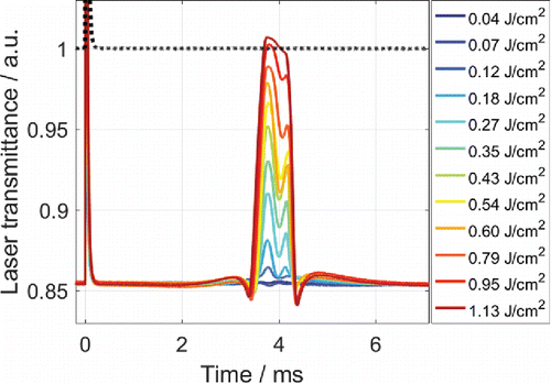
The LII affected region advects to the HeNe laser probe volume. This affected region shows three different features in the transmittance analysis. First, at the far downstream and upstream region of the heated volume, the transmittance signal gradually increases with the increasing pulsed-laser fluence. The transmittance at the upstream of the heated volume (the transmittance data between 4.6 and 6 ms) is higher and spread broader than the transmittance in the downstream (the transmittance data between 2.2 and 3.2 ms). The second feature is the decrease in laser transmittance in the laser-border regions. The decreased transmittance in the border regions indicates either an increase in soot volume fraction in these locations or an increase of the soot absorption function, E(m), that can be induced by graphitization of the soot. The third feature is the strong and sharp increase in the laser transmittance along the directly heated volume that can be associated to a sublimation-induced mass loss, hence reduction of the soot volume fraction. The increase of transmittance, thus reduction of soot volume fraction, is proportional to the pulsed-laser fluence. The variation of the transmittance signal along the heated volume is again due to the non-uniform spatial profile of the heating laser. In the case of the two highest laser fluences, the transmittance signal reaches up to the level of the incident beam. This means that the soot in the probe volume is (almost) completely sublimated and the HeNe laser light is not subjected to any attenuation. It also shows that the recondensation of sublimed carbon clusters and formation of new soot particles are negligible in the directly heated volume. The transmittance signal exceeding the incident beam signal is within the total system uncertainty.
Temperature
shows that after rapid heating, the temperature increase in the region directly contacted by the LII laser persists for several milliseconds. At high fluence heating, the residual temperature increase 200 µs after the laser pulse was measured as ∼400 K at the flame edge and ∼300 K at the flame center. The temperature gradually decreases as the heated volume advects towards the downstream. The persisting elevated temperature indicates that the energy transferred rapidly from the particles to the surrounding (via conduction and sublimed particles) causes a temperature increase of the bath gas, and the equilibrium between the particles and the bath gas is established at a higher temperature than without the laser. Nordström et al. (Citation2015) measured a 100 K temperature increase in the bath gas immediately after the LII pulse for low and mid-range fluence with a rotational CARS system. The measurements in this current work show this bath-gas heating at much longer time scales and higher laser fluences.
Figure 7. Flame temperature measurements were obtained using two different laser fluences (shown in the upper left corner) between approximately 50 and 60 mm HAB. Each image is time stamped at the bottom and a temperature scale is shown on the right side.
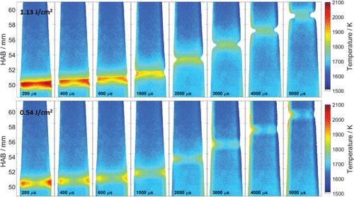
shows that the residual temperature increase is higher for the case of high-fluence laser heating at all-time delays compared to the mid-range fluence. This indicates that there is more energy pumped into the directly heated volume. In both laser fluences, the temperature increase is greater at the flame edge zone because the initial soot volume fraction is higher in this region and there is more soot heating the bath gas. At increased delays, the signal at these flame edge zones are completely lost due to the enhanced oxidation. This phenomenon will be explained further in detail in the discussion section (see and related explanation). The pyrometry images also show that the temperature at the laser-border zones increases after the LII pulse. The increase is more subtle compared to the directly heated volume, and dissipates completely after 3 and 5 ms for mid- and high-range fluences, respectively.
HRTEM
HRTEM analysis was completed using a TITAN Themis™ microscope. Micrographs of soot samples were extracted only from the grid center at various magnifications. Images were manually analyzed using the freeware ImageJ. In , HRTEM micrographs of soot sampled without laser heating (left image) and with laser heating (right image) are shown. Without the laser heating, several soot particles were found with amorphous structures and randomly oriented crystallites (left image). Such particles are known to be young and less graphitic. In the case where soot was sampled in the laser-border region, soot particles were found that were composed of thin graphite crystallites aligned parallel to the surface (and more randomly oriented near the core) (Michelsen Citation2017). These features can be seen in the right image in . For a better illustration of these well-ordered spherical crystallite layers, the area in the red square is further zoomed, converted into a binary image, and a sharpening filter was applied. The background was smoothed to give a higher contrast to the image. Changing of lattice structure and conversion of the graphite crystallites into well-ordered form are known to be results of aging and defect annihilation. Thermal annealing, which may be induced or accelerated by elevated temperature in the laser-border zones (see previous section), may be effective in such transitions. It should also be noted that the image on the left does not represent the entire micrograph set for the sampling without the laser. Particles with well-ordered peripheral lattice were also found. However, in the case of sampling with the laser, all imaged soot lacked fully random oriented structures. The full set of TEM images from both cases is available online and can be found in the Supplemental Materials section – Visualization 3 and 4 (with and without laser heating, respectively).
Discussion
Bath-gas heating
To understand the time scales of energy transfer mechanisms during LII, the energy- and mass-balance equations (Cenker and Roberts Citation2017) are solved for the flame edge zone at 50 mm HAB. In the simulations, the initial temperature of soot, hence the temperature of the bath gas, is set to 1750 K (López-Yglesias et al. Citation2014), the 1064-nm laser fluence is set to 0.3 J/cm2, and bath-gas heating is not considered. The particle reaches 99% of thermal equilibrium in less than 4 µs after the laser pulse. Two-color pyrometry imaging in the current work shows that the temperature increase in the LII affected zone persists up to several milliseconds. This indicates that the energy transferred from the particles to the surrounding actually heats up the bath gas, therefore the equilibrium between the particles and the bath gas is established at a higher temperature. Bath-gas temperature is crucial information in particle sizing with LII modelling. Will, Schraml et al. (Citation1998) showed that a 10% error in this model input can cause more than 30% error in the calculated particle size. Bath-gas heating is proportional to the soot volume fraction. In a case where the soot volume fraction in the measurement volume is extremely low, or with low fluence laser heating, the total energy transferred from the particle to the gas will be very low and bath-gas heating will not be substantial.
Pyrometry imaging in this work also shows that the temperature is partially responsible for the incandescence signal increase at the laser-border zones shown in and . The added energy into the volume directly contacted by the laser dissipates quickly into the neighboring regions and the temperature in these border zones increases. After a few milliseconds, the heat, hence the temperature, dissipates further down- and upstream in the flow. It should be re-emphasized that the temperature measurements in this work were performed with a constant E(m) (it only differs for different wavelengths). The temperature changes shown in may be partially due to an aftereffect of a change in the E(m) induced by thermal annealing and aging of soot. Ex-situ analysis of soot with the HRTEM shows evidences that the crystal structure of the soot tends to change at the elevated zone temperature and may be due to annealing (López-Yglesias et al. Citation2014; Michelsen Citation2003). We believe that both residual temperature increase and the emissivity change account for the increase in the incandescence signal.
Soot volume fraction
LEM measurements and high-speed images demonstrate that during the process of LII and afterwards, a much larger region is affected than the directly heated volume. shows that optical thickness of the flame increases and decreases at different distances to the directly heated volume. These variations in the optical thickness of the flame can be attributed to a change in the E(m) of the soot (Michelsen et al. Citation2010), a change in the soot volume fraction or a combination of both. In the previous section, it is mentioned that change of E(m) due to annealing can partially be a reason for these alterations.
When soot is rapidly heated with a high-fluence laser, a popping sound is generated due to the sonic expansion of the sublimed clusters as mentioned earlier. This expansion of the gas can deform the flow and transport the soot in the direction of expansion. Furthermore, rapidly heated soot can be pulled towards the neighboring zones, which are initially much cooler, via thermophoretic forces. These different mechanisms may cause a change of the soot volume fraction in the laser-border zones further up- or downstream of the directly heated volume. Measurements at different laser fluences show that the sublimation of soot, hence the increase of the transmitted signal in the directly heated volume, takes place even at 0.12 J/cm2. López-Yglesias et al. (Citation2014) measured values for sublimation thresholds in the same flame at 50 mm HAB (with 1064 nm laser heating) at 0.08 J/cm2 at the edge, and 0.1 J/cm2 in the center. A similar sublimation threshold was also measured previously by Saffaripour et al. (Citation2015) in a similar non-premixed ethylene/air flame.
Oxidation
Oxidation of soot was first considered in an LII model by (Michelsen Citation2003). Although it has a minor contribution to overall changes in the mass and energy of soot in conventional LII experiments, it may have a significant impact on soot over longer time scales. Oxidation rates are strongly correlated with the temperature. A soot particle (or an aggregate) remaining at elevated temperatures after the laser pulse undergoes more rapid oxidation.
The history analysis of the incandescence signal after a high fluence laser pulse (see ) shows that the signal at the flame edge is seen to fade out with time and eventually disappear completely after a few milliseconds. For a better visualization of this, the exercise of tracking soot particles is repeated for all the soot radially distributed in a narrow band (150 µm thick in the axial direction) in the middle of the directly heated volume after heating with 1.26 J/cm2 laser fluence. Compared to point-wise tracking, this analysis provides two-dimensional information. The information is again extracted from the high-speed images. The results are shown in . The absolute signal intensity is presented in false color scale and shown in the color bar on the right. White color represents absolute zero (the magnitude of the signal from the dark field). To compare the evolution of the laser-affected soot with the unheated soot, the convective boundary streamlines of the same region from the original flame are relayed on the plot (dashed black lines). To achieve this, the height of the flame (288 pixels) is scaled to the number of time points (166 frames) through linear interpolation.
Figure 9. Tracking of radially-distributed soot after a high-fluence laser pulse using high speed imaging. The x axis is radial distance and the y axis is time after the LII laser pulse. Particle tracking was performed in the center of the zone directly heated by the LII laser and the absolute signal intensity was mapped. The black dashed line represents the convective boundary streamlines of the flame without laser heating.
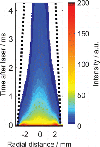
In , it can be seen that soon after the laser, the diameter of the heated soot band starts to narrow. At 4 ms after the pulse, a faint incandescence signal is detected only at the vicinity of the flame axis. The complete loss of the signal in an incandescence image can be due to a severe temperature drop (the radiation is roughly proportional to the fifth power of temperature in the Rayleigh approximation) or a mass loss that can be induced by sublimation or oxidation. The spatially- and temporally-resolved temperature information in shows that the temperature in the flame edge zone remains higher after the pulse (it is however not high enough to further sublime the soot). The higher temperature in this zone provides favorable conditions for enhanced oxidation. Furthermore the flame edges begin to curve inwards as the time scale increases forming a dent due to the enhanced oxidation. The oxidation can be seen at the edges due to the higher surface area at these locations. We, therefore, think that the signal loss in longer time scales is principally associated to oxidation of soot. Oxidation is an exothermic reaction and it can be partially responsible for the higher equilibrium temperatures in the LII-affected region. Oxidation begins at the outer most layer of the flame, where the oxygen-to-fuel ratio is highest. It then propagates toward the center of the flame as is the case for diffusion of oxygen into the flame.
Conclusion
Long-term and permanent effects of rapid heating on soot particles and carrying bath gas were investigated. High-repetition rate images of in-flame soot were acquired for a duration of 5 ms after volumetric heating with a pulsed laser at various fluences. Supplementary laser extinction measurements, two-color pyrometry imaging, and ex-situ characterization of soot were performed. The following conclusions are drawn from the measurements:
| 1. | In a conventional pulsed LII measurement, the sensitivity of the signal detection system is optimized for the peak signal which is significantly higher than the signal at or close to the thermal equilibrium. Such settings provide a signal lifetime on the order of hundreds of nanoseconds. A very high sensitivity setting was selected in this work at the expense of detector saturation around the peak signal timing allowing the LII signal to be detected at much longer time scales with a high-repetition rate camera. | ||||
| 2. | Heating of soot with a laser pulse results in a permanent marking, i.e., an altering of the incandescence signal, in a laminar flow. With high-speed imaging, the laser-affected zone can be tracked within the flow allowing flow velocity measurements to be made. It is shown that such permanent marking, through partial vaporization annealing, can be achieved with much lower laser fluences than the previously recommended fluence values in the literature. It is shown that this velocimetry method can also be applied with a high-repetition camera without intensifier. The method fails to extract velocity information in high Reynolds number turbulent flows. | ||||
| 3. | High-speed images show that at high fluence (>0.3 J/cm2) LII experiments, a much larger region is affected than the directly heated volume. Temperature, soot volume fraction and the emissivity of soot particles are subjected to change in the neighboring regions to the laser heated volume. | ||||
| 4. | The energy transferred from the particles to the surroundings heat up the bath gas, and the equilibrium between the particles and the bath gas is established at a higher temperature than the pre-laser state. The total energy transferred is proportional to the soot volume fraction. Therefore, the temperature gain of the bath gas is greater in the flame edge zone. | ||||
| 5. | The optical thickness of the directly heated soot starts to decrease at a fluences above 0.12 J/cm2, which can be attributed to sublimation of soot. The magnitude of this reduction gradually increases with increasing laser fluence. Elevated temperatures may change the emissivity of soot. This contributes to the uncertainty of the soot volume fraction and temperature measurements. | ||||
| 6. | After the rapid heating, the residual increased temperature enhances the oxidation of soot. It starts at the oxygen-rich outer edge of the flame and propagates towards the center of the flame. | ||||
| 7. | Thermophoretic sampling of soot was achieved with a TEM grid, which was gripped with a tweezer, moving in a single direction. The total sampling time (residence of the grid inside the sooting region) was around 1 ms. High-speed images show that sampling with this method limits flame perturbations. This extremely short sampling time (compared to previous exercises in literature) is still sufficient to collect soot for TEM analysis. | ||||
| 8. | The findings demonstrate that laser heating for LII measurements is invasive to the flame and soot and should be considered when analyzing LII data, particularly when coupling different measurements. | ||||
UAST 1368444 Supplementary File: Visualisation
Download Zip (15.5 MB)Acknowledgments
The research reported in this publication was supported by funding from King Abdullah University of Science and Technology (KAUST).
References
- Bambha, R. P., Dansson, M. A., Schrader, P. E., and Michelsen, H. A. (2013). Effects of Volatile Coatings on the Laser-induced Incandescence of Soot. Appl. Phys. B, 112:343–358.
- Cenker, E., Kondo, K., Bruneaux, G., Dreier, T., Aizawa, T., and Schulz, C. (2015). Assessment of Soot-particle Size Imaging with LII at Diesel Engine Conditions. Appl. Phys. B, 119:765–776.
- Cenker, E., and Roberts, W. L. (2017). Quantitative Effects of Rapid Heating on Soot Particle Sizing Through Analysis of Two-pulse LII. Appl. Phys. B, 123:74.
- Dankers, S., and Leipertz, A. (2004). Determination of Primary Particle Size Distributions from Time-resolved Laser-induced Incandescence Measurements. Appl. Opt., 43:3726–3731.
- De Iuliis, S., Cignoli, F., Maffi, S., and Zizak, G. (2011). Influence of the Cumulative Effects of Multiple Laser Pulses on Laser-induced Incandescence Signals from Soot. Appl. Phys. B, 104:321–330.
- Kuhn, P. B., Ma, B., Connelly, B. C., Smooke, M. D., and Long, M. B. (2011). Soot and Thin-filament Pyrometry Using a Color Digital Camera. Proc. Combust. Inst., 33:743–750.
- Liu, F., Snelling, D. R., Thomson, K. A., and Smallwood, G. J. (2009). Sensitivity and relative Error Analyses of Soot Temperature and Volume Fraction Determined by Two-color LII. Appl. Phys. B, 96:623–636.
- López-Yglesias, X., Schrader, P. E., and Michelsen, H. A. (2014). Soot Maturity and Absorption Cross Sections. J. Aerosol Sci., 75:43–64.
- Melton, L. A. (1984). Soot Diagnostics Based on Laser Heating. Appl. Opt., 23:2201–2208.
- Michelsen, H. A. (2003). Understanding and Predicting the Temporal Response of Laser-induced Incandescence from Carbonaceous Particles. J. Chem. Phys., 118:7012–7045.
- Michelsen, H. A. (2017). Probing Soot Formation, Chemical and Physical Evolution, and Oxidation : A Review of in Situ Diagnostic Techniques and Needs. Proc. Combust. Inst., 36:717–735.
- Michelsen, H. A., Liu, F., Kock, B. F., Bladh, H., Boiarciuc, A., Charwath, M., … Suntz, R. (2007). Modeling Laser-induced Incandescence of Soot: A Summary and Comparison of LII Models. Appl. Phys. B, 87:503–521.
- Michelsen, H. A., Schrader, P. E., and Goulay, F. (2010). Wavelength and Temperature Dependences of the Absorption and Scattering Cross Sections of Soot. Carbon N. Y., 48:2175–2191.
- Michelsen, H. A., Schulz, C., Smallwood, G. J., and Will, S. (2015). Laser-induced Incandescence: Particulate Diagnostics for Combustion, Atmospheric, and Industrial Applications. Prog. Energy Combust. Sci., 83:333–354.
- Michelsen, H. A., Tivanski, A. V., Gilles, M. K., van Poppel, L. H., Dansson, M. A., and Buseck, P. R. (2007). Particle Formation from Pulsed Laser Irradiation of Soot Aggregates Studied with a Scanning Mobility Particle Sizer, a Transmission Electron Microscope, and a Scanning Transmission X-ray Microscope. Appl. Opt., 46:959–977.
- Michelsen, H. A., Witze, P. O., Kayes, D., and Hochgreb, S. (2003). Time-resolved Laser-induced Incandescence of Soot: The Influence of Experimental Factors and Microphysical Mechanisms. Appl. Opt., 42:5577–5590.
- Musculus, M. P. B., and Pickett, L. M. (2005). Diagnostic Considerations for Optical Laser-extinction Measurements of Soot in High-pressure Transient Combustion Environments. Combust. Flame, 141:371–391.
- Musculus, M. P. B., Singh, S., and Reitz, R. D. (2008). Gradient Effects on Two-color Soot Optical Pyrometry in a Heavy-duty DI Diesel Engine. Combust. Flame, 153:216–227.
- Nordström, E., Olofsson, N.-E., Simonsson, J., Johnsson, J., Bladh, H., and Bengtsson, P.-E. (2015). Local Gas Heating in Sooting Flames by Heat Transfer from Laser-heated Particles Investigated Using Rotational CARS and LII. Proc. Combust. Inst., 35:3707–3713.
- Saffaripour, M., Geigle, K.-P., Snelling, D. R., Smallwood, G. J., and Thomson, K. A. (2015). Influence of Rapid Laser Heating on the Optical Properties of In-flame Soot. Appl. Phys. B, 119:621–642.
- Santoro, R. J., Semerjian, H. G., and Dobbins, R. A. (1983). Soot Particle Measurements in Diffusion Flames. Combust. Flame, 51:203–218.
- Seitzman, J. M., Wainner, R. T., and Yang, P. (1999). Soot-velocity Measurements by Particle Vaporization Velocimetry. Opt. Lett., 24:1632–4.
- Snelling, D. R., Liu, F., Smallwood, G. J., and Gülder, Ö. L. (2004). Determination of the Soot Absorption Function and Thermal Accommodation Coefficient Using Low-fluence LII in a Laminar Coflow Ethylene Diffusion Flame. Combust. Flame, 136:180–190.
- Snelling, D. R., Smallwood, G. J., Liu, F., Gülder, Ö. L., and Bachalo, W. D. (2005). A Calibration-independent Laser-induced Incandescence Technique for Soot Measurement by Detecting Absolute Aight Intensity. Appl. Opt., 44:6773–6785.
- Tea, G., Bruneaux, G., Kashdan, J. T., and Schulz, C. (2011). Unburned Gas Temperature Measurements in a Surrogate Diesel Jet Via Two-color Toluene-LIF Imaging. Proc. Combust. Inst., 33:783–790.
- Vander Wal, R. L., and Choi, M. Y. (1999). Pulsed Laser Heating of Soot: Morphological Changes. Carbon N. Y., 37:231–239.
- Vander Wal, R. L., and Jensen, K. A. (1998). Laser-induced Incandescence : Excitation Intensity. Appl. Opt., 37:1607–1616.
- Vander Wal, R. L., Ticich, T. M., and Stephens, A. B. (1998). Optical and Microscopy Investigations of Soot Structure Alterations by Laser-induced Incandescence. Appl. Phys. B, 67:115–123.
- Will, S., Schraml, S., Bader, K., and Leipertz, A. (1998). Performance Characteristics of Soot Primary Particle Size Measurements by Time-resolved Laser-Induced Incandescence. Appl. Opt., 37:5647–5658.
- Witze, P. O., Gershenzon, M., and Michelsen, H. A. (2005). Dual-Laser LIDELS: An Optical Diagnostic for Time-Resolved Volatile Fraction Measurements of Diesel Particulate Emissions. SAE Tech. Pap., 2005-01-3791: 1-23.
- Witze, P. O., Hochgreb, S., Kayes, D., Michelsen, H. A., and Shaddix, C. R. (2001). Time-resolved Laser-induced Incandescence and Laser Elastic-scattering Measurements in a Propane Diffusion Flame. Appl. Opt., 40:2443–52.
- Yang, P., and Seitzman, J. M. (2003). Soot Concentration and Velocity Measurement in an Acoustic Burner, American Institute of Aeronautics & Astronautics (AIAA) 2003-1014. In 41th Aerospace Science Meeting and Exhibition, Atlanta, GA.
- Yang, P., Seitzman, J. M., and Wainner, R. T. (2000). Particle Vaporization Velocimetry for Soot-Containing Flows. American Institute of Aeronautics & Astronautics (AIAA) 2000-0645. In 38th Aerospace Science Meeting and Exhibition, Reno, NV.
- Yoder, G. D., Diwakar, P. K., and Hahn, D. W. (2005). Assessment of Soot Particle Vaporization Effects During Laser-induced Incandescence with Time-resolved Light Scattering. Appl. Opt., 44:4211–4219.
- Zhao, H., and Ladommatos, N. (1998). Optical Diagnostics for Soot and Temperature Measurement in Diesel Engines. Prog. Energy Combust. Sci., 24:221–255.

