Abstract
One issue that has persisted since the beginning of what might be referred to as a modern era of biological aerosol research (since the 1990’s), is absence of reference biological aerosols that would permit quantitative comparison among different experimental studies. We believe sufficient technical progress has been made bridging the diverse fields of biology, chemistry, physics and engineering to consider implementing well-characterized biological aerosols as reference standards. Establishment of methods and procedures which result in reliable and repeatable generation of well-characterized biological aerosols would enhance a wide range of different topics under investigation, and permit wider utility for data acquired from individual efforts. In this article we discuss some of the challenges and limitations for two general approaches for biological aerosol generation: solvent evaporation from liquid suspension droplets, and dry powder dispersal. We provide detailed descriptions of an example for each of these two approaches in which sufficient control is demonstrated over particle size distribution, total particle composition, biological constituent quantification and biological state (viability or enzymatic activity) to serve as a comparison among different experimental investigations. These two specific cases are intended as examples, not necessarily as prescriptions.
EDITOR:
Introduction
A main objective of this article is to discuss and promote the concept of reference standards for certain biological aerosols that would permit comparison of experimental results across independent investigations. This discussion is motivated by two perceptions: first, a lack of uniformity of how experimental efforts involving biological aerosols are reported in scientific literature, and second, the absence of established sample preparation and aerosol generation methods permitting direct comparison of results among different experimental investigations. The topic concerns production (generation) of aerosol particles that have biological components: either microorganisms or biochemical materials, such as oligonucleotides, proteins, etc. Such aerosols are relevant to a broad range of applications including: improved understanding of naturally occurring bioaerosol generation, their transport and dispersion in the atmosphere, inhalational delivery and absorption of pharmaceuticals/medications, and development of detectors for pathogenic or hazardous biological aerosols. As a caveat to the reader, this manuscript was not intended as a comprehensive review, and while we attempt to reference representative examples, we make no claim to provide detailed comparisons among the very long list of specific bioaerosol generation methods, and/or prior studies.
A specific goal is to promote the concept of standardized biological aerosol(s) that would permit comparison among different study results. In this context, a reference standard implies extending deterministic control over sample selection, sample preparation and aerosol generation method, so that the net outcome is reliably repeatable as a well-characterized aerosol. A distinction can made between control and characterization of a resulting biological aerosol, and specific materials and methods for a given aerosol. Perhaps this is best expressed by an analogy: it would be difficult to imagine the current state of aerosol research without the availability of monodisperse micro- and nano-spheres as a reliable resource for well-characterized particles. Ubiquitous use of such microbead samples has made them de facto reference standards over the entire aerosol research enterprise. Nevertheless, it is clear that the numerical value of such a bead sample diameter, its molecular composition, and/or the specific aerosol generation method are not critical in order to achieve utility for aerosol research calibration and inter-comparison.
With regard to materials, any molecular vapors or gasses that result from biological processes (e.g., respiration), or a transient water vapor loss due to desiccation would be excluded since our focus is on aerosols. This distinction implies that nucleation, as a means for aerosol generation would be eliminated from consideration. Consequently, potential generation methods are narrowed down to essentially two broad categories: droplet dissemination (with subsequent solvent evaporation) or dry powder dispersal. In this article we discuss general considerations for both of these approaches, and illustrate each with a specific case we have developed and demonstrated as a precondition for a specific research objective. Consequently, the specific cases described here are not intended to be either exclusive or exhaustive, but rather, as examples to demonstrate feasibility. Establishing a perspective of controlled and characterized bioaerosol composition is, in our view, more important than specific sample materials.
Aerosol generation by droplet evaporation
Sample materials for biological aerosols are more commonly and perhaps most easily prepared as liquid suspensions. An important point in reporting bioaerosol study results is that the complete protocol and sample preparation process should be included or referenced. Very often, biological samples employ known standard recipes for additive materials to provide a benign environment for the biological element under study. Examples of such support materials include solutions such as phosphate buffered saline (PBS), normal saline, or growth media such as trypticase soy broth, or beef heart extract. These are just a few of many possible additives that can either be made from prescription or obtained from commercial sources. We recently experienced a case where commercially available culture media with the same listed ingredients and proportions, but from different vendors, showed significantly differing growth rates for the same organism sample aliquot and conditions. So, even seemingly small details such as specific sources can be important to report.
After sample preparation, the next step is selection and description of the droplet generation method. There is a very wide range of either commercially available devices, or descriptions for device fabrication, to achieve small droplet dissemination. Most of these methods will produce droplets with a broad range of diameters, but some may associate a specific range of droplet size with specific operating conditions. Spray devices with a broad continuum of initial droplet diameters frequently can be characterized with log-normal distributions (Grainger 2017). As one such example, the Collison nebulizer (CH Technologies, USA; [email protected]; http://chtechusa.com/products_tag_lg_collison-nebulizer.php; May Citation1973) has been popular, especially in aerobiological (animal exposure) studies for determining dose response to pathogens, partly because its primary droplet size is limited to relatively small droplets (< 10 µm) (Thomas et al. Citation2009) and because of its simple operation. When considering microorganisms as bioaerosols, another concern for pneumatic nebulizers such as the Collison is their damage to target microorganisms caused by high mechanical stresses (Reponen et al. Citation1997; Thomas et al. Citation2011). More recently, the degree of damage to organisms caused by aerosolization using a Collison has been quantified (Zhen et al. Citation2013); (Ibrahim et al. Citation2015) as well as comparison among selected devices (Zhen et al. Citation2014). Additionally, there are a number of methods (and some commercial devices) designed to provide very narrow or monodisperse (geometric mean standard deviation <1.25) initial droplets and provide a degree of control over the nominal droplet size (see, e.g., Lin, Eversole, and Campillo Citation1990; Kesavan et al. Citation2014; Waldrep and Dhand Citation2008; Kuo et al. Citation2019). Electrospray devices can also be operated in a monodisperse droplet mode (cone jet mode) (Tang and Gomez Citation1994; Eninger et al. Citation2009).
Regardless of which droplet generation method is chosen, the initial droplet dissemination is followed by a transient period in the flow where the solvent (usually water) evaporates from the droplet and a final residue particle is formed. The overall transformation from liquid suspension to droplet aerosol, and then to residual aerosol particles is depicted in . For the purposes of a general concept, we have illustrated our sample suspension by including a target microorganism (red dots), and potentially other suspended solids (green squares) as well as dissolved nonvolatile or low-volatility (persistent) chemicals that remain as liquid residue (yellow fill of the final particle) after the solvent evaporates. As a specific example, consider organisms suspended in PBS. In principle, the initial suspension would consist only of organisms in a solution; but in practice, there may be trace quantities of other solids such as cell fragments from the sample preparation process. Additionally, as droplets are formed and water evaporates from them, the salt concentration in the droplet increases, and completion of this process likely results in deliquescence where salts will precipitate to form new solid materials. The green squares in , then, represent either of these potential sources of solid materials in the final residue particle. This example also illustrates an additional point: sample constituents beneficial to the biological component initially, may become deleterious due to rapidly increasing salinity or pH as droplet evaporation occurs.
Figure 1. The process of bioaerosol generation from a liquid sample microdroplet dispersion is depicted schematically from left to right. Droplets formed from a homogeneous suspension, result in individual particle compositions that reflect the relative proportions of the original sample.

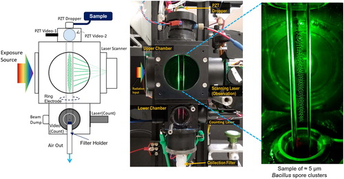
Since our objective is to highlight bioaerosol reference standards, it follows that achieving as much control as possible over the final aerosol composition and particle size distribution (PSD) is desirable. As one example of moving toward that objective, we briefly describe a recent research effort that required a simulant of biological agent, in which Bacillus anthracis Sterne strain (BA Sterne) spores were selected as the target biological aerosol component. As a brief overview, the experimental arrangement is depicted in as a composite set of three images. Starting on the left is a schematic drawing of the main apparatus components (from top to bottom): a piezoelectric transducer (PZT) droplet generator, an upper aerosol chamber with windows for observation and radiation exposure, a lower aerosol chamber for particle counting, and last, a filter-based particle sample collection assembly. The middle photo of the actual apparatus shows a close one-to-one correspondence with the elements in the schematic. At right is a close-up photo of the interior of the aerosol chamber showing a typical sample of Bacillus spore cluster aerosol particles. The assembled system is air-tight, and purged with a gas flow. Since this experiment is being used to illustrate biological aerosol preparation and characterization, only a brief description is provided here, as more detail and complete results will be reported separately in future reports. For this experiment it was necessary to generate and hold a population of biological aerosol particles to measure the effects of ultraviolet (UV) radiation exposure on their viability. Aerosol particles were charged and generated by droplet evaporation, and then held in the upper chamber using a linear electrodynamic quadrupole (LEQ) trap for specified periods of time while being exposed to UV radiation from sources outside the chamber. After exposure, the suspended particles were slowly released to the lower chamber while still confined by the LEQ electrodes where they were counted individually as they were collected onto a filter surface. Since the average number of spores per particle is determined from the sample spore concentration and the initial droplet volume, the total number of spores can be estimated from the particle count. Filters were subsequently removed for sample recovery into liquid, plating and culturing to determine survival fractions.
Figure 2. The schematic on the left shows the configuration of a piezoelectric transducer droplet generator, and a linear electrodynamic quadrupole trap aerosol chamber divided into upper and lower sections by an electric gate (ring electrode). A photo of the apparatus is shown in the middle. The photo on the right is an enlarged view of the interior of the upper chamber showing a suspended spore particle sample (between 200 and 300 particles).

Bacillus spores are atypical among bacterial organisms in that they offer perhaps the greatest degree of hardiness for a wide range of environmental factors, including aerosolization. Our spore samples were prepared at the U. S. Army Chemical and Biological Center (CBC), Edgewood, MD under the U. S. Army Combat Capabilities Development Command. The samples were delivered in a water suspension as washed, bright-phase spores with 0.01% (v/v) Tween 80 (polysorbate 80) added to mitigate coagulation and adherence to container walls. Each sample from CBC was provided with a specification sheet including a titer of the spore concentration, and these details are in the online supplementary information. To prepare samples for droplet generation the spore suspensions were centrifuged, supernatant removed and resuspended in 90% isopropanol, 10% water to reduce the initial droplets’ evaporation time, and the Tween concentration to 0.001%. (The effect of isopropanol on spore viability was determined to be insignificant before these experiments commenced.) Independent titers of sample solutions were performed for every experimental data collection. Stock sample titers also remained consistent, showing no degradation over the period of the experiment.
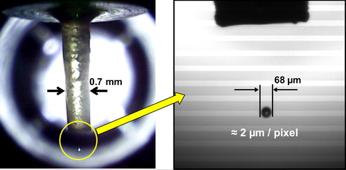
A PZT droplet generation method was chosen to provide highly monodisperse and reproducible droplets with minimal mechanical stress on the spores. While a complete PZT droplet generator system can be obtained commercially from more than one source, our specific system was a constructed from components mostly purchased from MicroFab Technologies Inc. (Plano, TX; http://www.microfab.com). We specified a 70 µm quartz capillary orifice with their (proprietary) hydrophobic coating on the nozzle tip. The system comes with a video camera to provide feedback and a back-pressure controller to improve droplet size stability. We customized our system, replacing the software generated pulse source with a standalone pulse generator, and installed a second camera with higher magnification and image resolution. Particle illumination is achieved with a 37 µsec white LED pulse synchronized to the PZT, so that the droplet images have no apparent motion blur. Images from both cameras are recorded for every run, and over a recent set of runs (≈ 47) at a constant spore sample concentration, the average droplet diameter was 68 µm with a standard deviation of ± 4 µm. This on-demand process can be used to generate either individual droplets or a series of droplets at a constant rate. In our case, a series of droplets are produced, usually in short bursts of about one second duration, to create an aerosol particle population in the upper chamber. The PZT pulse rate (droplet production rate) is nominally 2 kHz, so the chamber filling process is typically completed in a minute or so. Once the chamber is at capacity (typically 200 to 500 particles), droplet production is stopped until the next experimental run is initiated. During a given droplet production run, the drop-to-drop diameter variability is estimated to be less than 1% due to slowly changing environmental conditions. However, run to run variations often have larger absolute droplet diameter variation. shows typical images from each of the video cameras. The one on the left depicts the capillary nozzle (outside diameter ≈ 700 µm, inside diameter 70 µm), while the one on the right shows a higher resolution image with the nozzle silhouette above the droplet. Both these images are acquired at 30 Hz frame rates and each image is actually an overlay of about 66 droplets illustrating little jitter.
Figure 3. Two camera images of piezoelectric transducer droplet formation process are shown. The left photo shows the quartz capillary (70 µm ID), and the right photo shows a higher resolution image of typical droplets.

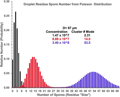
In these experiments the spore sample concentration is adjusted to effectively control the resulting aerosol particle size. Since the droplet size is directly measured during each experiment, it is straightforward to estimate the average number of spores per droplet by simply multiplying the spore concentration by the droplet volume. However, at low concentrations with such small volumes (nominally 1.65 × 10⁻⁷ ml), the statistical variation of the number of spores in any given droplet becomes an important consideration. Conceptually, for such a small volume within the total sample fluid volume, the number of spores inside that volume is continuously in flux as the spores undergo diffusion by Brownian motion. A basic underlying hypothesis is that all locations within the sample volume are equally likely to be occupied by a spore; i.e., the probability density for any spore in the sample vial is uniform. Under this condition the number of spores inside a small sub-volume is described by a Poisson probability. Consequently, the number of spores in each individual droplet as it detaches from the capillary tip varies around a mean value given by the concentration multiplied by the droplet volume. For large numbers of droplets, the numbers of spores in each droplet does converge to the predicted (Poisson) distribution. shows computational results of Poisson probability distributions for the number of spores in a 67 µm diameter droplet at three specific concentrations: 3.40 × 10⁸, 8.89 × 10⁷ and 1.47 × 10⁷ spores/ml (color-coded as blue, red and black respectively). These target spore sample concentrations effectively produced three distinct experimental PSDs.
Figure 4. Computed frequency of occurrence, or probability distributions, of the number of spores per droplet are shown for a 67 µm diameter droplet at three target concentrations as indicated in the color-coded legend.

As each droplet evaporates and dries, the spores will coalesce to a single aerosol particle (spore cluster). The probability distributions in appear broad because they are plotted versus spore number. Resultant spore cluster PSDs will, in fact, be relatively narrow since the equivalent volume of the spore cluster varies as the cube root of the spore number. As example, for an average cluster size with 54 spores, the Poisson standard deviation of about ± 7.3 spores results in a cluster diameter and variation of ≈ 4.3 ± 0.2 µm based on an assumed packing density of 0.55. Consequently, the three cluster size distributions will be well-separated in particle size. Concurrent work was performed at CBC using the same spore samples at similar concentrations and droplet sizes. For concentrations of 3.8 × 10⁸ and 8.9 × 10⁷ spores/ml, dried aerosol PSDs were measured using a TSI Aerodynamic Particle Sizer (APS; TSI Inc., Shoreview, Minnesota; https://www.tsi.com), and their results showed relatively narrow PSDs (geometric standard deviations ≈ 1.54) with modes at aerodynamic diameters of 4.0, and 2.5 µm respectively. For their small particle sample, they used a significantly lower concentration of spores to reduce the probability of particles with multiple spores, and their measured aerodynamic diameter was ≈ 1.2 µm, consistent with the size of a single spore. Moreover, representative SEM images of the larger spore clusters captured on polycarbonate track-etched filters exhibited an approximately spherical shape, as would be expected from droplet evaporation formation.
Where Poisson distribution statistics have greatest consequence is at the smallest, or “single-spore” cluster size. For the lowest concentration in , one can see that the frequency of occurrence for single spores has a probability of about 23% while the probability for doublets and triplets are roughly 27% and 21% respectively, and there is a 10% chance that a droplet will contain no spores. In order to achieve an aerosol population of predominantly single-spores (less than 1% doublets and higher multiples) would also require that ≈ 90% of the droplets generated would have no spores present. Experimentally, this is problematic because of the presence of 0.001% Tween in the original droplet solution. As shown in , the soluble, but nonvolatile, substances remain in the residue particle. An amount of Tween equivalent to 0.001% of 1.65 × 10⁻⁷ ml could leave a residue droplet of Tween that is ≈1.5 µm in diameter, and consequently “no-spore” droplets could still leave a Tween residue particle (microdroplet) that is about the same size and mass as an individual spore. This same amount of Tween is present with all larger spore clusters as well, but for clusters with modes at 14 and 54 spores, the Tween mass is a negligible fraction of the total. However, at low concentrations, our objective to have a sample of single-spore particles is thwarted by the presence of Tween droplets, since we have not found a simple way of differentiating between Tween-only and spore-containing particles. Fortunately, for our objectives, it was possible to use model predictions of photodegradation in spore clusters, to show that small clusters up to 4 or 5 spores will have equivalent absorption properties as single spores. Consequently, generating a distribution as shown in with a mode of 2.4 spores should be adequate for obtaining viability degradation data within our experimental uncertainties.
Dry powder aerosol generation
Association of powder dispersion with aerosol reference standards may at first invoke historical use of mineral dusts from specified geographical (geological) sources for application to industrial filter testing. This is an important commercial application which eventually became an established standard (ISO 12103-1:1997, 2016) for such “test dust” or “road dust” powder products (Powder Technology Inc.). However, such empirical methodology is not likely to be practical for biological aerosols since there are no consistent and abundant natural sources of biological powder.
At a high level, two main aspects of powder aerosolization can be differentiated: (1) dispersion methods and (2) sample powder preparation/characterization processes. These two aspects are not completely independent since, in general, different dispersion methods may be adapted specifically for different types of powder properties.
Regarding powder dispersion methods, aerosolization can be considered as a transformation of the powder primary PSD into a resulting aerosol PSD and concentration. If the powder material is conceptually regarded as having an intrinsic (“as received” or “as produced”) primary PSD, then in principle a mathematical transfer function can be invoked that maps the sample powder PSD into the final aerosol PSD. To illustrate this, consider as a special case, a powder composed of similar-sized basic units. Its primary PSD will consist of these fundamental units together with aggregates or agglomerates of basic units formed by physical contact. This is a main reason why powder materials typically exhibit very broad primary PSDs. If the dispersal mechanism(s) imparts sufficient energy to overcome aggregate/agglomerate binding forces, then the resulting aerosol PSD would have a lower median size than the powder PSD due to aggregate/agglomerate breakup. However, it is important to keep in mind that not all processes will reduce median particle size, and for some powders, milling or aerosolization mechanisms may exert mechanical compaction that tends to increase aerodynamic diameter, rather than decrease it.
While a transformation view of powder dispersal provides a useful conceptual framework for connecting powder particles to aerosol particles, generally, it is not possible to identify and assign a transfer function to a given dispersal method because of the interaction with, and dependence of, the final aerosol PSD on the powder sample properties. Additionally, for some designs (for example, the Pitt Generator [AlburtyLab, Inc., Model AG5025, Drexel, MO; http://alburtylab.com/our-expertise/our-products/]) and others with fixed powder samples, a time-dependent PSD for a given sample is a concern. Commercially available powder aerosol generators vary from simple Venturi-based eductor nozzles to fluidized beds to more complex devices such as the Vilnius Aerosol Generator or Wright Dust Feeder (CH Technologies Inc.) that collectively employ a wide range of different mechanisms to impart energy and gas to a powder sample to achieve aerosol dispersion. In some cases, multiple mechanisms are combined, and it is not obvious how much each contributes to the overall process. A common issue with powder dispersal is time-dependent aerosol concentration, and potentially concentration-dependent PSDs. Further detail regarding powder dissemination can be found in published literature such as Son, Worth Longest, and Hindle Citation2013; and Tiwari, Fields, and Marr Citation2013.
Preparation and processing procedures are important factors for powders in general, but especially so for biological materials. While most aerosol applications/investigations could be adequately characterized at some point in time by determination of a PSD and total concentration, aerosols of biological composition have the additional dimensions of biochemical or taxonomic identity, and viability (or biological activity) that will be necessary to measure for complete characterization. For living organisms, the powder preparation process will require freeze-drying, or lyophilization, and in most cases this will require additives to mitigate the degree of mortality from this step.
Given these complexities, the idea of a powder sample with well-controlled biological aerosol generation in the context of our original objectives might seem inherently difficult. For this reason, we thought it important to include one example in which well-characterized biological aerosols were achieved using powder dispersion. While there is normally a mortality penalty to freeze-drying (lyophilization), that can range from as little as 20%, up to 90% or more depending on the organism and procedures (e.g., Dewald Citation1974; Bircher et al. Citation2018), one exception to this effect is Bacillus spores which are metabolically inactive and inherently hearty. For the example that we describe below, we used Bacillus thuringiensis Al Hakam strain (BtAH) since, our research objectives were related to biological agent defense, and a simulant organism was required. In this example, sample preparation was carried out at the Naval Surface Warfare Center, Dahlgren, VA, and growth and sporulation protocol are available in detail elsewhere (Buhr et al. Citation2012). Bright-phase spore samples were washed in water, examined by SEM for quality control, and resuspended with a small amount of dispersant added. This slurry suspension was then lyophilized, and the resultant material was macerated and blended with sufficient silica additive to coat the individual spores in order to mitigate significant agglomeration. A final step of jet milling was used to increase the bulk powder density. Greater detail of the lyophilization and powder processing will be provided as a separate report which is in preparation. Once the powder was delivered to NRL, no further process steps were taken other than to check titer viability constancy of the spore powder.
For aerosol characterization, three commercially available powder generators initially were selected for evaluation as representing a range of dispersal mechanisms. They were: the Palas Rotating Brush Generator (RBG, Model 1000 LGD, Karlsruhe, Germany; https://www.palas.de/en/product/rbg. [U.S. Distributor: CH Technologies, Inc. http://chtechusa.com/products_tag_spg_rotating-brush-generator-rgb.php]), the Vilnius Aerosol Generator (VAG, single head) (http://chtechusa.com/products_tag_spg_vilnius-series-vag.php), and the TSI Small Sample Powder Disperser (SSPD 3343) (https://www.tse.com). While all of these devices produced similar PSDs for a given powder sample, they suffered from operational difficulties for bioaerosol generation that ultimately forced us to design and fabricate a custom dispersal device to carry out and complete our project. Our custom powder disperser mechanism was similar to the SSPD, and was referred to as the turntable Venturi disperser (TVD). All of these devices addressed the issue of time-dependent PSD and concentration uniformity adequately well. For the SSPD, the most serious issue was lack of containment; a non-negotiable flaw for BSL-2 aerosol generation. For the other two units the most serious issue was cross-contamination among samples, since they were extremely difficult to thoroughly clean. Consequently, the TVD was used to for final data collection since it was air-tight, very easy to clean and decontaminate compared to the commercial units, and capable of maintaining reliability, reproducibility, and low concentration temporal variability.
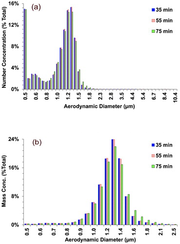
An objective of our study was to evaluate aerosol disperser output results under conditions similar to an outdoor or indoor setting with relatively low prevailing wind speeds. Since the disperser is using relatively high velocity gas flows to dilute the powder aerosols, characterization was conducted in a calm air chamber (CAC) (Feather and Chen Citation2003) in which the disperser output was allowed to equilibrate into a flow of HEPA-filtered room air of much larger volume, but much slower mean velocity. The low velocity aerosol/air mixture was completely contained inside a sealed chamber in which the output flow was double HEPA filtered. Measurements of the diluted disperser aerosol at a quiescent position in the chamber are shown in showing peak aerodynamic diameter at 1.2 to 1.3 µm which corresponds to the size of individual BtAH spores. These results are plotted from TSI APS data, and numerical analysis shows that 99% of the particle count is below an aerodynamic diameter of 2.0 µm; meaning the aerosol was essentially single spores. Data plots in show data integrated over five-minute intervals at three different times during a single continuous release: at 35, 55 and 75 min. These times were selected arbitrarily to illustrate consistency of the aerosol PSD over time. In , low, but finite, counts are seen from 2 µm out to 10 µm, showing that some agglomerate particles were produced. Using these particle counts, equivalent sphere volumes were computed from the measured data to calculate an estimated mass distribution, which is shown in , Integration of this distribution shows that 98% of the total aerosol mass is smaller than 2.0 µm, i.e., as single spores. Finally, notice that the count distribution is bimodal with a secondary peak at approximately 0.58 µm. This smaller peak matches independent measurements of the silica powder used to mitigate agglomeration in the spore powder sample. This suggests that a small fraction of the sample aerosol produced consists of excess silica particles that did not adhere to spore surfaces, but contributes a negligible amount to the total aerosol mass.
Figure 5. (a) Particle aerodynamic diameter distribution of the Bacillus thuringiensis Al Hakam powder sample material was measured with a TSI Aerodynamic Particle Sizer. Normalized data obtained with the Vilnius Aerosol Generator shows a consistent PSD shape at three arbitrary times up to 75 min. (b) Using the number count data, an equivalent sphere mass distribution vs. aerodynamic diameter was computed, showing that 98% of total aerosol mass is accounted for by aerodynamic diameters less 2.0 µm. An expanded horizontal scale allows a clearer view of data.

Summary and conclusions
We have described methods and results from two experimental investigations in which Bacillus spores were used to generate biological aerosols. Details relevant to aerosol generation and characterization have been included in an effort to promote/encourage consideration of practices that could be adopted to provide reference aerosols for comparison of biological aerosol data among different studies, experiments, investigations, tests and evaluations. The first example employed generation of liquid droplets of suspended Bacillus spores which subsequently dry by evaporation to produce a final aerosol. The second example prepared a powdered material of Bacillus spores and dispersed the powder to generate an aerosol. In both these cases the resultant aerosol was characterized, and demonstrated reliability and repeatability in terms of composition, PSD and viability. These examples lead us to believe that by exercising careful control over protocol for the preparation of Bacillus spores as well as subsequent steps in the sample preparation, a reliable and repeatable generation of a well-characterized bioaerosol can be achieved.
While this may seem like a relatively simple objective, the defense community concerned with development of biological warfare defense and sensor development has suffered from a lack of standardization in terms of materials, processes, procedures, and characterizations for biological aerosols necessary for test and evaluation since the mid-1990’s. Perhaps the most familiar example is the use of “BG” aerosols in outdoor releases for the purpose of evaluating developmental bioaerosol detection systems. The term “BG” is an acronym and was originally a reference to Bacillus globigii spores as aerosols. However, in a variety of official tests over time, from one year to the next, different species and strains or variants of Bacillus bacteria were used, procured from different vendors with a range of protocols for production, as well as procedures for processing the spore material into powders. These powdered samples were dispersed as simulant aerosols for biological agents. In hindsight, it is obvious that in order to make consistent progress in detection system development, a reference point in terms of a test aerosol would be necessary in order to obtain experimental results that could be compared with earlier biological aerosol test data. While there may have been legitimate technical barriers in establishing standards for Bacillus species selection, sporulation protocol, processing and dispersion 20 years ago, it is our opinion that it is overdue now.
At this point we have confidence that standardized processes for repeatable bioaerosol generation could be achieved. Important characteristics of such aerosols would include:
controlled, well-defined, and sufficiently narrow PSD,
controlled, well-defined particle composition and
quantified viability.
Within this context, the examples described in this article are sufficient. Here, the inherent hardiness of the Bacillus spores was used to provide essentially no loss of viability during sample storage, upon aerosolization, or as aerosols over periods of time consistent with conducting instrument tests or experiments on aerosol transport and dispersion. The composition of the aerosol particles should be simply the spores themselves with as minimal quantity of additives as practical. The resultant aerosol PSD will depend on whether droplet generation & evaporation, or dry powder dispersion, is chosen. For the powder sample, the resultant aerosol should be the same as the primary particle size of the powder sample, which in this case, would be essentially single spores as shown in . For the liquid droplet method of aerosol generation, an additional degree of control was demonstrated in terms of selecting a particle size (mode of the PSD) via spore concentration in the liquid sample. In this case, the width of the resultant PSD is limited by fundamental statistical properties, as discussed previously (see ). As a practical matter, for particles up to a few microns in diameter, this inherent variability will result in PSDs that, while perhaps not technically monodisperse, are nevertheless narrow. In the high concentration example shown in . the mean particle size has an SD of only ±4%. In conclusion, establishing reference standards will require a broad base of consensus and support, and confidence in a standardized method can only be established by demonstrating reliability and repeatability over time. While the examples described here may not constitute a final solution to bioaerosol generation standardization, we hope they represent a step forward on that issue, and provides a foundation for future discussion in the research community.
The authors also wish to acknowledge the efforts of the special edition editor, Dr. Tiina Reponen, of this issue: Bioaerosol Research: Methods, Challenges, and Perspectives, and also organizers of the Bioaerosol Working Group on which it is based, as well as a Bioaerosol Standardization Workshop at the International Aerosol Conference in St Louis, MO (September, 2018), with special thanks to Dr. Shanna Ratnesar-Shumate and Dr. Alex Huffman.
Supplemental Material
Download MS Word (15.8 KB)Additional information
Funding
References
- Bircher, L., A. Geirnaert, F. Hammes, C. Lacroix, and C. Schwab. 2018. Effect of cryopreservation and lyophilization on viability and growth of strict anaerobic human gut microbes. Microb. Biotechnol. 11 (4):721–33. doi:10.1111/1751-7915.13265.
- Dewald, R. R. 1966. Preservation of Serratia marcescens by high-vacuum lyophilization. J. Appl. Microbiol. 14 (4):561–67.
- Eninger, R. M., C. J. Hogan, Jr., P. Biswas, A. Adhikari, T. Reponen, and S. A. Grinshpun. 2009. Electrospray versus nebulization for aerosolization and filter testing with bacteriophage particles. Aerosol Sci. Technol. 43 (4):298–304. doi:10.1080/02786820802626355.
- Reponen, T., K. Willeke, V. Ulevicius, S. A. Grinshpun, and J. Donnelly. 1997. Techniques for dispersion of microorganisms into air. Aerosol Sci. Technol. 27 (3):405–21. doi:10.1080/02786829708965481.
- Waldrep, J. C., and R. Dhand. 2008. Advanced nebulizer designs employing vibrating mesh/aperture plate technologies for aerosol generation. Curr. Drug Deliv. 5 (2):114–9. doi:10.2174/156720108783954815.
- Buhr, T. L., A. A. Young, Z. A. Minter, C. M. Wells, D. C. McPherson, C. L. Hooban, C. A. Johnson, E. J. Prokop, and J. R. Crigler. 2012. Test method development to evaluate hot, humid air decontamination of materials contaminated with Bacillus anthracis ΔSterne and B. thuringiensis Al Hakam spores. J. Appl. Microbiol. 113 (5):1037–51. doi:10.1111/j.1365-2672.2012.05423.x.
- Feather, G. A., and B. T. Chen. 2003. Design and use of a settling chamber for sampler evaluation under calm-air conditions. Aerosol Sci. Technol. 37 (3):261–70. doi:10.1080/02786820300946.
- Grainger, R. G. 2017. Some Useful Formulae for Aerosol Size Distributions and Optical Properties. Accessed June 19, 2019. http://eodg.atm.ox.ac.uk/user/grainger/research/aerosols.pdf. [Notable for being both concise and clear: Log-normal – an expression for the frequency of occurrence or probability density function as a two-parameter model analogous to the classic normal, or Gaussian, distribution; but which avoids complications of truncated tails for zero and unmeaningful negative particle radius values.]
- Ibrahim, E., D. Harnish, K. Kinney, B. Heimbuch, and J. Wander. 2015. An experimental investigation of the performance of a Collison nebulizer generating H1N1 influenza aerosols. Biotech. Biotechnol. Equip. 29 (6):1142–8. doi:10.1080/13102818.2015.1059736.
- ISO 12103-1:1997. 2016. Accessed June 19, 2019. https://www.iso.org/standard/63386.html
- Kesavan, J. S., J. R. Bottiger, D. R. Schepers, and A. R. McFarland. 2014. Comparison of particle number counts measured with an ink jet aerosol generator and an aerodynamic particle sizer. Aerosol Sci. Technol. 48 (2):219–27. doi:10.1080/02786826.2013.868594.
- Kuo, Y.-M., W.-H. Chan, C.-W. Lin, S.-H. Huang, and C.-C. Chen. 2019. Characterization of vibrating mesh aerosol generators. Aerosol Air Qual. Res. 19 (8):1678–87. doi:10.4209/aaqr.2018.11.0436.
- Lin, H.-B., J. D. Eversole, and A. J. Campillo. 1990. Vibrating orifice droplet generator for precision optical studies. Rev. Sci. Instrum. 61 (3):1018–23. doi:10.1063/1.1141470.
- May, K. R. 1973. The Collison nebulizer: description, performance and application. J. Aerosol Sci. 4 (3):235–43. doi:10.1016/0021-8502(73)90006-2.
- Son, Y. J., P. Worth Longest, and M. Hindle. 2013. Aerosolization characteristics of dry powder inhaler formulations for the excipient enhanced growth (EEG) application: effect of spray drying process conditions on aerosol performance. Int. J. Pharmaceutics 443 (1-2):137–45. doi:10.1016/j.ijpharm.2013.01.003.
- Tang, K., and A. Gomez. 1994. Generation by electrospray of monodisperse water droplets for targeted drug delivery by inhalation. J. Aerosol Sci. 25 (6):1237–49. doi:10.1016/0021-8502(94)90212-7.
- Thomas, R. J., D. Webber, R. Hopkins, A. Frost, T. Laws, P. N. Jayasekera, and T. Atkins. 2011. The cell membrane as a major site of damage during aerosolization of Escherichia coli. Appl. Environ. Microbiol. 77 (3):920–5. doi:10.1128/AEM.01116-10.
- Thomas, R. J., D. Webber, W. Sellors, A. Collinge, A. Frost, A. J. Stagg, S. C. Bailey, P. N. Jayasekera, R. R. Taylor, S. Eley, et al. 2009. Characterization and deposition of respirable large- and small-particle bioaerosols. Appl. Environ. Microbiol. 74 (20):6437–43. doi:10.1128/AEM.01194-08.
- Tiwari, A. J., C. G. Fields, and L. C. Marr. 2013. A cost-effective method of aerosolizing dry powdered nanoparticles. Aerosol Sci. Technol. 47 (11):1267–75. doi:10.1080/02786826.2013.834292.
- Zhen, H. J., T. Han, D. E. Fennell, and G. Mainelis. 2013. Release of free DNA by membrane-impaired bacterial aerosols due to aerosolization and air sampling. Appl. Environ. Microbiol. 79 (24):7780–9. doi:10.1128/AEM.02859-13.
- Zhen, H. J., T. Han, D. E. Fennell, and G. Mainelis. 2014. A systematic comparison of four bioaerosol generators: Affect on culturability and cell membrane integrity when aerosolizing Escherichia coli bacteria. J. Aerosol Sci. 70:67–79. doi:10.1016/j.jaerosci.2014.01.002.
