Abstract
Objective. To analyse the outcome of exercise testing and myocardial perfusion scintigraphy (MPS) in primary care patients with chest pain of new onset. Design. Prospective, observational. Patients aged 20–79 years, consulting due to chest pain of new onset, were enrolled consecutively. Setting. Three primary care health centres in south-eastern Sweden. Patients. 191 patients where the possibility of stable ischaemic heart disease (IHD) could not be excluded by clinical examination alone. Main outcome measures. Exercise test results, when equivocal completed by MPS. Results. Exercise testing revealed IHD in 14 (7%) and no IHD in 134 (70%) of the cases. In 43 (23%) the exercise test results were equivocal. Thirty-nine of these patients underwent MPS, which showed IHD in 19 and no IHD in 20 cases. Among previously diagnosed cardiovascular disease and risk factors only atrial fibrillation in the male group showed a significant correlation to the outcome IHD. Conclusion. Exercise testing and MPS are both useful when investigating chest pain patients in primary care.
Exercise testing and myocardial perfusion scintigraphy (MPS) are both well established methods for diagnosing coronary artery insufficiency. The strength of these methods is well proven in patients with intermediate and high probability of coronary artery disease but little is known about the outcome of these methods in a primary care population with chest pain of new onset. Furthermore, the influence of cardiovascular risk factors and heart-related morbidity on the probability of ischaemic heart disease (IHD) has not been elucidated in this patient group.
In a previous report Citation[1] we have described a population of patients who consulted the primary care service for chest pain of new onset. In approximately 40% of cases, the general practitioner (GP) could not rule out stable IHD and this group therefore needed further investigation. The aim of this prospective observational study was to observe the outcome of exercise testing and, when required, MPS in this population. An additional aim was to investigate the impact of cardiovascular risk factors and previous manifestations of heart-related morbidity on the probability of IHD.
The assessment of patients with chest pain represents a daily challenge for the general practitioner. The outcome of exercise testing and myocardial perfusion scintigraphy is not well documented in primary care.
Both exercise testing and myocardial perfusion scintigraphy were indicated as being useful means of investigating chest pain patients.
In this setting, cardiovascular history and traditional risk factors were largely unhelpful in separating patients with and without IHD.
Material and methods
Study population
Patients aged 20 to 79 years, who consulted three primary care units in Sweden from May 1998 until April 2000, were consecutively included. All patients consulted due to chest pain of new onset as their chief presenting complaint. This was defined as pain, pressure, ache, burning, or stabbing sensation in the chest that had commenced during the last 6 months. Patients with previously confirmed stable coronary insufficiency were excluded, as were those who had, during the previous year, suffered an acute myocardial infarction or had been subjected to revascularization.
The general practitioners (GPs) reviewed the patients’ medical records with regard to risk factors (diabetes, smoking, hypertension, or hyperlipidemia) and other manifestations of cardiac-related morbidity (atrial fibrillation, congestive heart failure, heart valve disease, previous myocardial infarction or revascularization, peripheral or cerebrovascular disease manifestations). Patients where the GP's clinical judgement dismissed IHD as the reason for the chest pain were followed up by questionnaires Citation[1]. Cases of suspected unstable coronary heart disease were referred directly to hospital. Those presenting with symptoms of stable coronary disease were referred for an exercise test.
The study was approved by the ethics committee of the Faculty of Health Sciences of Linköping University.
Exercise testing
Exercise tests were performed using a bicycle ergometer. The maximal workload was appraised by calculating the percentage of normal workload specific for weight and gender Citation[2]. All exercise tests were conducted by the same clinical physiologist physician according to national guidelines Citation[1], Citation[3]. The results of the exercise tests were categorized into three groups:
IHD: ST-segment depression exceeding 0.1 mV (absolute depression) and angina-like chest pain in relation to exercise or pathological Q-wave on resting ECGs.
No IHD: Neither chest pain nor ECG changes.
Equivocal test result: Chest pain but no ST changes during or after exercise or vice versa – no chest pain but ECG changes. This group also included patients with non-assessable ECG reactions, due to e.g. left bundle branch block or digitalis medication.
Exercise myocardial perfusion scintigraphy
Patients with equivocal test result (group 3) were referred for MPS. The stress study was performed on day one, using a bicycle exercise test and technetium-99 m tetrofosmin as perfusion agent. On day two (within one week after stress MPS), the tetrofosmin preparation was administered to the resting patient. For gamma camera acquisition and post-processing, a GE STARCAM 3000XR/T was used. The acquisition was performed as a single photon emission computed tomography study. No scatter or attenuation correction was used.
Image interpretation
Two experienced observers made a blinded semi-quantitative visual interpretation of the stress and rest study, using a 13-segment heart model Citation[4]. After individual interpretation, consensus was achieved and used for evaluation of the present study. All patients with MPS demonstrating ischaemia and/or a myocardial scar were categorized as “IHD”, the remaining as “No IHD”.
Statistical methods
Student's t-test or chi-squared test was used to analyse continuous variables related to exercise test parameters and the main reasons for referral to MPS, respectively. Univariate logistic regression was used to test the relation of risk factors and details of cardiac-related morbidity to IHD, according to study endpoints. For controlling influence of gender the patients were divided into a male and a female group. Univariate logistic regression was used to select explanatory variables among risk factors and details of cardiac-related morbidity. If the explanatory variable was significant, it was included in multivariate logistic regression in the male and female group, respectively. The level of significance was taken as p<0.05. The analyses were conducted using StatView (version 5.0.1) and SPSS (release 11.5.1).
Results
Patient flow
Of 38 075 GP consultations during the study period, 577 consultations by 554 patients were included. In 295 consultations, the GPs ruled out IHD and 58 acute referrals to hospital were made on the basis of suspicion of acute coronary syndrome.
In 224 consultations, stable IHD could not be ruled out. The mean age of these patients was 60 years, range 26–79, only four patients being below the age of 40. In 10 of the consultations, the patient was judged unsuitable to undergo exercise testing and 16 did not attend when called. In seven consultations, there was no follow-up due to administrative failure. In total, 191 consultations by 191 patients were followed up by exercise testing.
Exercise testing
A total of 191 patients – of whom 106 were men – underwent an exercise test. At the exercise test, 44 patients (23%) were on treatment with beta-blocking agents. describes the exercise test parameters related to conclusive or equivocal exercise test results. No significant differences in the exercise parameters, age, or gender were present between these groups. After exercise testing, 134 patients (70%) were classified as “No IHD” and 14 (7%) as “IHD” (). In the remaining 43 patients (23%), the test results were categorized as equivocal. These patients were referred for myocardial scintigraphy. Thirteen individuals were referred because of chest pain in spite of normal exercise ECGs, 14 had pathological exercise ECGs but no chest pain, 10 patients had indeterminable exercise ECGs, and two were referred due to blood pressure drop during exercise test. In the first three groups, there was no statistical significance related to the final distribution between the “IHD” and the “No IHD” groups. Neither could any statistical significance be demonstrated when age, gender, and solitary parameters (maximal heart rate, maximal workload, ST depression, and occurrence of chest pain), from all the equivocal exercise tests, were tested against “IHD” and “No IHD”.
Figure 1. Results from exercise tests in 191 primary care patients consulting their GP for chest pain of new onset and results from myocardial perfusion scintigraphy in 39 patients with equivocal exercise test results.
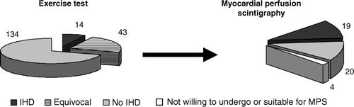
Table I. Bicycle exercise test parameters for patients with conclusive and equivocal exercise tests.
Myocardial perfusion scintigraphy
Of the 43 patients with indeterminable exercise tests, three patients were judged to be unsuitable for exercise perfusion imaging and one patient declined further testing. In the scintigraphy group, 10 patients (26%) were treated with beta-blocking agents. 87% of the patients reached a maximal heart rate of at least 85% of that expected according to age (220 – age in years).
After MPS, 20 (51%) patients fulfilled the criteria for “No IHD” while 19 (49%) were classified as “IHD” (see ): Six of these patients had ischaemia, two of whom also had myocardial scarring, and 13 patients had only myocardial scarring.
Risk factors and cardiac-related morbidity
Risk factors and cardiac-related morbidity details were extracted from the medical records and tested for their relation to IHD according to study endpoints in 187 patients. Univariate analyses showed that age, male gender, previous myocardial infarction, previous revascularization, and atrial fibrillation were significant according to the allocation to IHD or no IHD ().
Table II. Distribution of cardiac-related morbidity in the entire examined patient population (n=187) according to the results after exercise test and myocardial perfusion scintigraphy.
Only five female patients were classified as having IHD. Due to that, no further analyses were performed in the female group. Univariate analyses in the male group showed significance for age OR 1.06 (1.01–1.11) and atrial fibrillation OR 10.09 (1.90–53.59), according to the allocation to IHD or no IHD. Only atrial fibrillation remained significant in the multivariate analyses OR 6.17 (1.04–36.73).
Discussion
According to the results of the exercise tests there were no signs of IHD in 70% of chest pain patients; IHD was confirmed in 7% and was still in question in another 23%. In these 23%, MPS indicated IHD in half of the patients.
The primary strength of this study is the prospective setting and the large number of consultations during the two years of patient inclusion. The small number of IHD patients in the risk factor subgroups is in itself a finding that elucidates the epidemiology of chest pain in primary care.
In general, the sensitivity and specificity of the exercise test for the diagnosis of coronary artery disease are reported Citation[5] to be slightly less than 70% and 80%, respectively. However, in a recent study by Sumanen et al. Citation[6], patients with normal exercise tests conducted by GPs in primary care have proved to be associated with a favourable prognosis. Moreover, Niemann et al. Citation[7] have shown a significantly higher risk of cardiac events and death in patients with inconclusive exercise tests as compared to normals. This finding elucidates the necessity of further investigation in such patients. Furthermore, a correct diagnosis is of consequence when it comes to indications for pharmacological secondary prevention.
In this study we have not included any traditional gold standard method, such as coronary angiography. The main reason for this is that the aim of the study was not to test the accuracy of the exercise test or MPS but rather to investigate the prevalence of IHD in a primary care sample of chest pain patients, according to the results of exercise testing and, when needed, MPS. A bulk of literature has repeatedly warranted an excellent cardiac prognosis in patients with normal MPS Citation[8–10] even irrespective of the findings of coronary angiography Citation[11].
In our study, 43 exercise tests (23%) were designated as equivocal. This may seem to be a high number but in a comparable study by Mark et al. Citation[12] and in the multicentre study by Gibbons et al. Citation[8] the number of equivocals after exercise test was as high as 34–57%. Nearly half of the patients who underwent MPS showed signs of IHD. This is noteworthy but we judge it to be the result of the extensive selection process these patients went through. Gibbons Citation[8] and his group also found an abnormal MPS in 47% of their patients with an equivocal exercise test. The “IHD” group consists of patients with signs of ischaemia (pathological stress test, respective reversible defects on MPS), as well as myocardial scarring (pathological Q-wave on rest ECG or fixed defects on MPS). We chose to include patients with scarring in the “IHD” group because of the negative prognostic prospects of this group. Elhendy et al. Citation[13] have shown increased risk of death in patients with suspected IHD and fixed defects on MPS. Furthermore, several studies have shown viability by Positron Emission Tomography (PET) Citation[14–16] in a considerable number of patients with fixed defects on MPS.
With regard to risk factors and cardiac-related morbidity, only one factor showed statistically significant correlation to IHD among men: atrial fibrillation. We assume that the explanation for this finding is the low cardiovascular morbidity in a primary care milieu.
Limitations
A much larger number of patients would undoubtedly have yielded a significant correlation between risk factors for or history of cardiovascular disease and the outcome IHD. Nevertheless, our results underline the limited usefulness of this for decision-making in the individual primary care patient with chest pain in a patient population with low prevalence of both IHD and risk factors. Although not performing coronary angiography for anatomical confirmation of IHD may seem a limitation the rationale for our design has been described above. The healthcare system concerned, and to what extent patients primarily seek hospital or primary care, would influence the inclusion of patients and the outcome; otherwise there was no selection bias.
Conclusion
In patients with chest pain of new onset in whom the GP could not rule out coronary insufficiency as the reason for their symptoms, scarcely 20% showed signs of IHD. In addition, a cardiovascular history or traditional risk factors were largely unhelpful in separating patients with and without IHD due to the low prevalence of IHD as well as the risk factors. This stresses the importance of testing the diagnostic procedures and their outcome in the setting in which they are to be applied. These findings indicate the usefulness of exercise testing in primary care, with referral for supplementary myocardial perfusion scintigraphy when the exercise test is equivocal.
Acknowledgements
The authors would like to thank colleagues and staff at the Departments of Clinical Physiology, Norrköping County Hospital and Linköping Heart Centre, University Hospital of Linköping, at the primary health centres of Valdemarsvik, Vikbolandet and Åby, and at the Research & Development Unit, Local Health Care, County of Östergötland, for their contribution to the study. This study was supported by grants from the County Council of Östergötland, Stig and Ragna Gorthon's Foundation and from the Swedish Heart and Lung Foundation.
References
- Nilsson S, Scheike M, Engblom D, Karlsson LG, Molstad S, Akerlind I, et al. Chest pain and ischaemic heart disease in primary care. Br J Gen Pract 2003; 53: 378–82
- Nordenfelt I, Adolfsson L, Nilsson JE, Olsson S. Reference values for exercise tests with continuous increase in load. Clin Physiol 1985; 5: 161–72
- Brauer K, Jorfeldt L, Brudin L. Det kliniska arbetsprovet [The clinical exercise test]2nd ed. Studentlitteratur, Lund 2003
- Svane B, Bone D, Holmgren A, Landou C. Polar presentation of coronary angiography and thallium-201 single photon emission computed tomography: A method for comparing anatomic and pathologic findings in coronary angiography with isotope distribution in thallium-201 myocardial SPECT. Acta Radiol 1989; 30: 561–74
- Gibbons RJ, Balady GJ, Bricker JT, Chaitman BR, Fletcher GF, Froelicher VF, et al. ACC/AHA 2002 guideline update for exercise testing: Summary article. A report of the American College of Cardiology/American Heart Association Task Force on Practice Guidelines (Committee to Update the 1997 Exercise Testing Guidelines). J Am Coll Cardiol 2002; 40: 1531–40
- Sumanen M, Jussila M, Mattila K. Exercise treadmill test may predict clinical outcome among working-aged patients suspected of coronary heart disease in general practice. Scand J Prim Health Care 2005; 23: 47–51
- Niemann T, Labouriau R, Sorensen HT, Thorsgaard N, Nielsen TT. Five-year cardiovascular and all-cause mortality, and myocardial infarction in all subjects referred for exercise testing in two Danish counties. Scand Cardiovasc J 2004; 38: 137–42
- Gibbons RJ, Hodge DO, Berman DS, Akinboboye OO, Heo J, Hachamovitch R, et al. Long-term outcome of patients with intermediate-risk exercise electrocardiograms who do not have myocardial perfusion defects on radionuclide imaging. Circulation 1999; 100: 2140–5
- Shaw LJ, Hendel R, Borges-Neto S, Lauer MS, Alazraki N, Burnette J, et al. Prognostic value of normal exercise and adenosine (99m)Tc-tetrofosmin SPECT imaging: Results from the multicenter registry of 4,728 patients. J Nucl Med 2003; 44: 134–9
- Beller GA, Zaret BL. Contributions of nuclear cardiology to diagnosis and prognosis of patients with coronary artery disease. Circulation 2000; 101: 1465–78
- Brown KA, Rowen M. Prognostic value of a normal exercise myocardial perfusion imaging study in patients with angiographically significant coronary artery disease. Am J Cardiol 1993; 71: 865–7
- Mark DB, Shaw L, Harrell FE, Jr, Hlatky MA, Lee KL, Bengtson JR, et al. Prognostic value of a treadmill exercise score in outpatients with suspected coronary artery disease. N Engl J Med 1991; 325: 849–53
- Elhendy A, Schinkel AF, van Domburg RT, Bax JJ, Poldermans D. Prognostic significance of fixed perfusion abnormalities on stress technetium-99m sestamibi single-photon emission computed tomography in patients without known coronary artery disease. Am J Cardiol 2003; 92: 1165–70
- Yoshinaga K, Katoh C, Noriyasu K, Yamada S, Ito Y, Kuge Y, et al. Low-dose dobutamine stress gated SPET for identification of viable myocardium: comparison with stress-rest perfusion SPET and PET. Eur J Nucl Med Mol Imaging 2002; 29: 882–90
- Bonow RO, Dilsizian V, Cuocolo A, Bacharach SL. Identification of viable myocardium in patients with chronic coronary artery disease and left ventricular dysfunction: Comparison of thallium scintigraphy with reinjection and PET imaging with 18F-fluorodeoxyglucose. Circulation 1991; 83: 26–37
- Dilsizian V, Arrighi JA, Diodati JG, Quyyumi AA, Alavi K, Bacharach SL, et al. Myocardial viability in patients with chronic coronary artery disease: Comparison of 99mTc-sestamibi with thallium reinjection and [18F]fluorodeoxyglucose. Circulation 1994; 89: 578–87