Abstract
Malignant mixed mullerian tumors (MMMTs), also known as carcinosarcoma because they contain both carcinomatous and sarcomatous elements are aggressive tumors, which usually arise in the uterus and ovary. Extragenital carcinosarcomas are extremely rare and most cases develop from the peritoneum. To our knowledge, only 29 cases have been described in English literature. Here we report a case of a primary carcinosarcoma of the pelvic peritoneum with five-year disease-free survival after managing the patient with surgery, chemotherapy and radiotherapy.
Case report
A 45-year-old woman, Gravida 5 Para 2 Abortion 3 presented with fullness of abdomen, lower abdomen and left gluteal pain for 3 months. Her medical history included a right salpingo-oophorectomy for an ovarian endometrioma ten years ago. Her sister died of advanced stage epithelial ovarian carcinoma; and the daughter of this sister also has ovarian cancer. Her two other sisters had mastectomy for breast cancer. She had no prior history of radiation. She was not on hormonal replacement therapy. Physical examination revealed a large, firm, non-tender mass in lower abdomen and pelvis. Uterine cervix and upper vagina were pushed anteriorly by the mass. Ultrasonographic examination revealed a large, solid pelvic mass, measuring 10 cm in diameter with resistance, and pulsatility indices of 0.40 and 0.59, respectively. A CT scan and a MRI of the abdomen confirmed a large pelvic mass displacing and compressing the uterus, rectum, and bladder. Her serum CA 125 was 31.7 U/ml. The preoperative diagnosis was pelvic sarcoma of undetermined origin.
Exploratory laparotomy disclosed a large neoplasm behind the uterus. The tumor filled the entire pelvis and extended to the abdominal cavity. It invaded the lower uterine segment, the serosa of the descending colon, the pelvic sidewall, and other peritoneal surfaces. The tumor was resected (), and total hysterectomy, left salpingo-oophorectomy, partial omentectomy, pelvic and para-aortic lymphadenectomy were performed.
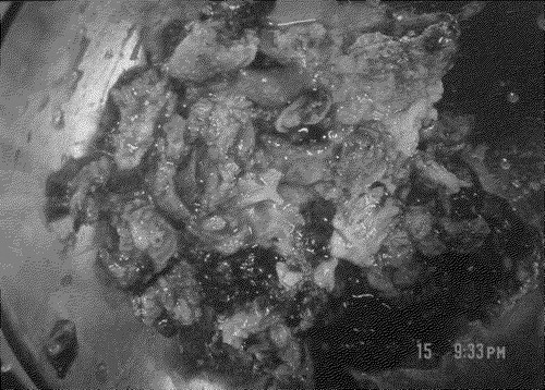
Figure 1. Primary peritoneal carcinosarcoma/ MMMT excised from the pelvic cavity. More than a hundred pieces of tumorous tissue measuring up to 5.3 x 4.1 x 3.5 cm in size and 223 g in weight was submitted for pathologic study. They were yellow to white in color and soft in consistency, with hemorrhage and necrosis.
Histologically, the tumor showed a picture of carcinosarcoma characterized by a mixture of malignant epithelial and sarcomatous elements (). The epithelial component was composed of neoplastic cells arranged in endometrioid type glandular pattern. The sarcomatous component showed spindle cells and bizarre multinucleated giant cells. There was also MMMT involving the serosal surface and the outer corpus wall of the lower uterine segment. There was no primary carcinosarcoma in the endometrium and the left ovary. The outer cervical wall contained both MMMT and endometriosis. Focal adenomyosis of the uterus was also noted. The left fallopian tube, left ovary, omentum, and all 33 lymph nodes were free of tumor.
Figure 2. Peritoneal Malignant mixed mullerian tumor characterized by admixture of epithelial (endometrioid type gland) [arrow] and non-epithelial (spindle cells) sarcomatous elements [arrowhead].
![Figure 2. Peritoneal Malignant mixed mullerian tumor characterized by admixture of epithelial (endometrioid type gland) [arrow] and non-epithelial (spindle cells) sarcomatous elements [arrowhead].](/cms/asset/2272e8b5-a9b0-4108-b449-36ecafeabb81/ionc_a_125184_f0002_b.jpg)
She received postoperative Ifosfamide (5 mg/m2) and Cisplatin (75 mg/m2 initially then 40 mg/m2) chemotherapy every 3 weeks for 6 cycles, then radiotherapy (whole pelvis 38 Gy in 25 fractions). Tri-monthly follow-up physical examinations and pelvic CT scan demonstrated no evidence of disease. CA 125 levels also stayed normal. The patient remains disease-free 60 months following the surgery.
Three years after the diagnosis of pelvic carcinosarcoma, the patient felt a mass in her right breast. A mammography revealed heterogeneous dense breast tissue with no definite microcalcifications. A biopsy of the breast tumor revealed a well-differentiated invasive ductal carcinoma. A week after the biopsy, she underwent lumpectomy of the right breast and axillary lymph node dissection. Pathology revealed a well-differentiated and invasive ductal carcinoma. The axillary lymph nodes were free of tumor. An immunohistochemical study detected no estrogen and progesterone receptors and no HER-2/neu protein.
Due to strong family history of breast and ovarian cancer, the patient underwent genetic testing BRCA1 tumor suppressor-gene mutation using her peripheral blood. She was found positive for the BRCA1 mutation.
Most peritoneal carcinosarcomas originate in the pelvic peritoneum, followed by decreasing frequency in the serosal surface of the colon, retroperitoneum, anterolateral abdominal peritoneum, and omentum Citation[1]. They generally occur in the elderly postmenopausal women, and usually present with abdominal pain, abdominal distention or pelvic mass Citation[2–4].
The etiology and pathogenesis of peritoneal carcinosarcoma are unknown. However, its association with endometriosis has been suggested Citation[2]. Occurrence of carcinosarcoma with previous radiation exposure or post radiation therapy has also been reported Citation[1], Citation[3], Citation[5]. Unopposed estrogens may also result in tumor development from polypoid endometriosis Citation[6]. This patient denied any intake of HRT following menopause.
Anatomically, the secondary mullerian system consisting of the abdominal and pelvic peritoneum and its underlying mesenchymal tissue is derived from the coelomic epithelium. As a derivative of coelomic epithelium, it retains its potential to differentiate into mullerian tissue. These mullerian type tumors may therefore originate directly from totipotential cells or from the transformation of neoplastic epithelial cells into sarcomatous cells Citation[7]. Pelvic and abdominal peritoneum may also differentiate into lesions known as endosalpingiosis, endometriosis, and endocervicosis, which in turn, may undergo malignant transformation. Interestingly, our patient had a prior operation for ovarian endometriosis. Endometriosis and MMMT of the outer cervical wall were noted on pathology. It is most plausible that the tumor in our patient arose from a preexisting endometriosis found on the cervical wall, especially since the glandular component is of endometrioid type. However, the MMMT found on the serosal surface of the uterus and the pelvic peritoneum may have originated from the totipotential cells of the peritoneal mesenchyme.
Mullerian carcinocarcinomas may be categorized as homologous or heterologous, depending on the histologic characteristics of the sarcomatous elements. Homologous neoplasms show no differentiated sarcomatous elements, nor differentiation toward endometrial stroma or smooth muscle. Heterologous neoplasms, on the other hand show malignant cartilage or skeletal muscle Citation[8]. It has been suggested that the presence of stromal cartilaginous differentiation has relatively more favorable outcome compared to the presence of skeletal muscle Citation[9]. In the case described here, the epithelial component is composed of neoplastic cells arranged in endometrioid type glandular cells. Hence, it may be considered as a homologous type of carcinosarcoma that originated from the secondary mullerian system.
Malignant mixed mullerian tumors of the peritoneum are aggressive tumors. The prognosis is poor, and most patients follow a rapidly fatal course, regardless of the initial tumor stage Citation[10]. A review of the primary carcinosarcoma of the peritoneum done by Garamvoelgyi et al. Citation[3] showed that most patients died within one year, with median postoperative survival time of 14 months, and a range of 7 days to 73 months. The MMMT in the patient followed up for 73 months appeared to represent a second primary tumor after being treated for disseminated ovarian serous papillary carcinoma. It was during the fourth laparotomy for recurrent ovarian serous papillary carcinoma that carcinosarcoma was discovered. The patient died of disease 11 months after being diagnosed to have carcinosarcoma Citation[11]. The longest disease-free survival was reported by Rose et al. Citation[12] in which the patient was followed up to 42 months.
Owing to the rarity of the disease, limited data regarding the management of peritoneal MMMT exists. Recommendations for the treatment of MMMT are based on individual cases only Citation[4], Citation[7], Citation[8]. Since the uterus is the most common site of gynecologic sarcoma, treatment of other gynecologic sarcomas is often based on prior experience and outcomes with uterine sarcoma Citation[12]. Recent reports have identified Ifosfamide and Cisplatinum as the most active agents in MMMT of the female genital tract. A prospective study by the GOG noted the response rate of 25% for Ifosfamide and 17% for Cisplatin as single agent for uterine carcinosarcomas Citation[13]. The role of radiotherapy remains controversial Citation[14].
We conducted a comprehensive review of primary peritoneal MMMT (). A review of these cases and our current case provides the following information:
Table I. Primary peritoneal MMMT reported in English literature.
A complete cytoreduction, with resection of cancer to a status of no evidence of disease by the surgeon's unaided eye should be attempted. Several studies have emphasized the presence of residual disease as an important significant prognostic factor for peritoneal MMMT Citation[15], Citation[16].
Ifosfamide and Cisplatin combination therapy may play a role in the management of both advanced and localized disease. It appears that those patients treated with chemotherapeutic agents other than Ifosfamide and Cisplatin had lower survival rate. Rose et al. Citation[12] reported a case that received post-operative Ifosfamide and Cisplatin chemotherapy. The patient was disease free for 42 months.
It has been suggested that the presence of an epithelial component makes the tumor favorable for adjuvant chemotherapy Citation[14].
Adjuvant radiotherapy after surgery in combination with chemotherapy may be considered for patients who have small residual disease after cytoreduction. Garamvoelgyi et al. Citation[3] reported a patient who received postoperative radiotherapy that survived for 8 months. Callister et al. Citation[17], after a study of 300 patients with uterine MMMT, reported that adjuvant pelvic radiotherapy decreased the risk of pelvic recurrence. It may delay the appearance of distant metastasis after surgery for uterine MMMT.
In the recent decade, inherited mutations in the BRCA1 gene confer increased susceptibility to breast and ovarian carcinomas. It has been observed that the presence of BRCA mutations may predispose to primary peritoneal cancers, and this neoplasm could be a part of the hereditary breast ovary cancer syndrome Citation[18–20]. A strong family history of breast and ovarian cancer led us to take genetic testing for this patient for the possibility of BRCA 1 and 2 mutations. It turned out that she carries BRCA 1 mutation.
In the present case, the patient underwent optimal tumor debulking and a combination of chemotherapy with Ifosfamide and Cisplatin, followed by pelvic irradiation. There were no signs of tumor recurrence after 48 months. In conclusion, this patient has the longest disease-free survival in English literature. Multimodality treatment strategies may be an effective treatment for primary peritoneal carcinosarcoma.
Related Research Data
References
- Shen DH, Khoo US, Xue WC, Ngan HY, Wang JL, Liu VW, et al. Primary peritoneal malignant mixed mullerian tumors. A clinicopathologic, immunohistochemical and genetic study. Cancer 2001; 91: 1052–60
- Dincer AD, Timmins P, Pietrocola D, Fisher H, Ambros RA. Primary peritoneal mullerian adenosarcoma with sarcomatous overgrowth associated with endometriosis: a case report. Int J Gynecol Patho 2001; 21: 65–8
- Garamvoelgyi E, Guillou L, Gebhard S, Salmeron M, Seematter RJ, Hadji MH. Primary malignant mixed mullerian tumor (metaplastic carcinoma) of the female peritoneum. A clinical, pathologic, and immunohistochemical study of three cases and a review of literature. Cancer 1994; 74: 854–63
- Wei L, Wang J, Zhang X, Cui H, Shen D, Qian H. Primary omental malignant mixed mullerian tumor in a 67-year-old woman. Chin Med J 2002; 115: 138–40
- Garde JR, Jones MA, McAfee R, Tarraza HM. Extragenital malignant mixed mullerian tumor: review of literature. Gynecol Oncol 1991; 43: 186–90
- Parker RL, Dadmanesh F, Young RH, Clement PB. Polypoid endometriosis. A clinicopathologic analysis of 24 cases and a review of the literature. Am J Surg Pathol 2004; 28: 285–97
- Mira JL, Fenoglio-Preiser CM, Husseinzadeh N. Malignant mixed mullerian tumor of the extraovarian secondary mullerian system. Report of two cases and review of English literature. Arch Pathol Lab Med 1995; 119: 1044–9
- Sumathi VP, Murnaghan M, Dobbs P, McCluggage WG. Extragenital Mullerian carcinosarcoma arising from the peritoneum: report of two cases. Int J Gynecol Cancer 2002; 12: 164–7
- McCluggage WC. Malignant biphasic uterine tumors: carcinosarcomas or metastatic carcinomas?. J Clin Pathol 2002; 55: 321–5
- Shintaku M, Matsumoto T. Primary Mullerian carcinosarcoma of the peritoneum: report of a case. Int J Gynecol Pathol 2001; 20: 191–5
- Chen KT, Wolk RW. Extragenital malignant mixed mullerian tumor. Gynecol Oncol. 1988; 30: 422–6
- Rose PG, Rodriguez M, Abdul-Karim FW. Malignant mixed mullerian tumor of the female peritoneum: treatment and outcome of three cases. Gynecol Oncol 1997; 65: 523–5
- Thigpen JT, Blessing JA, Beecham J, Homesley H, Yordan E. Phase II trial of cisplatin as first-line chemotherapy in patients with advanced or recurrent uterine sarcomas: a Gynecologic Oncology Group study. J Clin Oncol 1991; 9: 1962–6
- Topuz E, Eralp A, Aydiner A, Saip P, Tas F, Yavuz E, et al. The role of chemotherapy in malignant mixed tumors of the female genital tract. Eur J Gynecol Oncol 2001; 22: 469–72
- Inthasorn P, Carter J, Valmadre S, Beale P, Russel P, Dalrymple C. Analysis of clinicopathologic factors in malignant mixed Mullerian tumors of the uterine corpus. Int J Gynecol Cancer 2002; 12: 348–53
- Arrastia C, Fruchter R, Clark M, Maiman M, Remy JC, Macasaet M, et al. Uterine carcinosarcomas. Incidence and trends in management and survival. Gynecol Oncol 1997; 65: 158–63
- Callister M, Ramondetta LM, Jhingran A, Burke TW, Eifel PJ. Malignant mixed Mullerian tumors of the uterus: Analysis patterns of failure, prognostic factors and treatment outcome. Int J Radiat Oncol Biol Phys 2004; 58: 786–96
- Goshen R, Chu W, Elit L, Pal T, Hakimi J, Ackerman I, et al. Is uterine papillary serous adenocarcinoma a manifestation of the hereditary breast-ovarian cancer syndrome?. Gynecol Oncol 2000; 79: 477–81
- Menczer J, Chetrit A, Barda G, Lubin F, Fishler Y, Sltaras M, et al. Frequency of BRCA mutations in primary peritoneal carcinoma in Israeli Jewish women. Gynecol Oncol 2003; 88: 58–61
- Khoo US, Ngan HY, Cheung AN, Chan KY, Lu J, Chan VW, et al. Mutational analysis of BRCA1 and BRCA2 genes in Chinese ovarian cancer identifies 6 novel germline mutations. Hum Mutat 2000; 16: 88–9