To the Editor
The results of conventional x-ray radiotherapy for loco-regionally advanced esophageal carcinoma have been disappointing because it is usually difficult to administer a sufficiently high radiation dose to the tumor located adjacent to organs with lower tolerance dose; the 5-year survival rate is only 6 – 10% Citation[1–7].
The potential advantages of proton beam therapy over conventional x-ray radiotherapy have been demonstrated in the treatment of a number of cancers Citation[8–17]. In a recent study from a group of Swedish radiation oncologists, about 80 patients with esophageal cancer per year in their country (population 9 million) are judged eligible for proton beam therapy Citation[18], Citation[19].
In a previous investigation of 46 esophageal cancer patients treated with protons between 1985 and 1998, the local control rate of all patients was 57%, and was 29% for stages T2 – T4 Citation[17]. We have started a twice-a-day irradiation regimen to patients with advanced esophageal cancer since 1998. Below, we report a case of a patient with advanced esophageal cancer who has survived for eight years after radiotherapy with a combination of proton and x-ray beams.
A 58-year-old man was admitted to University of Tsukuba Hospital in February 1998. Barium contrast esophagography showed a well-defined, irregularly shaped, protruding lesion, 6 cm in maximal length, in the cervical esophagus (). CT of the neck and chest showed that the tumor had directly invaded the thyroid gland and the outer layer of the trachea. It also showed swelling of the cervical paraesophageal lymph nodes, which were attached to the primary tumor. The diagnosis was a squamous cell carcinoma of the cervical esophagus (cT4N1M0, stage III) Citation[20].
Figure 1. Barium contrast esophagogram before radiotherapy showing a protruding tumor with ulceration, 6 cm in length, in the cervical esophagus (arrow).

X-ray radiotherapy was administered from weeks 1 to 5 for a total dose of 41.4 Gy in 23 fractions. Proton beam therapy was administered for the first two weeks and on one day in week 5 for a total dose of 10.8 Gy in 9 fractions, and from weeks 5 to 8 for a total dose of 26 Gy in 13 fractions. Proton irradiation was performed concomitantly with the x-ray irradiation, at an interval of six hours in the first two weeks and of one day in week 5. The total proton dose was 36.8 Gy, and x-ray dose was 41.4 Gy. The combined x-ray and proton dose was 78.2 Gy. The radiation dose was the physical dose using relative biological effectiveness of unity.
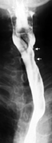
Barium contrast esophagography in June 1999 (one year after radiotherapy) showed that the tumor was well controlled (). A biopsy specimen in May 2004, five years after the radiotherapy, revealed no evidence of recurrence. Neck, chest, and abdominal CT scans in November 2003, 4.5 years after radiotherapy, showed no evidence of tumor relapse. The patient was well as of February 2006.
Figure 2. Barium contrast esophagograms 1 year after the radiotherapy showing a slight deformity of the wall in only the upper portion of the cervical esophagus (arrow), but no tumor.
Narcotic analgesics were required for 21 days during the treatment because of pharyngeal pain (Grade 2 esophagitis according to the RTOG scoring criteria Citation[21]). But no pain on swallowing attributable to the esophagitis was observed during or immediately after the course of irradiation therapy. No other treatment-related sequelae were observed in the lung, spinal cord, or skin.
Combinations of chemotherapy and x-ray radiotherapy (chemoradiotherapy) have recently become common in the management of patients with advanced esophageal cancer Citation[22]. A randomized trial of chemoradiotherapy showed that the estimated local control rate at two years and survival rate at five years were 55% and 27%, respectively Citation[23], which are comparable to those obtained with the combination of x-ray and proton beam therapy without chemotherapy at our hospital Citation[17], Citation[23]. Cooper et al. reported that 8% of the patients who underwent chemoradiotherapy in their series experienced acute Grade 4 toxic effects, an additional 2% experienced acute Grade 5 toxic effects, and 4% experienced late Grade 4 toxic effects Citation[22]. In our own institution, 39 patients with esophageal cancer treated with combined proton and x-ray irradiation were followed up for a minimum of six months after treatment between November 1985 and February 1998. None had acute Grade 4 or 5 toxic effects, and two (5%) had late Grade 5 toxic effects Citation[17].
Possible reasons for a high incidence of radiation ulcer in the esophagus include a high total dose of combined protons and x-rays and a larger proton dose per fraction. To reduce the incidence of radiation ulcer in the esophagus, we used a twice-a-day irradiation regimen for this patient, which allowed us to shorten the treatment period. This is the first report of a patient with advanced esophageal carcinoma in whom twice-a-day combined proton and x-ray radiotherapy can be effective.
References
- Earlam R, Cuhana-Melo JR. Oesophageal squamous cell carcinoma. II. A critical review of radiotherapy. Br J Surg 1980; 67: 457–61
- Okawa T, Kita M, Tanaka M, Ikeda M. Results of radiotherapy for inoperable locally advanced esophageal cancer. Int J Radiat Oncol Biol Phys 1989; 17: 49–54
- Herskovic A, Martz K, Al-Sarraf M, et al. Combined chemotherapy and radiotherapy compared with radiotherapy alone in patients with cancer of the esophagus. N Engl J Med 1992; 326: 1593–8
- Smalley SR, Gunderson LL, Reddy EK, Williamson S. Radiotherapy alone in esophageal carcinoma: Current management and future directions of adjuvant, curative and palliative approaches. Semin Oncol 1994; 21: 467–73
- Al-Sarraf M, Martz K, Herskovic A, et al. Progress report of combined chemoradiotherapy versus radiotherapy alone in patients with esophageal cancer: An intergroup study. J Clin Oncol 1997; 15: 277–84
- Chan A, Wong A. Is combined chemotherapy and radiation therapy equally effective as surgical resection in localized esophageal carcinoma?. Int J Radiat Oncol Biol Phys 1999; 45: 265–70
- Cooper JS, Guo MD, Herskovic A, et al. Chemoradiotherapy of locally advanced esophageal cancer. Long-term follow-up of a prospective randomized trial (RTOG 85-01). JAMA 1999; 281: 1623–7
- Lee M, Wynne C, Webb S, Nahum AE, Dearnaley D. A comparison of proton and megavoltage x-ray treatment planning for prostate cancer. Radiother Oncol 1994; 33: 239–53
- Isacsson U, Montelius A, Jung B, Glimelius B. Comparative treatment planning between proton and X-ray therapy in locally advanced rectal cancer. Radiother Oncol 1996; 41: 263–72
- Isacsson U, Hagberg H, Johansson KA, Montelius A, Jung B, Glimelius B. Potential advantages of protons over conventional radiation beams for paraspinal tumours. Radiother Oncol 1997; 45: 63–70
- Bush DA, Slater JD, Bonnet R, et al. Proton-beam radiotherapy for early-stage lung cancer. Chest 1999; 116: 1313–9
- Suit H. The Gray Lecture 2001: Coming technical advances in radiation oncology. Int J Radiat Oncol Biol Phys 2002; 53: 798–809
- Kagei K, Tokuuye K, Okumura T, et al. Long-term results of proton beam therapy for carcinoma of the uterine cervix. Int J Radiat Oncol Biol Phys 2003; 55: 1265–71
- Shioyama Y, Tokuuye K, Okumura T, et al. Clinical evaluation of proton radiotherapy for non-small-cell lung cancer. Int J Radiat Oncol Biol Phys 2003; 56: 7–13
- Tokuuye K, Akine Y, Kagei K, et al. Proton therapy for head and neck malignancies. Strahlenther Onkol 2004; 180: 96–101
- Igaki H, Tokuuye K, Okumura T, et al. Clinical results of proton beam therapy for skull base chordoma. Int J Radiat Oncol Biol Phys 2004; 60: 1120–6
- Sugahara S, Tokuuye K, Okumura T, et al. Clinical results of proton beam therapy for cancer of the esophagus. Int J Radiat Oncol Biol Phys 2005; 61: 76–84
- Ask A, Johansson B, Glimelius B. The potential of proton beam radiation therapy in gastrointestinal cancer. Acta Oncol 2005; 44: 896–903
- Glimelius B, Ask A, Bjelkengren G, Björk-Eriksson B, Blomquist E, Johansson B, et al. Number of patients potentially eligible for proton therapy. Acta Oncol 2005; 44: 836–49
- Sobin LH, Wittekind CH. ”TNM classification of malignant tumours6th ed. John Wiley & Sons, Inc, New York 2002
- Cox JD, Stetz J, Pajak TF. Toxicity criteria of the Radiation Therapy Oncology Group (RTOG) and the European Organization for Research and Treatment of Cancer (EORTC). Int J Radiat Oncol Biol Phys 1995; 31: 1341–6
- Cooper JS, Guo MD, Herskovic A, et al. Chemoradiotherapy of locally advanced esophageal cancer: Long-term follow-up of a prospective randomized trial (RTOG 85-01). Radiation Therapy Oncology Group. JAMA 1999; 281: 1623–7
- Al-Sarraf M, Martz K, Herskovic A, et al. Progress report of combined chemoradiotherapy versus radiotherapy alone in patients with esophageal cancer: An intergroup study. J Clin Oncol 1997; 15: 277–84