To the Editor
The clinical application of F-18-fluoro-deoxyglucose (FDG) positron emission tomography (PET) for mucosa-associated lymphoma (MALTomas) has not been well defined. Although FDG-PET is a well-known imaging modality for the management of many kinds of lymphomas, a few cases has been reported in the literature with regard to gastric MALTomas owing to variable FDG avidity in those tumors Citation[1–4]. We found a patient with low-grade early-stage gastric MALToma showing intense uptake of FDG into the tumors. Additionally, the images could be used as an earlier predictor of disease remission during the follow-up period.
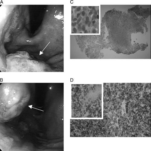
One and half years ago, an upper gastrointestinal endoscopic examination was carried out on a 51 year old male who complained of long-term intermittent postprandial fullness. Two polypoid tumors with surface nodularity and ulceration at the mid-body and upper-body were found. (A, B) Microscopic features showed small atypical lymphoid cells diffusely infiltrated with fibrinoid debris and purulent inflammation in the biopsied gastric mucosa (C); a few bacilli were also found. The immunohistochemical staining using L-26 (B-cell) and UCHL-1 (T-cell) was diffusely positive for B-cell and focally positive for T-cell (D). A low-grade B-cell lymphoma of the MALT type (MALToma) was identified.
Figure 1. Endoscopic examination found two polypoid tumors with surface nodularity and ulceration at the (A) mid-body and (B) upper-body. (C) Microscopic features and (D) immunohistochemical staining revealed a low-grade B-cell MALToma.
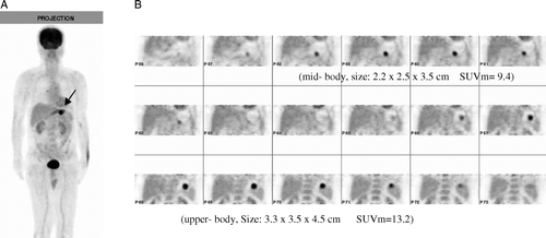
FDG-PET was arranged for initial staging. Whole-body imaging, reconstructed iteratively with attenuation correction, was performed 60 min after intravenous injection of 278 MBq (7.5 mCi) of FDG on a Siemens ACCEL PET scanner. An additional 3 h delayed imaging after having a meal to distend the stomach was carried out. The projection view showed a focal area with intense FDG uptake in the inner left upper quadrant of the abdomen (A). Serial coronal views of the delayed images revealed two separate tumors in the stomach, corresponding to the lesions seen on endoscopy. The maximal standard uptake value (SUVm) of the lesions on the delayed images was 9.4 and 13.2, respectively (B). Additional endoscopic ultrasound (EUS) and computed tomography (CT) examinations were negative on perigastric lymph node and elsewhere. A case of a low-grade gastric MALToma with glucose hypermetabolism in stage I was diagnosed.
Figure 2. (A) The projection view of FDG-PET showed a focal area with intense FDG uptake in the inner left upper quadrant of the abdomen. (B) Serial coronal views of the delayed images with distend stomach revealed two separate tumors. The SUVm of the lesions was 9.4 and 13.2, respectively.
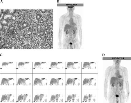
After diagnosis, the patient began to receive Helicobacter pyloric (HP) eradication therapy using the regimen of esomeprazole (Nexium; 40 mg/day), clarithromycin (Klaricid; 1 000 mg/day), and amoxicillin (Hiconcil; 2 000 mg/day) Citation[5]. For therapeutic evaluation, endoscopic examinations accompanied by tissue biopsies were carried out at two month intervals. In the sixth month, slight swelling of the gastric mucosa at the mid-body was visualized, and a residual MALToma still existed in the biopsied tissue (A). However, FDG-PET failed to delineate the lesion (B: projection view and C: serial coronal views). It was not until the tenth month after therapy, that an endoscopic examination revealed gastric ulcer scar, no residual tumor was demonstrable in the biopsied specimen. Meanwhile, FDG-PET remained negative (D). Remission of the disease has being reached.
Figure 3. (A) In the 6th month after therapy, a residual MALToma still existed in the biopsied tissue. (B) Projection view and (C) coronal views of FDG-PET failed to delineate the lesion. (D) FDG-PET remained negative in the 10th month after therapy.
To our knowledge, FDG-PET has been reported in a few cases of gastric lymphoma. Earlier reports indicated that FDG-PET was undetectable in extranodal marginal zone lymphoma Citation[1], Citation[3]. In a recent report of 42 cases of MALTomas which underwent FDG-PET study, tracer uptake was observed in 81% of the cases. However, a relative lower positive rate of 67% (4/6) was found in gastric MALT Citation[4].
In this case, intense FDG uptake into gastric MALToma manifested with high SUVm level was demonstrated. Malignant grading was discrepant between glucose metabolism and histopathology, physiological vs. morphological. Whether association of infectious/inflammatory process in the tumor enhanced higher FDG uptake was undetermined. An additional finding showed that negative FDG-PET preceded the resolution of histopathology, indicating that the residual malignant tissue was biologically inactive or that limited numbers of malignant cells which were not delineated on the imaging remained in the regressed tumor. FDG-PET may act as a predictor of disease remission.
References
- Hoffmann M, Kletter K, Diemling M, Becherer A, Pfeffel F, Petkov V, et al. Positron emission tomography with fluorine-18-2-fluoro-deoxyglucose (F18-FDG) dose not visualize extranodal B-cell lymphoma of the mocosa-associated lymphoid tissue (MALT)-type. Ann Oncol 1999; 10: 1185–9
- Aigner RM, Schwarz T, Wurzinger G, Karpf E. Detection of unsuspected gastric MALT-lymphoma with F-18-FDG-PET. Nuklearmedizin 2000; 39: N107
- Hoffmann M, Kletter K, Becherer A, Jäger U, Chott A, Raderer M. 18F-fluorodeoxyglucose positron emission tomography (18F-FDG-PET) for staging and follow-up of marginal zone B-cell lymphoma. Oncology 2003; 64: 336–40
- Beal KP, Yeung HW, Yahalom J. FDG-PET scanning for detection and staging of extranodal mariginal zone lymphomas of the MALT type: A report of 42 cases. Ann Oncol 2005; 16: 473–80
- Fischbach W, Goebeler-Kolve M-E, Dragosics B, Greiner A, Stolte M. Long term outcome of patients with gastric marginal zone B cell lymphoma of mucosa associated lymphoid tissue (MALT) following exclusive Helicobacter pylori eradication therapy: Experience from a large prospective series. Gut 2004; 53: 34–7