To the Editor
Pemphigus vulgaris (PV) is a rare autoimmune muco-cutaneous bullous disease with potentially significant morbidity and mortality. Mean age at presentation is 40–50 years, with increased incidence in Jews and other Mediterranean peoples Citation[1]. The etiology is idiopathic in most cases but can be induced by many triggers, including drugs, physical agents (burns), infections (herpes virus), contact dermatitis, neoplasm, emotional stress, and ultraviolet radiation Citation[2], Citation[3]. In exceptional instances, PV can also be provoked by ionizing radiation therapy. Recently, a systematic review determined that PV might be considered as a radiotherapy-induced side effect Citation[4]. We report a case of mucosal and esophageal PV without skin manifestations induced by radiotherapy. To the best of our knowledge, this is the first report of this rare manifestation of PV as a side effect of radiotherapy.
A 49-year old, previously healthy, married mother of four presented with a 4×3 cm lump in the inner upper quadrant of her left breast. Biopsy revealed invasive breast carcinoma with positive estrogen and progesterone receptors and negative HER2 receptor. Sentinel lymph node biopsy showed one positive lymph node with capsular extension. After treatment with neo-adjuvant chemotherapy [5-flourouracil, doxorubicin and cyclophosfamide combination] for three cycles without response, treatment was changed to paclitaxel once weekly for 12 weeks, with partial response.
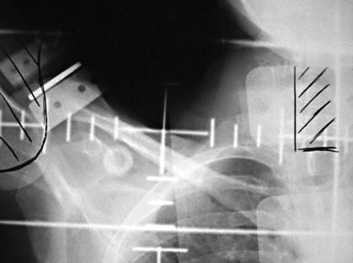
The patient then underwent left lumpectomy and axillary lymph node dissection. Pathology revealed a 1 cm invasive carcinoma without other lymph-node involvement. Adjuvant radiotherapy of 50 Gy was given to the whole breast with two tangential fields, 16 Gy boost to the tumor bed and 50 Gy to whole axilla by one anterior field (). The radiation treatment was completed without any specific side effect. Tamoxifen 20 mg daily was started a few days before radiation.
Figure 1. The radiation field “whole axilla” of the patient. The field included a small part of the larynx and esophagus.
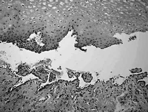
Two weeks later, the patient developed hoarseness and throat pain. These symptoms were suspicious for late mucositis in the radiated volume and she was treated symptomatically, with some improvement. Two weeks later, after one episode of fever, she began to suffer from severe mucositis, difficulties in food intake, loss of weight and severe hoarseness. Infection disease was suspected and she was treated with antibiotic and antifungal treatment, without improvement. Biopsies taken from the mouth and the esophagus revealed PV by histology and immunofluorescence (). There were no suspicious skin lesions. Prednisone 60 mg daily was started, with good clinical response, and was tapered down, together with 15 mg of methotrexate weekly.
Figure 2. Biopsy specimen of oral mucosa reveals intra-epidermal bulla with supra-basal separation and acantholitic cells, consistent with pemphigus vulgaris (×40).
The bullae that characterize the lesions of pemphigus are due to the loss of intercellular keratinocyte adhesion, a process known as acantholysis. There is strong evidence that intercellular autoantibodies directed against keratinocyte cell-surface antigens are pathogenic and cause these bullae by disrupting desmosomes. The desmosomal transmembrane adhesion molecules, desmogleins 1 and 3, are the best-characterized target antigens. In PV, antibodies against desmolglein 3 predominate in mucous membrane disease and antibodies against both desmogleins 1 and 3 are usually present in mucocutaneous disease Citation[5], Citation[6].
Most patients with pemphigus present with oral lesions and more than 90% of cases with skin lesions have oral lesions as well Citation[5]. Clinically, these lesions are fragile painful blisters, which are rarely intact in the oral cavity. They usually rupture, producing ulcerations with gray membranes. The most frequently involved areas are the buccal mucosa, palate, and gingiva, but rarely the pharynx. Nasal, laryngeal, and esophageal manifestations are extremely rare Citation[7], Citation[8]. In the current case, lesions were present from the oral cavity mucosa to the esophageal-gastric junction. Clinically, the first area involved was in the larynx-upper esophagus, which was included in the whole axilla field.
In a recently published systematic review of radiation-induced bullous pemphigoid Citation[4], 27 cases were found. The majority (89%) experienced blistering confined to the irradiated area, and there was no report of mucosal pemphigus. As in this case, most of the reported cases were breast carcinoma (78%). The use of hormonal therapy was reported in five cases. Although a possible correlation between PV and tamoxifen cannot be excluded, there is no reported case of tamoxifen-induced PV without radiotherapy. In the current case, the patient continued the tamoxifen treatment, which did not interfere with the PV recovery.
It is unclear how radiation therapy acts as a triggering mechanism to induce PV. Several theories have been proposed. One is that radiotherapy itself changes antigenic properties and induces autoantibody formation through the alteration of the basal membrane with unmasking of the antigen Citation[4], Citation[9]. Another possible explanation is that patients who developed PV after radiation may already have circulating low-titer anti-basement membrane antibodies, and the tissue damage through radiotherapy may enhance the deposition of antibodies Citation[4], Citation[9]. This may explain the rarity of this radiation-induced side effect. The unusual development of mucosal ulcers after radiotherapy should raise a suspicion of this diagnosis.
References
- Uzun S, Durdu M, Akman A, Gunasti S, Uslular C, Memisoglu HR, et al. Pemphigus in the Mediterranean region of Turkey: A study of 148 cases. Int J Dermatol 2006; 45: 523–8
- Orion E, Barzilia D, Brenner S. Pemphigus vulgaris induced by diazinon and sun exposure. Dermatology 2000; 201: 378–9
- Ruocco V, Wolf R, Ruocco E, Baroni A. Viruses in pemphigus: A casual or causal relationship?. Int J Dermatol 1996; 35: 782–4
- Mul VE, Van Geest AJ, Pijls-Johannesma MC, et al. Radiation-induced bullous pemphigoid: A systematic review of an unusual radiation side effect. Radiother Oncol 2007; 82: 5–9
- Dick SE, Werth VP. Pemphigus: A treatment update. Autoimmunity 2006; 39: 591–9
- Amagai M, Tsunoda K, Zillikens D, Nagai T, Nishikawa T. The clinical phenotype of pemphigus is defined by the anti-desmoglein autoantibody profile. J Am Acad Dermatol 1999; 40: 167–70
- Vasiliou A, Nikolopoulos TP, Manolopoulos L, Yiotakis J. Laryngeal pemphigus without skin manifestation and review of the literature. Eur Arch Otorhinolaryngol 2007; 264: 509–12
- Frangogiannis N, Gangopadhyay S, Cate T. Pemphigus of the larynx and esophagus. Ann Intern Med 1995; 122: 803–4
- Cliff S, Harland CC, Fallowfield ME, Mortimer PS. Localised bullous pemphigoid following radiotherapy. Acta Derm Venereol 1996; 76: 330–1