To the Editor
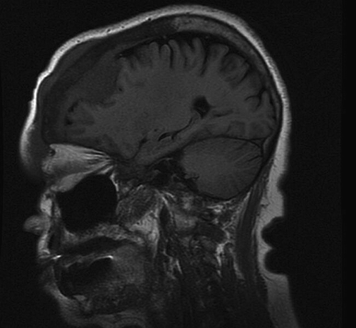
A seventy year old female presented to our surgery department with a one year history of an expanding forehead mass which was initially thought to be a lipoma. A CT scan was performed which demonstrated a soft tissue mass within the ethmoid air cells with intracranial extension and compression of the right frontal lobe along with erosion of the right frontal bone (). Subsequent MRI showed a 3.1×3.4×2.2 cm mass in the right frontal bone with intracranial and extracranial extension. The mass involved the nasal bone as well. Patient underwent tumor embolization, bi-frontal craniotomy, complete resection of mass, involved bone and dura followed by reconstruction with a calvarial graft.
Figure 1. CT scan demonstrating soft tisue mass within right frontal bone with intracranial and extracranial extension.
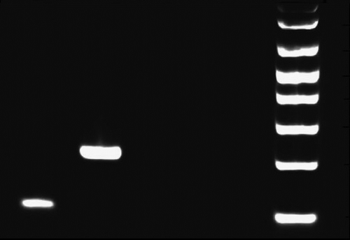
Pathology sections demonstrate a dense, vaguely nodular infiltrate of small angulated centrocytes admixed with fewer centroblasts (seven centroblasts/hpf) in a background of sclerosis and focal necrosis (). The frontal bone and duramater are also infiltrated. Immunohistochemical stains demonstrate these infiltrating cells to be B-cells (CD20 positive; ) with CD10 and BCL2 co-expression. CD3, CD5 and CD43 stain the fewer accompanying T-cells. Proliferation index marker (Ki 67) is low (1%). These findings are consistent with follicular lymphoma (FL), grade 2. DNA was extracted from frozen brain tissue with the Puregene system (Gentra) and analyzed for MBR and MCR BCL2-IgH fusions by nested PCR using published primers (Gribben JG et. al, Blood 78(12):3275–80, 1991) and a clinically validated assay. The MBR BCL2-IgH fusion was detected in the patient specimen (). The MCR fusion was not detected.
Figure 2. Hematoxylin and Eosin stained section of the lymphoma demonstrating a nodular infiltrate of centrocytes admixed with fewer centroblasts (original magnification×100).

Figure 3. Follicular lymphoma cells positive for B-cell marker CD20 immunostain. (original magnification×400).
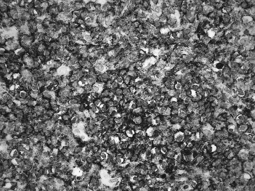
Figure 4. Nested PCR demonstrating presence of MBR BCL2-IgH fusion. Lane 1: Patient sample; Lane 2: Positive MBR control; Lane 3: Polyclonal DNA; Lane 4: water; M-1kb ladder.
Staging bone marrow biopsy and whole body CT scans were negative. Given the very limited nature of the disease, systemic therapy was not given and patient received localized radiation therapy which she tolerated well. Post therapy scans showed no residual lymphoma.
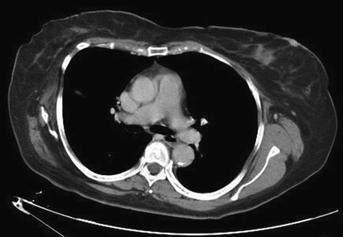
Twenty one months after she first noticed the tumor, patient re-presented to her primary care physician with complaints of intermittent pain in her right forearm. An x-ray and MRI were done which revealed a mass in the right radius raising concern for recurrent lymphoma. Whole body CT scans and MRI were performed which showed bilateral breast and lung masses () and lymphomatous infiltration of the right iliac bone with soft tissue extension. Biopsy of the breast mass was performed and confirmed recurrent low grade FL. She is currently on chemotherapy (CVP-R) of which she has received six cycles. She is doing well and recent CT scans show good response to the chemotherapy.
Figure 5. CT scan demonstrating relapse in bilateral lungs and breasts.
Discussion
Primary bone non-Hodgkin's lymphoma (PBL) is a rare entity accounting for approximately 3% of all primary bone malignancies and is defined as a solitary bone lymphoma without evidence of systemic dissemination found within 6 months of detection of the tumor Citation[1]. Bone involvement is not uncommon in advanced stage lymphoma originating in other sites, but PBL constitutes less than 5% of extranodal lymphomas Citation[2] and less than 2% of all lymphoma in adults Citation[3]. Only 15 cases of primary cranial vault lymphoma have been reported in the literature so far. Large B-cell lymphomas account for the majority of cases of PBL Citation[4].
Extranodal, extracutaneous FL's are rare and have been reported in ocular adnexa, gastrointestinal tract, testis, epididymis, thyroid and salivary glands. Most of these extranodal, extracutaneous FL's except for the primary gastrointestinal FL's are different from nodal FL in being usually BCL2 negative and t(14;18) negative Citation[5]. We document the first case of primary cranial vault BCL2 positive and t(14;18) positive low grade follicular lymphoma.
PBL usually presents with localized/radiating pain and a palpable mass or soft-tissue swelling. The peak incidence of PBL is the fifth decade with a male/female ratio of 1.8:1. Majority of PBL's are large B-cell type or, in the pediatric population, the lymphoblastic type. Only a few cases of low-grade histology have been described in the literature. The exact etiology of PBL is not known, but a few cases have been described in patients with chronic osteomyelitis. Some authors suggest that chronic suppurative inflammation and production of immunosuppressive cytokines create a microenvironment which is less accessible to T-cell surveillance, allowing clonal proliferation of Epstein-Barr virus (EBV) infected B cells. Few cases of high-grade B-cell lymphoma arising in chronic inflammatory peripheral skeletal sites have also been described. A similar mechanism has been described in EBV-associated pleural cavity malignant lymphoma. Rare cases of PBL developing in sites of previous trauma have also been described Citation[6].
FL is a neoplasm of follicle center cells. It commonly involves lymph nodes, spleen, bone marrow, peripheral blood and Waldeyer's ring. Involvement of extranodal sites such as gastrointestinal tract, soft tissue, skin and other sites may occur, but this is usually in the setting of widespread nodal disease. Primary extranodal, extracutaneous FL is rare and most examples are reported within larger cohorts analyzing the distribution of all lymphoma subtypes at extranodal locations or as case reports or small series. These reports include ocular adnexa, thyroid, gastrointestinal tract, salivary gland, testis and epididymis as the involved primary extranodal sites Citation[6].
The differential diagnosis of a solitary mass lesion of the cranial vault usually includes metastatic carcinoma, meningioma, osteomyelitis, Ewing's sarcoma and multiple myeloma Citation[7]. Although primary malignant lymphomas of the cranial vault are rare, they should also be included in this differential.
Patients with extranodal FL demonstrate a high relapse rate but are more likely to achieve complete remission and may ultimately have a more favorable long-term prognosis than those with equivalent nodal disease Citation[8]. Our patient presented with an unusual BCL2 positive and t(14;18) positive primary low grade follicular lymphoma of the cranial vault that relapsed 9 months later in extranodal soft tissue locations.
References
- Agbi CB, Bannister CM, Turnbull IW. Primary cranial vault lymphoma mimicking a meningioma. Neurochirurgia (Stuttg) 1983; 26: 130–2
- Dubey P, Ha CS, Besa PC, Fuller L, Cabanillas F, Murray J, et al. Localized primary lymphoma of bone. Int J Radiat Oncol Biol Phys 1997; 37: 1087–93
- Finn LS, Viswanatha DS, Belasco JB, Snyder H, Huebner D, Sorbara L, et al. Primary follicular lymphoma of the testis in childhood. Cancer 1999; 85: 1626–35
- Lonjon M, Hofman P, Thyss A, Paquis P, Roche JL. Primary lymphoma of the cranial vault. Apropos of a case. Neurochirurgie (in French) 1993; 39: 50–3
- Pardhanani G, Ashkan K, Mendoza N. Primary non-Hodgkin's lymphoma of the cranial vault presenting with unilateral proptosis. Acta Neurochir (Wien) 2000; 142: 597–8
- Sato M, Saito T, Yamaguchi K. Primary malignant lymphoma of the skull presenting a huge mass lesion: Case report. No Shinkei Geka (in Japanese) 1993; 21: 1061–4
- Stein ME, Kuten A, Gez E, Rosenblatt KE, Drumea K, Ben-Shachar M, et al. Primary Lymphoma of bone-A retrospective study. Experience at the Northern Israel Oncology Center (1979–2000). Oncology 2003; 64: 322–7
- Vergier B, Belaud-Rotureau MA, Benassy MN, Beylot-Barry M, Dubus P, Delaunay M, et al. Neoplastic cells do not carry bcl2-JH rearrangements detected in a subset of primary cutaneous follicle center B-cell lymphomas. Am J Surg Pathol 2004; 28: 748–55