Abstract
The purpose of this study was to clarify outcome for concurrent chemoradiation (CT-RT) in locally advanced cervix cancer in Japan. This is a non-randomized retrospective analysis of 226 patients treated with definitive CT-RT or radiotherapy alone (RT alone) in nine institutions between 2001 and 2003. External irradiation consisted of whole pelvic irradiation and pelvic side wall boost irradiation, using a central shield during the latter half of the treatment with the anteroposterior parallel opposing technique. The external beam irradiation was performed with 1.8 or 2 Gy per fraction. High-dose-rate intracavitary brachytherapy (HDR) was performed in all cases. In chemotherapy, platinum based drugs were used alone or in combination with other drugs such as 5FU. Grade of late complications was scaled retrospectively with CTCv2.0. Overall survival rate at 50 months of stage Ib, II and III, IV was 82% and 66% in CR-RT and 81% and 43% in R alone, respectively. Disease-free survival rate at 50 months of stage Ib, II and III, IV was 74% and 59% in CR-RT and 76% and 52% in R alone, respectively. There was no significant difference between CT-RT and RT for overall survival and disease free survival. Univariate analysis suggested that loco-regional control was better with CT-RT, but multivariate analysis could not confirm this finding. Compared to RT alone, CT-RT caused significantly more acute and late complications. Thus, late complication (grade 3-4) free survival rate at 50 month was 69% for CT-RT and 86% for RT alone (p<0.01). The therapeutic window with concomitant radiochemotherapy and HDR brachytherapy may be narrow, necessitating a close control of dose volume parameters and adherence to systems for dose prescription.
Since the recommendation of the NCI alert in 1999 to use platinum based concurrent chemotherapy with radiation in the treatment of cervical cancer Citation[1], a change in patterns of practice has occurred in the treatment of cervical cancer. Concurrent chemoradiation (CT-RT) is now considered the standard care for locally advanced cervical cancer. Cisplatin with or without 5FU is the most frequently used regimen. Japanese institutions have also begun to use CT-RT in the treatment of cancer of the uterine cervix.
However, data on late complications of CT-RT is lacking Citation[2], Citation[3] although Eifel et al. has updated the RTOG data with regard to late toxicity Citation[4] and Potter et al. has recently published data with MRI guided HDR brachytherapy combined with radiochemotherapy showing excellent outcome Citation[5]. Besides, it is not clear if it is safe to give chemotherapy concurrently with HDR (high-dose-rate intracavitary brachytherapy) as HDR was not used in the brachytherapy regimens of the randomized trials of CT-RT Citation[6–10].
As HDR is used in over 80% of Japanese institutions, the JROSG (Japan Radiation Therapy Oncology Group) determined to clarify the treatment results of CT-RT in cervical cancer, focusing especially on acute and late complications. The JROSG was founded in Japan to perform multi-institutional trials in the field of radiation oncology, as does the RTOG.
There has been a significant increase in late complications with CT-RT recently in results of JROSG when compared with RT alone, even though the follow-up period is rather short. Therefore, we report here these results.
Materials and methods
Patient characteristics
This is a retrospective analysis of 226 patients with cancer of the uterine cervix treated by nine institutions of JAROSG that are representative for Japan between 2001 and 2003.
To be eligible for inclusion in this study, patients had to be stage Ib1-IVa and exclusion criteria consisted of preoperative or postoperative radiotherapy and neoadjuvant chemotherapy. Patients were staged clinically on palpation, cystoscopy, and proctoscopy by both gynecologist and radiation oncologist without general anesthesia. CT was used to evaluate the size of primary tumor and the metastasis to pelvic or paraaortic lymph nodes in all patients. MRI was also used in some patients.
Characteristics of patients in radiotherapy alone (R alone) or CT-RT group are listed in . All patients had no evidence of para aortic disease with CT or MRI. We used 5 cm to classify the tumor diameter considering reports that the locoregional control rates for tumors were different between tumors (<5 cm) and tumors (>5 cm) Citation[11].
Table I. Characteristics of the patients.
Treatment
Characteristics of radiotherapy are listed in . The total dose to point-A given in is only for HDR. Treatment was usually performed according to the protocol that Arai et al. proposed Citation[12]. In this protocol, a central shield was used in whole pelvic irradiation from the beginning or during the latter half of the treatment. The weight of external irradiation was increased and that of intracavitary irradiation decreased for patients of a more advanced stage. So, doses of external irradiation and intracavitary irradiation had the large variation in this study.
Table II. Treatment for cervical cancer.
The target for external irradiation was the primary tumor and the regional lymph nodes. 3D target definition was not usually performed. The superior margin of external irradiation was usually placed on upper margin of L5 and the lateral margins were 2 cm lateral to the obturator foramen. The inferior margin was put on inferior margin of the obturator foramen but extended inferiorly when there was vaginal invasion. We used multi-leaf collimators to block upper corners and lower corners of the radiation field. The external irradiation was performed with free set up in supine position. The external irradiation was delivered with a 6 or 10-megavolt (MV) x-ray with the anteroposterior parallel opposing technique with 1.8 or 2 Gy per fraction and 5 fractions per week.
HDR was performed once a week in most cases with a 60CO or 137Cs remote-controlled afterloading radiation machine. Insertion of the intracavitary apparatus, consisting of a polyethylene nonrigid tandem and two non-shield colpostats, was carried out without general anesthesia. A Henschke-type metal applicator was usually used. Vaginal packing was used to maintain the position of the tandem and ovoids. The patients were treated in the lithotomy position. In Japan, the method of intracavitary irradiation is based on the modified Manchester system Citation[13] and the concept of ICRU Report 38 Citation[14] is not in widespread use. So, doses to the ICRU bladder and rectum points were not always calculated. This study consisted of data of nine institutions that had different RI such as 60CO or 137Cs and the radioactivity of RI was not same. So, the total reference air kerma (TRAK) is not same but not calculated in all institutions.
In chemotherapy, platinum based drugs were used alone or in combination with other drugs in all patients. Twenty nine patients were treated with 20 – 40 mg/m2 of cisplatin weekly, usually for 5 weeks. Ten patients received 2 AUC (area under the curve) of carboplatin weekly for 5 – 7 weeks. Twelve patients received 2 cycles of 70 – 80 mg/m2 of nedaplatin every 4 weeks.
Six patients received 2 cycles of CDDP- 5FU regime (50 mg/m2 CDDP on day 1 and 500 mg/m2 5FU on day 1–4 every four weeks) during radiotherapy and additional 2 cycles after radiotherapy. Five patients received 2 cycles of mBOMP (nedaplatin 70 mg/m2 on day 1, vincristine 1 mg/body on day 1, mitomycin C 7 mg/m2 on day 1, and pepleomycin 40 mg/body on day 1–5) during radiotherapy and additional 3 cycles after radiotherapy. One patient received 2 cycles of CDDP- irinotecan regime (50 mg/m2 CDDP on day 1 and 50 mg/m2 irinotecan on day 1–3 every four weeks).
Statistical analysis
The follow-up period for the living patients ranged from 9 to 67 months and the median follow-up for the living patients was 36 months. We followed the patient periodically with physical examination, tumor markers such as SCC, and CT and MR scanning.In analysis of treatment results of cervix cancer, it is important to analyze the influence of doses delivered by external irradiation and HDR. We used the tumor and late tissue equivalent doses (Deq) to obtain the cumulative dose at point A administered by whole pelvis external irradiation and HDR. To express the BED in terms more familiar to clinicians, BED was converted to equivalent doses (DEq) as though given at 2 Gy/day for tumors and late effects respectively using the formula Citation[15].We assumed a value of α/β = 3 Gy for acute complications and α/β = 10 Gy for late complications Citation[16].
For calculation of disease-free survival, treatment failure was defined as locoregional recurrence, para-aortic recurrence, distant progression, second cancer diagnosis, or death as a result of any cause. Overall, disease-free survival, locoregional-recurrence-free survival, and distant metastasis-free survival rates were calculated from the start of treatment. Late complication-free survival rates were estimated from the end of radiotherapy.
The difference of characteristics of patients was examined with the chi-square test. Overall, disease-free survival, locoregional-recurrence-free survival, distant metastasis-free, and late complication-free survival rates were estimated using the Kaplan-Meier method Citation[17]. Survival curves and curves for actuarial morbidity were compared with a log-rank test.
In order to compare various kinds of survival between CT-RT and R alone, the univariate analysis was performed. When there was significance with univariate analysis, multivariate analysis was also performed using Cox’ s proportional hazard regression model Citation[18]. Complications of treatment that occurred within 90 days of the start of radiotherapy were considered acute complications, and those occurring or persisting more than 90 days after the start of radiotherapy were considered late complications. The severity of acute and late complication was classified according to the National Cancer Institute -Common Toxicity Criteria (version 2). Scoring of morbidity also was done retrospectively based on the patient chart.
Results
CT-RT was utilized more in patients who were younger, had better Karnofsky performance score, larger tumor diameter and more positive pelvic lymph nodes, compared with R alone. Patient distribution in tumor diameter and pelvic node status was significantly different between CT-RT and R alone (p < 0.01).
shows overall survival rates curves of patients treated with CT-RT or RT alone according to stage. shows locoregional failure-free survival rates curves of patients treated with CT-RT or RT alone according to stage. demonstrates various kinds of survival rates at 50 months and results of univariate analysis. We could not calculate 5 year survival rates due to shortness of the follow-up period. There were no significant differences in overall survival or disease-free survival rates between CT-RT and R alone. CT-RT had the significantly higher locoregional-free survival than R alone in Stage I and II. There were no significant differences in distant metastasis-free survival rates between CT-RT and R alone.
Figure 1. Overall survival rates in CT-RT and R alone; solid thick line (Stage Ib, II, CT-RT), solid thin line (Stage Ib, II, RT alone), dotted thick line (Stage III, IV, CT-RT), dotted thin line (Stage III, IV, RT-alone).
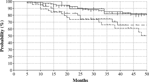
Figure 2. Locoregional failure-free survival rates in CT-RT and R alone; solid thick line (Stage Ib, II, CT-RT), solid thin line (Stage Ib, II, RT alone), dotted thick line (Stage III, IV, CT-RT), dotted thin line (Stage III, IV, RT-alone).
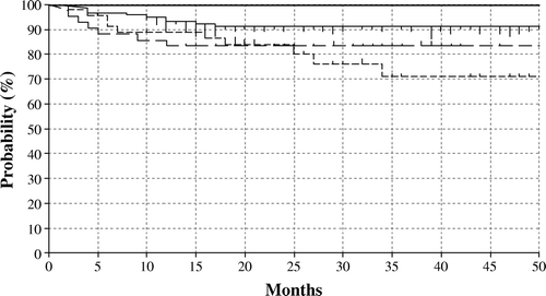
Table III. Survival Rates at 50 months.
shows acute complications in CT-RT and R alone. CT-RT or R alone, age, stage, doses of external radiation, and total doses to point A were analyzed for prognostic significance for various types of acute complications in by univariate analysis and multivariate analysis.
Table IV. Acute complications.
CT-RT was the only prognostic factor in acute complications in leukocytes regardless of whether the comparison was between all grades of patients (Grade 1 to 4), or between those with Grade 2 or higher (p < 0.01) in univariate analysis and multivariate analysis.
CT-RT was the only prognostic factor in acute complications in platelets whether the comparison was between all grades of patients (Grade 1 to 4) (p < 0.01) in univariate analysis. However, there was no prognostic factor in acute complications in platelets in multivariate analysis.
CT-RT was the only prognostic factor in diarrhea whether the comparison was between all grades of patients (Grade 1 to 4) (p < 0.05) in univariate analysis. However, there was no prognostic factor in acute complications in diarrhea in multivariate analysis. There was no prognostic factor in urinary frequency in unitivariate analysis.
We then analyzed late complications Grade 2 or higher in rectum, small or large bowel, and bladder.
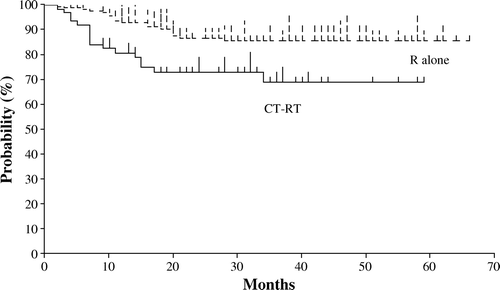
demonstrates late complications (rectum, small or large bowel, and bladder)-free survival rates in CR-RT and R alone. Late complications-free survival rate at 50 months was 69% in CR-RT and 86% in R alone. There was the significant difference between CR-RT and R alone (p < 0.01).
Figure 3. Late complications-free survival rates in CT-RT and R alone. Patients who had late complications Grade 2 or higher in rectum, small or large bowel, and bladder were analyzed.
CT-RT or R alone, age, doses of external radiation, and doses of HDR (DEq) were analyzed for prognostic significance for late complications (rectum, small or large bowel, and bladder)-free survival by multivariate analysis using Cox's proportional hazard regression model. There was no significant prognostic factor in late complications -free survival.
We compared late complications (small or large bowel) free survival rates between CR-RT and R alone. Colon fiberscopy and CT were commonly used to find the origin of bowel symptoms. Late complications-free survival rate at 50 months in small or large bowel was 86% in CR-RT and 95% in R alone. There was the significant difference between CR-RT and R alone (p < 0.05).
We compared late complications (rectum) free survival rates between CR-RT and R alone. Late complications-free survival rate at 50 months in rectum was 87% in CR-RT and 90% in R alone. The difference between CR-RT and R alone was not significant (p = 0.73).
We compared late complications (bladder) free survival rates between CR-RT and R alone. Late complications-free survival rate at 50 months in bladder was 93% in CR-RT and 96% in R alone. There was the significant difference between CR-RT and R alone (p < 0.05).
Discussion
Here we reported results the treatment results of CT-RT in cervical cancer, focusing especially on acute and late complications conducted by the JROSG. In this study, CT-RT was employed more in patients who had the larger tumor diameter and more positive pelvic lymph nodes, as compared with RT alone. However, locoregional failure-free survival in patients treated with CT-RT was significantly better than in patients with R alone in Stage Ib or II, indicating that CT-RT overcame disadvantages for local control such as larger tumor diameter and more positive pelvic lymph nodes. These results concurred with randomized trials Citation[4], Citation[7], Citation[9], Citation[10] which reported that local failures were significantly decreased in the cisplatin arms of these studies, suggesting that chemotherapy was acting as a radiation sensitizer Citation[19].
There were no significant differences in overall survival and disease-free survival rates between CT-RT and R alone in our study. These results did not agree with the randomized trials Citation[4], Citation[7], Citation[9], Citation[10]. The prevalence of larger tumor diameters and more positive pelvic lymph nodes in CT-RT patients may have influenced our results.
Concomitant chemotherapy did not decrease distant metastasis in our study. The prevalence of disadavantageous factors working against systemic control in CT-RT patients, such as larger tumor diameters and more positive pelvic lymph nodes were perhaps too great for the chemotherapy to produce systemic benefit.
CT-RT significantly increased late complications in rectum, small or large bowel, and bladder in our study. Actually, 31% of patients treated with CT-RT suffered complications higher than Grade 2 in rectum, small or large bowel, and bladder at 50 months after treatment. However, CT-RT was no prognostic factor in multivariate analysis, indicating that the interpretation of results obtained by non-randomized trials should be careful due to bias. CT-RT was employed more in patients who had the larger tumor diameter and more positive pelvic lymph nodes in this study. Besides, the heterogeneity of the schedules used for concurrent chemotherapy and the wide spread use of combination chemotherapy should be recognized as a problem in analyzing the influence on late complications.
HDR was used in all patients in this study. It is not clear if it is safe to give chemotherapy concurrently with HDR. In the randomized trial conducted by Canadian institutions, low dose rate, medium dose rate, and high dose rate techniques were all allowed. Only 38 of the 253 patients treated had HDR, with a schedule of 8 Gy times 3 fractions. There was no subset analysis done to determine any difference in toxicity between LDR, MDR, and HDR Citation[20]. There are only a few reports in which HDR was used in CT-RT Citation[21–24]. Besides, the number of patients in their reports was small and their results are conflicting. Souhami et al. reported an increased incidence of GI toxicity (26% with cisplatin versus 7.5% without) in patients receiving cisplatin-based chemotherapy along with external beam and HDR, which occurred rather early at a median follow-up of 11 months Citation[24]. Sood et al. reported increased acute toxicities with chemotherapy and radiation using HDR techniques, but no increase in late complications Citation[23]. It is unclear if the use of HDR in this study was related to increased late complications because CT-RT and R alone showed no differences in late complications in rectum or bladder, which are influenced to a greater degree than small or large bowel by the intracavitary irradiation.
Mature analysis confirms that the addition of fluorouracil and cisplatin to radiotherapy using LDR significantly improved the survival rate of women with locally advanced cervical cancer without increasing the rate of late treatment-related side effects Citation[4].We are planning the randomized trials to elucidate optimal radiation doses when HDR is used. Recently, Potter et al. reported that combined intracavitary and interstitial MRI-based brachytherapy in patients with significant residual disease after external-beam therapy is feasible and allows excellent local control and a low rate of morbidity Citation[5]. Dimopoulos et al. reported that MRI-based MRI-based 3D conformal HDR and cisplatin appeared to be safe and effective Citation[25]. We are also considering to use these high quality HDRs.
In summary, CT-RT significantly improved local control of patients with Stage Ib and II in univariate analysis. CT-RT caused significantly more acute complications such as leukopenia, thrombocytopenia, and diarrhea. Late complications (rectum, small or large bowel, and bladder) in CT-RT increased significantly, as compared with R alone in univariate analysis. These results were obtained by using non-randomized data and the interpretation of these results should be careful due to bias in data. However, our results indicate that the optimal radiation doses for CT-RT remain to be resolved. Further randomized trials in which strict dose guidelines are provided in these protocols are necessary to elucidate optimal radiation doses for CT-RT.
Related Research Data
References
- NCI Clinical Announcement R, MD. UD Dept of Health and Human Services Public Health Service. National Insitututes of Health 1999.
- Green JA, Kirwan JM, Tierney JF, Symonds P, Fresco L, Collingwood M, et al. Survival and recurrence after concomitant chemotherapy and radiotherapy for cancer of the uterine cervix: A systematic review and meta-analysis. Lancet 2001; 358(9284)781–6
- Kirwan JM, Symonds P, Green JA, Tierney J, Collingwood M, Williams CJ. A systematic review of acute and late toxicity of concomitant chemoradiation for cervical cancer. Radiother Oncol 2003; 68: 217–26
- Eifel PJ, Winter K, Morris M, Levenback C, Grigsby PW, Cooper J, et al. Pelvic irradiation with concurrent chemotherapy versus pelvic and para-aortic irradiation for high-risk cervical cancer: An update of radiation therapy oncology group trial (RTOG) 90-01. J Clin Oncol 2004; 22: 872–80
- Potter R, Dimopoulos J, Bachtiary B, Sissolak G, Klos B, Rheinthaller A, et al. 3D conformal HDR-brachy- and external beam therapy plus simultaneous cisplatin for high-risk cervical cancer: Clinical experience with 3 year follow-up. Radiother Oncol 2006; 79: 80–6
- Keys HM, Bundy BN, Stehman FB, Muderspach LI, Chafe WE, Suggs CL, 3rd, et al. Cisplatin, radiation, and adjuvant hysterectomy compared with radiation and adjuvant hysterectomy for bulky stage IB cervical carcinoma. N Engl J Med 1999; 340: 1154–61
- Morris M, Eifel PJ, Lu J, Grigsby PW, Levenback C, Stevens RE, et al. Pelvic radiation with concurrent chemotherapy compared with pelvic and para-aortic radiation for high-risk cervical cancer. N Engl J Med 1999; 340: 1137–43
- Peters WA, 3rd, Liu PY, Barrett RJ2nd, Stock RJ, Monk BJ, Berek JS, et al. Concurrent chemotherapy and pelvic radiation therapy compared with pelvic radiation therapy alone as adjuvant therapy after radical surgery in high-risk early-stage cancer of the cervix. J Clin Oncol 2000; 18: 1606–13
- Rose PG, Bundy BN, Watkins EB, Thigpen JT, Deppe G, Maiman MA, et al. Concurrent cisplatin-based radiotherapy and chemotherapy for locally advanced cervical cancer. N Engl J Med 1999; 340: 1144–53
- Whitney CW, Sause W, Bundy BN, Malfetano JH, Hannigan EV, Fowler WC, Jr, et al. Randomized comparison of fluorouracil plus cisplatin versus hydroxyurea as an adjunct to radiation therapy in stage IIB-IVA carcinoma of the cervix with negative para-aortic lymph nodes: A Gynecologic Oncology Group and Southwest Oncology Group study. J Clin Oncol 1999; 17: 1339–48
- Teshima T, Chatani M, Inoue T. Remote afterloading high-dose rate intracavitary therapy of carcinoma of the uterine cervix. I. Survival, prognostic factors, cause of death and patterns of failure and complication. Nippon Igaku Hoshasen Gakkai Zasshi–Nippon Acta Radiologica 1987; 47: 821–8
- Arai T, Nakano T, Morita S, Sakashita K, Nakamura YK, Fukuhisa K. High-dose-rate remote afterloading intracavitary radiation therapy for cancer of the uterine cervix. A 20-year experience. Cancer 1992; 69: 175–80
- Shigematsu Y, Nishiyama K, Masaki N, Inoue T, Miyata Y, Ikeda H, et al. Treatment of carcinoma of the uterine cervix by remotely controlled afterloading intracavitary radiotherapy with high-dose rate: A comparative study with a low-dose rate system. Int J Radiat Oncol Biol Phys 1983; 9: 351–6
- 38 IR. Dose and volume specification for reporting intracavitary therapy in gynecology. 1985.
- Nag S, Gupta N. A simple method of obtaining equivalent doses for use in HDR brachytherapy. Int J Radiat Oncol Biol Phys 2000; 46: 507–13
- Dale RG. The application of the linear-quadratic dose-effect equation to fractionated and protracted radiotherapy. Br J Radiol 1985; 58(690)515–28
- Kaplan EL, Meier P. Non-parametric estimates from incomplete observations. J Am Stat Assoc 1953; 53: 457–80
- Cox DR. Regression models and life tables. J R Stat Soc B 1972; 34: 187–220
- Rose PG, Eifel PJ. Combined radiation therapy and chemotherapy for carcinoma of the cervix. Cancer J 2001; 7: 86–94
- Pearcey R, Brundage M, Drouin P, Jeffrey J, Johnston D, Lukka H, et al. Phase III trial comparing radical radiotherapy with and without cisplatin chemotherapy in patients with advanced squamous cell cancer of the cervix. J Clin Oncol 2002; 20: 966–72
- Clark BG, Souhami L, Roman TN, Evans MD, Pla C. Rectal complications in patients with carcinoma of the cervix treated with concomitant cisplatin and external beam irradiation with high dose rate brachytherapy: A dosimetric analysis. Int J Radiat Oncol Biol Phys 1994; 28: 1243–50
- Malviya VK, Han I, Deppe G, Malone JM, Jr, Christensen CW, Kim Y, et al. High-dose-rate afterloading brachytherapy, external radiation therapy, and combination chemotherapy in poor-prognosis cancer of the cervix. Gynecol Oncol 1991; 42: 233–8
- Sood BM, Gorla G, Gupta S, Garg M, Deore S, Runowicz CD, et al. Two fractions of high-dose-rate brachytherapy in the management of cervix cancer: Clinical experience with and without chemotherapy. Int J Radiat Oncol Biol Phys 2002; 53: 702–6
- Souhami L, Seymour R, Roman TN, Stanimir GW, Trudeau M, Clark BG, et al. Weekly cisplatin plus external beam radiotherapy and high dose rate brachytherapy in patients with locally advanced carcinoma of the cervix. Int J Radiat Oncol Biol Phys 1993; 27: 871–8
- Dimopoulos JC, Kirisits C, Petric P, Georg P, Lang S, Berger D, et al. The Vienna applicator for combined intracavitary and interstitial brachytherapy of cervical cancer: Clinical feasibility and preliminary results. Int J Radiat Oncol Biol Phys 2006; 66: 83–90