To the Editor
Accurate imaging is important in nasopharyngeal carcinoma as tumours are located in a confined space with nearby critical normal tissues such as optic nerve and chiasm, brain stem and temporal lobes.
Case report
A 36-year-old woman with T2N2M1 undifferentiated carcinoma of the nasopharynx underwent CT simulation in a thermoplastic mask. Images were acquired using a slice thickness of 2.5mm. An FDG-PET scan was performed that day under simulation conditions and using the mask constructed earlier. An MRI scan was performed separately. Contrast enhanced T1 images were used for analysis. The three image sets were fused within the radiation treatment planning system. A rigid method of co-registration was used.
The GTV on each modality was contoured without reference to the other images sets. GTV-CT and GTV-MR were delineated visually using level and window settings that provided optimal soft tissue contrast. GTV-PET was defined as the volume of tissue in the nasopharyngeal region with a Standardized Uptake Value (SUV) of 2.5 or greater ().
Figure 1. GTV-CT, GTV-MR and GTV-PET on blended CT/MR image

Three new contours were then defined and constructed.
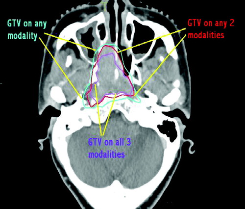
GTV – (1 modality). The volume of tissue defined as tumour on at least 1 modality.
GTV – (2 modality). The volume of tissue that was defined as tumour on at least 2 of the 3 modalities.
GTV – (3 modality). The volume of tissue that was defined as tumour on all 3 modalities.
Figure 2. Derived GTVs viewed on CT image.
The volume measurements for the three imaging modalities were:
GTV-CT 41.1 cm3
GTV-MR 32.3 cm3
GTV-PET 49.7 cm3
The volume measurements according to whether 1, 2, or 3 imaging modalities suggested tumour were:
GTV – (1 modality) 60.5 cm3
GTV – (2 modality) 41.3 cm3
GTV – (3 modality) 24.3 cm3
Discussion
In this patient a GTV that was the sum of CT, MR and PET volumes was 47% larger than the original GTV-CT. Conversely a GTV that required all three modalities to agree was 41% smaller than the original GTV-CT. If a criteria of any 2 of the 3 modalities was used the GTV was similar in volume measurement to the GTVs defined on the individual imaging modalities.