Abstract
This is an “Opinion” or “Commentary” text to support the Invited Opening Plenary Lecture of the NACP2008 Conference. It is an outrageously broad title, that I have been given, and I have made selections for the focus of the lecture and for this report. I observe a research methodology in medical physics whereby key developments have come from standing on the shoulders of those who have come before and I illustrate this by the invention of x-ray computed tomography and the development of intensity-modulated radiation therapy. The equally key role of somewhat maligned incremental science is also discussed. Some commentary is made on the enormous range of activities in medicine to which medical physicists have contributed. Conversely, future gazing is a totally unscientific process. Nevertheless I add my thoughts in broad generalities and also in specifics for the field (radiotherapy physics) in which I work and might be expected to be more accurate in my proposals. I conclude with some remarks on the conditions needed to achieve good scientific outcome.
At the outset it should be said that this is not a paper in the traditional style. It does not “state a problem to be solved”; it has no “materials and methods”; it has no “results”; it does have a conclusion but not in the usual sense. It is instead a “Commentary” or “Opinion paper” and represents the transcript corresponding to the invited Opening Plenary Lecture of the NACP2008, the Nordic Medical Physics meeting which took place in June 2008, in Aarhus, Denmark.
Overview of medical physics
The development and implementation of modern clinical medicine owes a great deal to the contribution from physics. Physics as Applied to Medicine or Medical Physics is now a recognised, well respected, sub-specialty of Physics. Medical physicists are employed to do research, to implement the fruits of research and development in the clinic (sometimes known as clinical physics or hospital physics) and to teach the new generations of medical physicists both at academic specialty level and also with “hands-on” practical training. When I began my career in the subject (in the early 1970s) it was possible and encouraged to combine all these roles. It was also expected (probably unrealistically even at that time) that one could turn ones hand to almost any branch of the subject. There was in the UK very little grant-driven research and generally medical physicists were very free to be inventive, creative and expansive and to change direction whenever this was appropriate. That was a good thing. The funding for this just “seemed to be available”.
Very soon afterwards, imported from the USA, a culture of grant-driven research emerged in the UK and throughout Europe and medical physics itself developed many sub-specialties with somewhat artificial, yet very real, boundaries between them through which passage was not at all easy. It also became necessary to be more proscriptive as to what research would be done and then to stick to those proposals. I felt that was not wholly such a good move. I might smile a little to see now the emergence of grant calls for cross-disciplinary research which, to my somewhat older mind, is a call to do what we were naturally able to do before these boundaries ever existed. However, that may be to start on too negative and cynical a note because the culture of grant-driven research has also had the immense benefit of providing increased funding for the subject, ever expanding departments and the influx of a wealth of talent that, at least in principle (through the Bologna process), knows no geographical boundaries. Also it has to be acknowledged that each branch of medical physics has now become enormously complicated and that understanding the long-term goals and the pathway to them is a necessary part of the research process.
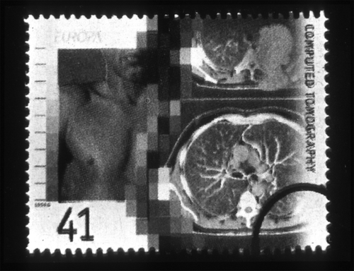
Figure 1. A UK postage stamp of 1994, value 41p, showing CT scans.
It might also be observed that, whereas in the early days of medical physics there was almost no formal induction to the subject, with the passage of time have emerged superb training schemes that not only teach the formal basics of the subject but also ensure regulatory compliances and the maintenance of high professional standards. In most developed countries there are now professional organisations whose remit it is to foster the development of medical physics. In the UK this is the Institute of Physics and Engineering in Medicine (IPEM) Citation[1]; throughout Europe it is the European Federation of Organisations for Medical Physics (EFOMP) Citation[2] and Internationally it is the International Organisation for Medical Physics (IOMP) Citation[3]. These training programmes address the above mentioned complexity of modern medical physics. However, they must not be allowed to become so lengthy and so stifling as to throttle the budding initiative of young trainee minds especially those anxious to make definitive contributions rather than have yet more training. Also it is necessary to ensure that, as medical physicists necessarily focus narrowly on a complex topic, they do not overlook the possibility of lateral thinking from other fields and even those that are not primarily aimed at medicine.
How it was and how it is and how it will be
When can we regard the subspecialty of medical physics as having been born? At the risk of making an over-restrictive generalisation, most physicists today work on two broad areas: (i) imaging and diagnosis in relation to disease and (ii) therapy in relation to disease, where the main disease speciality is cancer. So, it might be said that medical physics began in 1895 when, on November 8th, the then 50-year-old German professor Wilhem Conrad Röntgen discovered, in Wurzburg, the x-ray in the early evening, probably the only major event of its kind not just to be dated but timed for its place in history. However, physicists were plying their science (and sometimes almost art) to medicine for centuries before this. See for example the interesting developments in attempting to harness physics to benefit medicine in the early 1800s Citation[4].
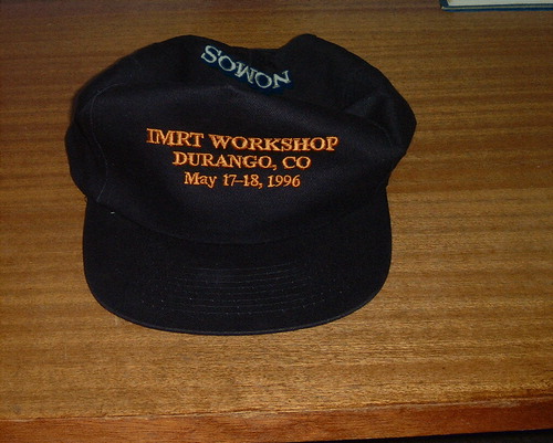
Figure 2. The hat won by delegate to the world's 1st IMRT School in Durango, Co in May 1996.
Those who look backwards are sometimes tempted to regard early days as better days. Certainly if freedom from associated burdens of grant writing, report constructing, audit, too much burocracy and too many committees, etc is valued, then better times they were in that medical physicists were largely 100% doing medical physics. But times were not so good in other ways. We must recall that for the first half of the 1900s medical physicists were numerically few and often working in totally uncharted territory. Imagine trying to do radiotherapy in the days when the unit of dose was hardly defined and very primitive ways to measure dose were in vogue. Imagine having to define the Röntgen and the rad. Imagine having only primitive x-ray equipment available that could only take planar x-rays and even then over-irradiated the patient's skin in the process. Imagine having no functional imaging and having to invent it. Imagine no artificial radioisotopes or their use in nuclear medicine. Imagine … imagine … the list is endless. The early medical physicists were like the early mariner explorers seeking new continents. The early medical physicists made what today seem fundamental discoveries and clarifications but they had little but their intuition and skills from studying mainstream physics to guide them. There were very few professional societies and those there were were largely offshoots of the medical profession. The British Institute of Radiology was an exception that in 1920 gave physicists equal professional status with medical practitioners and even organised some simple training programmes. The UK Hospital Physicists Association (HPA) was not formed until 1943 when there were just 53 medical physicists in the UK Citation[5], Citation[6]. Imagine also the much more primitive communication channels – no commercial aircraft, the need to go to the USA or Far East or Australia by ship, no email (though sometimes nowadays, on bad overloaded days, we may think that was not such a bad thing), no internet. There are some quaint stories of slow professional travel around the world in a book Citation[7] written by a UK professor of medical physics (John Roberts) who was also the 1st Editor of Physics in Medicine and Biology, the journal of which I am currently the 11th Editor. A fascinating retrospective has also been recently written by Alan Jennings, one of the only four surviving Founder members of the HPA Citation[8]. Another fascinating retrospective was the Wellcome Witness on the development of physics as applied to medicine in the UK between 1945–1990 which also references many more substantial tomes on the history of our subject Citation[9]. There is a mini industry of publishing memories of the early days of medical physics and here it is impossible to do justice to that. Those who grow old in their subject tend to be those with greatest interest in retrospective study.
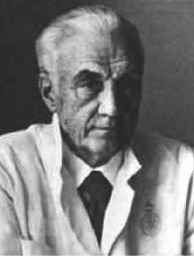
Figure 3. The pioneering Lars Leksell.
It is far harder to write about, and there is far less literature on, the future of medical physics. It is quite fashionable to ask (sic) luminaries to prophesy the future. “They” usually ask you when you are near career-end and may not be around long enough to see if you were correct. I have been asked 4 times before to act in this capacity so this is the 5th such request Citation[10–13]. Some might argue that this is a fruitless effort. Scientists are trained to hypothesise, investigate, report their findings and stop at that. Prophesying the future is a totally unscientific process. It can however provide insight into future research channels and can be entertaining; at worst writers and lecturers can look egocentric and possibly ridiculous. An innovative approach was taken for the 50th anniversary of the UK National Health Service in which a Symposium bringing together forward thinkers was followed up by a book Citation[14].
Two example key contributions of physics to medicine
Rarely can or do any medical physicists set out to make key discoveries, much as they would like to. That is not an approach likely to work. Rather, it is with hindsight that one can look back and identify that certain key discoveries or inventions or key understanding of a phenomenon came about by people, whose names are now forever remembered and associated with those events, “standing on the shoulders of giants” as Newton so eloquently put it. “Big hit science”, the science that has changed the world forever has generally evolved this way. Most physicists, conversely, have performed important “incremental science”, sadly a phrase much disliked by grant-funding bodies; yet it is this very incremental science that has often led to the big-hit landmarks. Let us look at some examples.
Commercial x-ray computed tomography (CT) has been hailed as the greatest revolution in radiology since the discovery of the x-ray. The first scanner, invented and built by the British EMI Company, was announced in April 1972. Whilst work leading up to this announcement was joint between several scientists in London Hospitals, the DHSS (Department of Health and Social Security) and EMI it is the name of Sir Godfrey Hounsfield (28.8.1919–12.8.2004) which will always be associated with the CT scanner. He made a reality what many others had worked on and even “nearly achieved”. He was knighted, became an FRS (Fellow of the Royal Society), received the 1979 Nobel Prize for Medicine and was feted with honours. His original CT scanner stands in the London Science Museum alongside the Apollo-10 lunar module that circled the moon, a surely not un-deliberate juxtaposition. He has been cited as one of the 1 000 most influential people of the 20th century Citation[15]. Yet if one looks back at the history it can be seen that many shared his vision. Alan Cormack (23.3.1924–7.5.1998) performed pioneering laboratory experiments in 1963 and shared the Nobel Prize. Gabriel Frank patented the (nearly) CT principle in 1940. A CT scanner was reputedly built in Kiev for medical purposes in 1957. David Kuhl made a CT scan in 1965 Citation[16].
Another key development in medical physics is the invention of techniques to deliver intensity-modulated radiotherapy for curing cancers Citation[17–20]. Most acknowledge that Anders Brahme of Stockholm, Sweden published the first paper explaining primitive techniques for achieving this Citation[21]. He visited a number of other cancer Centres and work in these may have been stimulated to commence by his visits: (DKFZ-Heidelberg: Schlegel and Bortfeld; MSKCC-New York: Mohan and Mageras; ICR-RMH-London: Webb). Mark Carol, CEO of the NOMOS Corporation announced the invention of the World's first commercial IMRT delivery equipment, the MIMiC, in October 1992 and this dominated American delivery of IMRT for about 3 years before multileaf-collimator-based methods caught up and probably overtook. Yet this development also did not come out of nowhere. An American mathematician, George Birkhoff explained in 1940 the principles whereby any drawing could be composed of the superposition of a series of straight lines from different directions and of different blackness, including negative blackness, the addition of erasure components. Apart from the un-physicality of negative x-rays this is the IMRT principle if the lines are interpreted as x-rays and the picture is interpreted as a dose distribution Citation[22]. Basil Proimos introduced the concepts of gravity blocking to produce dose distributions with concave isodoses as long ago as 1940 Citation[23].
Figure 4. A famous quotation and Tenniel illustration from Lewis Carroll's book “Alice in Wonderland”.

Key being stressed here is that these are landmarks that most would agree “matter”. They have changed the way patients are diagnosed and treated. Let us not perhaps argue too strongly over which names should be linked to them. The award of prizes and honours can be a divisive business (witness the quite high profile writing about Damadian over the non-award of the Nobel prize for magnetic resonance imaging Citation[24]). The key issue for the future and well-being of mankind is the landmark itself and, as seen in these two examples, it stands firmly on the shoulders of the giant work preceding. Finally, it should be clearly said that many other similar examples to these could have been cited. I am not wishing to say these are the two most important landmarks; they are two I understand. The next section introduces a much wider set of landmarks.
A broad-brush overview of some other past contributions of medical physics to medicine
In this section we make some brief remarks about the whole field of contributions of medical physics to medicine. This is an almost impossible task given that whole books are given to each topic, that this paper can have only 25 references and that the stories behind these contributions are huge. However, let us try.
Concentrating initially on the disease of cancer, physicists invented many ways of diagnosing disease by imaging. The earliest was x-radiology (1895), simple xerography (1907), then classical tomography (blurred slice imaging) (1920), optical diaphanography (1929), stereoscopy, analogue tomosynthesis leading to x-ray CT in 1972 as already discussed. Nuclear medicine imaging started in the 1940s with the use of simple Geiger counters (1948), scintillation scanning (1950), Anger camera imaging (1957), the technetium generator (1960), rotating SPET scanner (1963), analogue single photon tomography (1967) and the commercial gamma camera SPECT (1978). Positron emission tomography (PET) started in 1951 and simple PET scanners were invented by 1953. The principles of magnetic resonance were developed in 1946 and the first in vivo MR imaging was around 1976, pathological MRI following around 1980. Ultrasonic scanners date back to the 1930s but the WWII produced the impetus for technological development and the first human ultrasound pictures were made around 1950. Imaging of pregnancy by ultrasound started in 1961. Leksell in Sweden was working on ultrasound imaging in the 1950s. Electrical impedance tomography started in 1982.
The detection of gamma-ray photons owes much to the development of the Crookes’ spinthariscope (1903), the Wilson cloud chamber (1895), the gold-leaf electroscope and the Geiger counter (1929). Artificial radioisotopes were first produced by the Lawrence cyclotron (1931) and nuclear reactors at Oak Ridge and Brookhaven and AERE (Atomic Energy Research Establishment) Harwell in the 1940s.
Radiotherapy for treating cancer owed its success initially to the development of big particle-accelerating machines Citation[17]. Gustaf Ising proposed the idea for the linear accelerator in Sweden in 1924. For a while other methods were used however, e.g. Coolidge used cascaded tubes to obtain 900 keV (1927). Rolf Widerøe from Norway invented the Betatron (1928) and got the linac concept to work; Van der Graff built his prototype generator in 1929. The cyclotron was invented in 1931 by Lawrence. Vickers developed the Cockroft-Walton generator in 1937 in London. The Varian brothers invented the klystron in 1937 and a magnetron was developed by Randall and Boot in 1939. The Kerst Betatron was available for medical use in 1943. Grimmett produced the first practical proposal for patient irradiation using cobalt-60 in 1948 after Mayneord had brought radioactive cobalt back to the UK from AEC Chalk River and Oak Ridge in 1946. The first medical cobalt unit was in Saskatoon in 1951. Leksell introduced radiosurgery in 1951. The first medical linear accelerator was in London in 1953 and the first in the USA in 1954. The first medical proton irradiation was in 1954 at Berkeley Radiation Laboratory and the first cancer patient was treated at the Gustaf Werner Institute (now the Svedberg Laboratory) in 1957 in Uppsala, Sweden. The first computerised treatment-planning system was in 1958. The multileaf collimator was patented in 1959 but not commercially available until 1984. The gamma knife was first used in Stockholm in 1958. Note that all these important inventions and applications of technology in radiotherapy were the work of physicists and engineers (who maybe did not all think of themselves as medical physicists) and also all these developments long predate IMRT which stands on the shoulders of these technologies.
The majority of medical physicists work in imaging and therapy for cancer, but not all. Medical physicists play an important role in cardiology, in surgery, in understanding the forces on and within the human body. They work on understanding the physics of lungs and breathing, the behaviour of electricity within the human body, the understanding of pressure in the body, audiology, optical behaviour in the eyes and the mechanics of vision.
If we can also include people who would rather call themselves medical engineers then there are yet more who design and build equipment for physiological measurement and physiological correction of disfunction. There are those who work to improve all manner of devices for the disabled, the scope far exceeding mobility devices such as wheelchairs. There is physics and engineering in anaesthetics, in sports medicine and treating sports injury. Medical physicists have made contributions to the study of the effects of air travel, of space travel, of the use of mobile phones, of electricity pylons, of domestic electrical equipment, of other environmental pollutants and concerns.
Medical physicists and engineers have built equipment for DNA sequencing, for a host of laboratory physics and chemistry associated with drug discovery and refinement. It might be hard to identify areas of medicine where medical physicists do not play some role.
Before leaving landmarks we might note there is a significant difference between output and outcome. Output is very easy to quantitate through numbers of papers, citation indices, h-indices of authors, lists in reports etc. Outcome is much harder to quantitate. Usually there is a very long time lag between the fundamental inventions and their impact on society.
Some key Nordic (medical) physicists
Grouping, somewhat artificially, together a number of physicists whose work has influenced the development of medicine, we may identify several key Nordic contributors. Anders Brahme (Sweden) has already been mentioned in connection with IMRT. The Norwegian: Niels Henrik Abel (August 5, 1802 – April 4, 1829) has given his name to an integral (Abel's integral) which arises in the only analytically soluble IMRT problem which is an analytically soluble inverse problem first studied by Brahme et al. Citation[21]. His name is also given to a branch of mathematics and much more. Lars Leksell (Sweden) (1907–1986) was the pioneer of radiosurgery, stereotaxy and the Gamma Knife. Bjarne Stroustrup, born December 30, 1950 in Aarhus as you must surely know, is the inventor of C + +. Many inverse-planning codes and other codes in medical physics are written in C + +. The Norwegian-American Ernest O. Lawrence, born August 8, 1901, died August 27, 1958 was a physicist and inventor of the Cyclotron, much used in nuclear medicine. Rolf Sievert (1896–1966) (Sweden) showed how to calculate the absorbed dose to tumours from intracavitary and interstitial sources. He also invented a number of ingenious instruments for dose measurements, among other the world-wide known Sievert chamber. He gave us the unit for equivalent dose, the sievert. Some other key Nordic contributors are (i) Niels Finsen (Denmark): light treatment, (ii) Søren Bentzen (Denmark): radiobiology, (iii) Anders Ahnesjö (Sweden): radiotherapy treatment planning, (iv) Hans Svensson (Sweden): IAEA contribution, (v) Jens Overgaard (Denmark): radiotherapy (and the “green journal”. And (dare we add?) the “adopted Nordics” (i) Pedro Andreo (Spain/Sweden): Monte Carlo and dosimetry, (ii) Alan Nahum (UK/Sweden): dosimetry.
Can I add a disclaimer please? The writers of history tread dangerously and compiling a list such as this will exclude some people who may then be intensely irritated at their omission. Please forgive me. I write in good faith, being somewhat selective.
Regarding elements much used in medicine we may note some major Nordic discoveries. Some of the discoverers were from Scandinavia and some discovered in other countries have been given Nordic names with reference to http://home.clara.net/rod.beavon/elements.htm: A slide of these was shown in the Lecture but the full lengthy list is not given here.
Thoughts on the general goals of cancer research
Whilst not the only focus of physics applied to medicine, cancer research is a key area. We might observe that the goal of cancer research as it was enunciated early in the 1900s was to cure cancer i.e. arrest tumour growth with possibly little attention played to the sparing of dose to normal structures (largely physically impossible anyway) or to the preservation of a high quality of life. People died relatively young and so keeping them alive was the primary goal. With much increased life expectancy, modern man may expect to live well into his 80s and maybe 90s with cancer-incidence age not having changed much. The goal of much cancer treatment is therefore to put the patient into largely symptom-free remission and expect that tumours may require to be re-treated later in life. To that end the preservation of normal function and minimisation of dose to organs at risk becomes a major focus of attention in addition to treating the primary tumour. This is one of the main achievements of IMRT, that it can be performed with an eye to the future. The goal is to ensure that man lives long and dies quickly in contrast to the situation today where there is much chronic disease as health may gradually deteriorate.
The future for medical physics in cancer
If the future of research in cancer-applied medical physics were certain we should be little more than implementational technicians. We cannot know, just as our forebears did not know, the complete course of evolution of medical physics. Indeed, many of the key past developments have come by lateral thinking from other fields. Predicting the future is a totally unscientific process whereby hope, promise and expectation rule at the expense of cold logical and rational scientific argument. Nevertheless there can be goals and key areas in which to work can be identified.
Undoubtedly research will continue in the main areas of medical imaging e.g. SPECT, PET, MRI and MRS, ultrasound and optics. High-speed digital x-ray imaging and CT imaging will make further progress. The current focus of attention in both the imaging and therapy field on understanding and compensating for tissue and organ motion on many different timescales will continue to optimise diagnosis and therapy. Whilst the human genome mapping project is complete, exploiting this knowledge is in its infancy and the development of gene-based therapies will continue. Nanotechnology has been identified as a key focus with the expectation that nanoparticles could act as contrast agents, be vehicles for drug delivery, act as microsensors of local pH, temperature, drug concentration, dose, DNA damage and cell survival. Nanocrystals of semiconductor material coated with a shell to form so-called quantum dots can fulfil these roles. Already ultrasound microbubbles are in use and could be further exploited to deliver drugs locally to tissues.
It is claimed that high-intensity pulsed electric fields applied to cancer cells can enhance the uptake of cytotoxic drugs. It is possible that photochemical internalisation can treat cancer by releasing drugs internally by shining light on selected regions. There may be a link between late radiation damage and genetic profile, still to be explored.
What is inevitable is that the trends in development in computing will make it possible to solve today's problems faster and also open up the possibility to solve problems quite impossible to address today. In my paper Citation[13] I gave the data, derived from help by Gerry Battista at the 15th ICCR in Toronto, on the doubling times for many computer benchmarks. Also DNA computing, now in its infancy, may deliver orders of magnitude more computing power Citation[25].
The future for medical physics in radiotherapy
I can be more specific in my “prophetic remarks” concerning the future of medical physics in my own discipline of improving the physical basis of radiation therapy. I can see further ahead, be less vague and also create “wish lists for the 3rd millennium”. Some thoughts are little more than extensions of today's activities; others are more contentious and may even be outrageously unthinkable.
Regarding improving diagnosis, more thorough screening programmes should lead to a shift in stage profile and a greater focus on cancer patient survival. Biochemical tests and genetic tests should be added to the information from 3D multimodality imaging. A move further away from anatomical imaging to functional imaging will occur. Telemedecine, which was much talked about years ago, seems to have failed to develop widely; yet it is just this technology which could bring the medicine of the highly developed world to the less-well-developed world. Even in areas with equal development, telemedicine could overcome inequalities in health care provision on a geographical basis (postcode medicine) There may be no need at all to have the doctor in the same place as the patient. Another early prediction Citation[14] that mankind will somehow carry its own biosensors, remotely and automatically linked to room-based diagnostic devices, summoning medical help even to asymptomatic patients, does not seem to have taken off a decade later; but it might. It may appear science fiction to those waiting to see their GP (General Practitioner i.e. family doctor) in today's cash-strapped medical economy.
Radiotherapy treatment planning should be patient-individualised and based on measuring radiosensitivity, response to tissue assays and 3D functional as well as anatomical imaging. The bottleneck of tissue contouring by automation never seems to go away, yet needs to. Each patient treatment plan, delivered data and therapy outcome should be entered into a database to form the basis in the future of elaborate correlation studies to determine what really does influence the outcome of radiotherapy. The use of single-modality treatments may become obsolete as the possibility to deliver multi-modality treatments (combined photon, proton, heavy-ion therapy with brachytherapy, radionuclide therapy, targeted drug therapy and focused ultrasound). Multiple plans might be prepared to select an optimum plan. Planning should rely increasingly heavily on 4D data with multiple imaging throughout the course of therapy and adaptive readjustment of plans to cater for changed functional status, the discrepancy between delivered and planned dose and other factors. Finally, regarding planning, MonteCarlo methods should render obsolete the rather quaint terminology of 20th century radiotherapy physics dosimetry.
Radiotherapy treatment will also greatly change. 3D and 4D medical imaging will be used to adaptively adjust the plan both for inter-fraction motion and intra-fraction motion. As well as simple re-positioning, the fractionally incremented delivered dose could be computed to form the basis for adaptation. Predict-ahead techniques will be required for overcoming system latency between making a measurement of patient motion and correcting for it. Regarding technology, why should therapy be limited to the use of one delivery machine at a time. Maybe multiple robots could be used just as they are in the car-manufacturing industry. Whilst the role and importance of hadron therapy is still debateable (for some), it is not arguable that a country without a hadron facility cannot sensibly contribute to the scientific debate. For this reason every country should be so equipped by at least one Facility to act as a National Referral Centre. Micro-beam radiotherapy is just starting and as yet unevaluated. Is there also the possibility that gene therapy and radiotherapy could be combined in some synergistic way.
Finally regarding radiotherapy the assessment of response to treatment is still in its infancy. Post-treatment data collection must become the normal practice and include all the scientific planning and delivery data as well as the patient's observed and also self-reported response. Were such data centrally recorded, it would be possible to conduct trans-national studies with large cohorts of patient data. The biological outcome can then be much more clearly understood.
Establishing the conditions for good progress
As possibly overemphasised here, “future gazing” is an unscientific yet required process and some opinions have been expressed. It is a little more straightforward to expound some principles on which the performance of good science should be based. In staccato form we may say:
The supply of medical physicists must be ensured by educating schoolchildren to find science, which is difficult to do, rewarding; specialisation is useful but medical physicists are first and foremost physicists and many breakthroughs have come from untrained medical physicists who have thought laterally. Despite increased professionalisation there must be a route into medical physics for such people without too great a re-training impediment.
Training schemes are, however, vital and need to be (and are) well supported by National and International organisations. They should not, however, become too long or stifle creative initiative. The Bologna process should widen.
Medical physicists are often required to do 3 simultaneous jobs – research, teach and implement clinical service. Doing all three fulltime generally doesn't work. Conditions must be engineered so that there is a suitable balance for the individual as well as satisfying the “business model” of the employing organisations.
Research failure should be allowed and tolerated. At present I submit there is too risk-averse a culture.
Too much grant-driven research will kill the initiative process. It should be kept in proportion.
Attention to historical knowledge might avoid unwitting re-invention of topics and problems.
The clutter of employment and institutional diversions, such as committees, scientific politics, administration, business and some unnecessary professional work should be minimised. Those who serve in these capacities do so because they either were, or hopefully still are, medical physicists first and foremost. They should remain such.
Conclusions
In his seminal book for children “Alice in Wonderland” the professor of Mathematics at Oxford University, Charles Dodgson, who wrote under the name Lewis Carroll, describes a meeting between Alice and the Cheshire Cat. The meeting is immortalised in the drawing of Sir John Tenniel. Alice inquires of the Cat “Would you tell me please which way I ought to go from here?” “That depends a great deal on where you want to get to” replied the Cat before disappearing leaving only its smile. This wise answer clarifies the need for goals albeit we may not have available waymarked routes. I have attempted to document some goals on a variety of timescales and with a variety of confidence. I have also tried to indicate the constructional nature of medical physics research and the need to study history and relate this to key landmarks.
Acknowledgements
I should like to thank Dr Vibeke Hansen (Denmark and UK) and Ms Maria Holstensson (Sweden and UK) and Dr Phil Evans (UK) for help with some of the “Nordic associations”. In view of the fact that the invitation to lecture at the NACP2008 Conference in Aarhus with this title was almost identical to the invitation to lecture at the EFOMP Congress in Pisa in September 2007, the two lectures had much in common. Consequently there is an overlap of some ideas expressed in this paper and that in Citation[13] which is in the proceedings from Pisa. I have, however, attempted to re-write those thoughts in a different way, to also substantially add to them and to show the Nordic contribution.
References
- http://www.ipem.ac.uk/ipem%5Fpublic/.
- http://www.efomp.org/.
- http://www.iomp.org/.
- Duck FA. Nineteenth century medical physics. Scope 1994; 3: 32–5
- Collation: no stated author. The history of the Hospital Physicists’ Association 1943–1983. London: HPA; 1943.
- SCOPE. The heritage of the institute of physics and engineering in medicine (Special Issue). York: IPEM; 2006.
- Roberts JE. Meandering in medical physics. IOPP, Bristol 1999
- Jennings A. The early days of medical physics. Article in December 2007 on the IOPP Site Medical Physics Web http://medicalphysicsweb.org/cws/article/opinion/32305; 2007.
- Christie DA, Tansey EM. Wellcome Witnesses to twentieth century medicine Vol 28. The development of physics as applied to medicine in the UK, 1945–1990, 2006.
- Webb S. The future of photon external-beam radiotherapy: The dream and the reality. Physica Medica 2001; 17: 207–15
- Webb S. Radiotherapy physics: The next ten years of technical development. Imag Oncol 2005; 1: 43–50
- Webb S, Evans PM. Innovative techniques in radiation therapy editorial, overview and crystal ball gaze to the future. In: Innovative techniques in radiation therapy. Webb S, Evans PM, Sem Rad Oncol 2006;16:193–8.
- Webb S Combatting cancer in the third millennium – the contribution of medical physics. Physica Medica (EJMP) 2008;42:42–8.
- Marinker M, Peckham M. Clinical futures. British Medical Journal Books, London 1998
- 1000 makers of the 20th century. Sunday Times book. Section on Godfrey Hounsfield written by S.Webb. 1990.
- Webb S. From the watching of shadows – the origins of radiological tomography. IOPP, Bristol 1990
- Webb S. The physics of three-dimensional radiation therapy – conformal radiotherapy, radiosurgery and treatment planning. IOPP, Bristol 1993
- Webb S. The physics of conformal radiotherapy – advances in technology. IOPP, Bristol 1997
- Webb S. Intensity modulated radiation therapy. IOPP, Bristol 2000
- Webb S. Contemporary IMRT – developing physics and clinical implementation. IOPP, Bristol 2004
- Brahme A, Roos JE, Lax I. Solution of an integral equation encountered in rotation therapy. Phys Med Biol 1982; 27: 1221–9
- Birkhoff GD. On drawings composed of uniform straight lines. J Mathematiques Pures Appliqués 1940; 19: 221–36
- Proimos BS. Synchronous field shaping in rotational megavolt therapy. Radiology 1960; 74: 753–7
- http://whyfiles.org/188nobel_mri/ and http://wwwtalkorigins.org/indexcc/CA/CA320_2.html and http://wwwfonar.com/nobel.htm and http://wwwanswersingenesis.org/creation/v16/i3/science.asp and countless other web references.
- Kari L. See website: http://www.csd.uwo.ca/~lila/.