Abstract
Purpose. To quantify by means of cone beam CT the random and systematic uncertainty involved in radiotherapy, and to determine if this information can be used for e.g. technical quality assurance, evaluation of patient immobilization and determination of margins for the treatment planning. Patients and methods. Eighty four cancer patients have been cone beam CT scanned at treatment sessions 1, 2, 3, 10 and 20. Translational and rotational errors are analyzed. Results and conclusions. For the first three treatment sessions the mean translational error in the AP direction is 1 mm; this indicates a small error in the calibration of coronal isocentric laser. The observed SD of the systematic error in each direction is 1 mm if a correction is made after the third fraction with an action limit of 4 mm. The SD of the random errors of the patient group is approximately 1 mm in each direction. The rotational errors have a vanishing mean and a systematic error of 0.5–1.2 degrees and a random error of 0.4–0.7 degrees. The uncertainties from the first three treatment sessions (disregarding rotations) lead to a margin of 4 mm from ITV to PTV for Head-and-Neck patients (all directions) and Thorax patients (AP and lateral directions). In the CC direction, the margin has to be 5 mm for the Thorax patients. The total uncertainty on the patient position grows during the treatment course, especially in the CC direction for patients receiving thoracical irradiation. This may stem from problems in the immobilization of these patients. Consequently, it may be necessary to increase the margins in the CC direction. Once the CBCT scans have been made, the information is available for off-line analysis without any extra workload. Thus, the CBCT data can supplement scheduled QA checks.
In modern radiotherapy (RT) there is a focus on conformity in order to deliver a high radiation dose to the tumour target without compromising nearby critical organs. The aim of highly conform treatments lead to increased demands for geometric precision in the planning as well as in the delivery of the treatment.
Geometric uncertainties enter at different stages in the process. Computed tomography (CT) scans have a finite voxel resolution, which in turn leads to a discretization of the delineation of target volumes. Linear accelerators are mechanical constructions with backlash and calibration uncertainties in all the moving parts. Even though immobilization equipment is used, there will be small differences in the patient position at each scanning and treatment occasion. There are uncertainties in the positioning laser system and temporal variations in the anatomical geometry of the patient.
All these uncertainties can be estimated as can the combined total uncertainty, but it is not easy to make these estimates with high precision. However, the combination of cone beam (CB) CT and image registration software makes it possible to measure the positional error in the treatment situation directly. By means of a number of CBCT scans of each patient, it is possible to obtain a sampling of the empirical positional distribution. In this way, it is possible through evaluation of the errors to use CBCT as an efficient and important quality assurance (QA) tool of the geometric precision of the entire chain:
The aim of this study is to use the patient positioning accuracy data, which is available with no extra workload and no QA time on the machine, to supplement the clinical and technical QA procedures applied in our department. Specific areas of interest include:
Technical treatment machine QA. Standard QA procedures for a treatment machine take time, which could otherwise be used for patient treatment. Therefore, machine QA is often limited to weekly, monthly, quarterly or annual system checks. It is of interest to use all available information to assure the technical quality of the radiotherapy treatment in the interval between scheduled QA checks.
Evaluation of patient immobilization. The quality of the immobilization techniques can be inferred from patient positioning accuracy.
Treatment planning margins. In our department, we use standard margins in the treatment planning. A QA system must be used to demonstrate the continued validity of the standard margins.
Patients and methods
This study includes 84 cancer patients whom have been treated with radical radiotherapy on an Elekta Synergy accelerator, see . All patients have been immobilized by an individualized VacFix bag and an AquaPlast cover. Image co-registration of CBCT and the planning CT scan has been performed at sessions 1, 2, and 3, and subsequently it has been determined whether the patient marking needed correction. Corrections are verified by a new CBCT at the following treatment session, and reproducibility of patient positioning is verified by CBCT at sessions 10 and 20 of the remaining treatment course for each patient. The image registration is performed automatically by the X-ray Volume Imaging (XVI) software and verified by the treatment staff. The grey-value matching algorithm offered by the XVI system is used as this algorithm appears more robust than the bone match that is also included in the software. The automatic registration minimizes the inter observer variations and thereby increases the validity of the data.
Table I. Patient population.
The record and verify system used at our facility is Mosaiq (Elekta, Sweden) which employs a Microsoft SQL database. All translational and rotational errors in patient position are registered automatically in another Microsoft SQL database by in-house developed software, and relations to the patient table in the Mosaiq database are made. Thus, the positional errors can be analyzed based on the stratifications, which can be extracted from the Mosaiq database, e.g. patient diagnosis.
In this study the CBCT scans obtained at the first three treatment sessions are considered separately in order to estimate systematic translational error ∑, which can be corrected by a repositioning of the patient, and to estimate rotational and random translational (σ) errors (for details on systematic and random errors, see Citation[1]). In this way changes in patient geometry due to e.g. weight loss or tumour shrinkage can be disregarded. Patients are stratified according to the anatomical region of the tumour (Head-and-Neck, Thorax and Abdomen). Subsequently, the positional errors at sessions 10 and 20 are considered in order to evaluate the reproducibility of patient positioning over a longer period.
Results
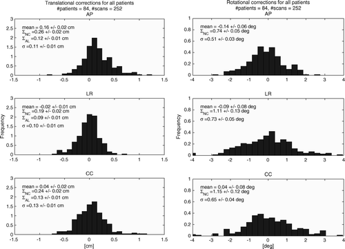
The translational and rotational corrections found for the 84 patients on the first three treatment sessions are illustrated in . Means and standard deviations (SD) of the errors are included in the figure, and the uncertainties on these have been found by bootstrapping Citation[2]. Two values for the systematic translational error are given. ∑NC is the systematic error if the patient position is not corrected (NC) after the third treatment session. ∑AL is the systematic error if the patient position is corrected by the mean error with an action limit (AL) of 4 mm after the third treatment session.
Figure 1. Histograms of the corrections found by co-registration of the CBCT with the planning CT. Translations are in the left column (bin size 1 mm), rotations in the right column (bin size 0.3 deg); AP direction in top row, LR direction in the middle row, CC direction in bottom row. For rotations, the direction indicates the direction of the rotation axis. The histograms are normalized to unity area. σNC is the standard deviation of the systematic error when no correction is taken; σAL is the standard deviation of the systematic error when a correction is made whenever the mean deviation over the 3 first fractions exceeds 4 mm; σ is the SD of the random errors of the patient group.

The mean translational error in the AP direction is non-zero, and the observed SD of the systematic error in each direction is 2–3 mm if no correction is made and 1 mm if a correction is made after the third fraction with an action limit of 4 mm. The SD of the random errors of the patient group is approximately 1 mm in each direction.
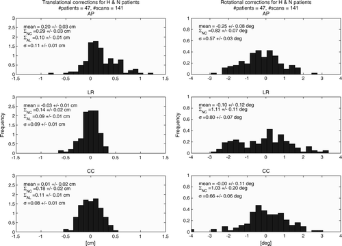
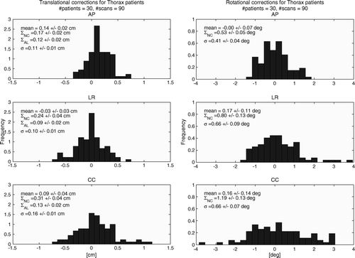
Histograms for the two largest groups of patients in this study, Head-and-Neck and Thorax (C. pulmonis and C. oesophagis), are shown in and , respectively, where the notation is identical to the notation of .
In the overall patient population inaccuracy ∑eff at sessions 10 and 20 are reported. This can be approximated by , and one notes that the combined uncertainty grows in the course of the treatment, especially for the treatment of Thorax patients.
Table II. Patient population standard error (measured in cm).
Discussion and conclusions
Image registration with CBCT combined with automatic storing of the observed positional errors is an easy and efficient way to collect information on the geometric accuracy of the complete set of actions involved in radiotherapy. Once the automatic storing of errors is set up, the quantitative information on treatment accuracy in the form of a large number of CBCT scans is available for analysis in order to reduce the systematic positional errors in planning, plan transfer and treatment. Small errors, which are notoriously difficult to identify on the individual patient, become obvious when a large number of patients enter the statistical analysis.
Technical quality assurance
Radiotherapy treatment machines must be subjected to a range of quality assurance (QA) procedures Citation[3–5]. Standard QA procedures for a treatment machine take time, which could otherwise be used for patient treatment. Therefore, machine QA is often carried out in limited time slots on a weekly, monthly, quarterly or annual basis. It is of interest to use all available information to assure the technical quality of the radiotherapy treatment in the interval between scheduled QA checks.
The patient position data can supplement the scheduled technical quality assurance. Small errors in the mechanical calibration of the various machine parts may produce small systematic errors in the patient positioning. The sudden appearance of a systematic error for the patient population, or a subgroup hereof, is an indication that something in the treatment planning and execution chain goes wrong. Obviously, it must be determined whether any systematic error stems from the CBCT imaging system. If this is not the case, the error must be introduced somewhere else.
Through evaluation of the mean and standard deviation of the observed errors (which can be done off line), it was realized that the coronal laser in the treatment room should be adjusted by 1 mm. The means of the rotations in the three directions are less than 0.24 degrees, which indicates that the directions of the isocentric lasers are well calibrated.
This error was only realized after a large number of scans were available for analysis. The individual scans were performed with another purpose, i.e. to position the individual patient correctly, but subsequently the data were available for technical QA at no extra cost or workload.
Quality of immobilization
The quality of the immobilization techniques used can also be evaluated. Systematic translational errors can be corrected by re-marking the patient. Systematic rotational errors could be corrected by means of a treatment couch with more angular degrees of freedom (such as the HexaPod), but without such a couch all rotational errors as well as random translational errors must be avoided by careful positioning and immobilization of the patient.
If a patient is well immobilized, the typical rotational error will be small. An error of one degree corresponds to a translational error of 1.7 mm in a distance of 10 cm from the axis of rotation, which passes through the isocenter. Thus, a typical displacement due to a rotational error varies from zero at axis of rotation to a magnitude similar to the typical displacement due to a translational error. The exact dosimetric effect of a rotational error is difficult to analyze in simple terms as it depends heavily on geometric factors, e.g. for a spherical target rotations are insignificant, but for a highly elongated target it may be extremely important. Similar considerations apply to organs at risk.
This study reports uncertainties in patient positioning that are similar in magnitude to previously published studies on the quality of immobilization devices, see e.g. Citation[6] and Citation[7].
The total uncertainty on the positioning of the patients increase in the course of the treatment, especially for the patients receiving thoracical irradiation. This indicates problems in the immobilization of these patients, such as a slight softening of the vacuum bag due to incoming air or mechanical stress of the thermoplast. Further studies are needed in order to be more specific.
Planning margins
From the systematic error ∑ and the random error σ it is possible to estimate the margins which must be added to the clinical target volume in order to ensure sufficient target dose coverage (▵ = 2.5× ∑AL +0.7×σ) Citation[1]. Furthermore, since ∑ and σ can be calculated for the individual patient groups, this margin can be individualized to these groups. The uncertainties from the first three treatment sessions (disregarding rotations) lead to a margin of 4 mm from ITV to PTV for Head-and-Neck patients (all directions) and Thorax patients (AP and lateral directions). In the CC direction, the margin has to be 5 mm for the Thorax patients. Thus, our clinically applied standard margin of 5 mm from ITV to PTV is reasonable for our treatment setup, but for the Head-and-Neck patients the present results suggest that the margin could be decreased to 4 mm if the rotational error is ignored.
If the entire treatment series is considered, there is some evidence that the margin may have to be increased for the Thorax patients. From the results presented in one finds that the typical error increases as the treatment progresses. However, since no consecutive scans have been made at this point in the treatment it is not possible to obtain ∑AL and σ separately.
Acknowledgements
Declaration of interest: The authors report no conflicts of interest. The authors alone are responsible for the content and writing of the paper.
References
- van Herk M, Remeijer P, Rasch C, Lebesque JV. The probability of correct target dosage: Dose-population histograms for deriving treatment margins in radiotherapy. Int J Radiat Oncol Biol Phys 2000; 47: 1121–35
- Petruccelli JD, Nandram B, Chen M. Applied statistics for engineers and scientists. Prentice Hall; 1999.
- Kutcher GJ, Coia L, Gillin M, Hanson WF, Leibel S, Morton RJ, et al. Comprehensive QA for radiation oncology: Report of AAPM Radiation Therapy Committee Task Group 40. Med Phys 1994; 21: 581–618
- Purdy JA, Harms WB. Quality assurance for 3D conformal radiation therapy. Strahlenther Onkol 1998; 174(Suppl 2)2–7
- Williamson JF, Dunscombe PB, Sharpe MB, Thomadsen BR, Purdy JA, Deye JA. Quality assurance needs for modern image-based radiotherapy: Recommendations from 2007 interorganizational symposium on “quality assurance of radiation therapy: Challenges of advanced technology”. Int J Radiat Oncol Biol Phys 2008;71 (1 Suppl) :S2–S12.
- Cambria R, Cattani F, Ciocca M, Garibaldi C, Tosi G, Orecchia R. CT image fusion as a tool for measuring in 3D the setup errors during conformal radiotherapy for prostate cancer. Tumori 2006; 92: 118–23
- Zeidan OA, Langen KM, Meeks SL, Manon RR, Wagner TH, Willoughby TR, et al. Evaluation of image-guidance protocols in the treatment of head and neck cancers. Int J Radiat Oncol Biol Phys 2007; 67: 670–7

