Abstract
The diagnostic medical image contains, apart from the pixel data, a detailed description of how the image was produced. The information reveals details on image geometry, radiation data as well as more complex quality index in a varying degree, mostly dependent on the age of the equipment. There is no simple way to retrieve, process and display this data in a general image workstation.
Material and Methods. Since November 2004 a DICOM metadata repository has been used to record image header parameters. The automated data extraction, storage and display are based on simple standard programming and have performed without malfunction since the start, today containing metadata from 18 million images.
Results. The data in the metadata repository has been used in dose optimization for a Computed Radiography image plate system, analyzing the exposure index and making use of the possibilities to organize the data in examinations, projections as well as examination rooms. Analysis of exposure index in the context of these parameters shows promising qualities as it makes detection of dosimetric problems as well as follow-up of dose adjustments simpler.
Current work is aimed at creating a vendor independent platform and to further develop methods to support dose optimization for flat panel direct digital detectors and computed tomography (CT) systems. The possibilities to detect equipment malfunction will be further investigated.
The radiology departments in county Dalarna, Sweden, are fully digital since May 2001. They provide service to 280 000 inhabitants from 4 hospitals and 7 primary healthcare units. When establishing a central PACS (Picture Archiving and Communication System, AGFA Healthcare), the possibility to collect, process and display information from the DICOM Citation[1] (Digital Imaging and Communications in Medicine) image files in the county was recognised. The detector systems in use today includes conventional digital techniques from computed radiography (CR) image plates to flat panel direct digital detectors and other digital modalities such as computed tomography, magnetic resonans imagers, gamma cameras and ultrasound equipment.
Digital modalities generate information on performed exams that can be used to monitor patient dosimetry, radiographic methodology and image quality. Important parameters such as image processing parameters, detector dose, patient dose and geometric information are generated by the modality and transferred from the modality to the PACS database Citation[2]. Even though the information is important, the effort spent exceeds the gain of knowledge when retrieving the information manually, image by image. Many authors have described methodologies to extract DICOM header information for particular studies Citation[3].
The DICOM image file consists of a header, containing patient information and image metadata, and the pixel data i.e. the image itself. Most PACS systems make it possible to view more details from the image header by use of special tools. These tools are limited to handling one image at a time and the image information can not easily be retrieved for further processing.
The PACS database is optimised for clinical usage. Retrieving information that is not used in normal clinical viewing presents a challenge due to the fact that the information is dumped in one field of the PACS database. The structure of the database is another obstacle as it is highly complicated and often considered a commercial secret by the vendor. Access to database query tools, the skills and information needed to retrieve information is normally available only to system administrators.
In November 2004 a data repository for modality parameters from image DICOM headers was created. An automated workflow for web presentation of the information was put in practice at the same time.
This article describes the technique used to extract, process and display the information. The impact of information access for all images produced in the county on radiation protection and dose optimization is discussed. The use of the information is indicated by two examples.
Material and methods
Retrieving Modality parameters from the DICOM header in the PACS database ()
The workflow implemented uses the information in the DICOM header of the image file to create a separate meta data repository at a separate location in the network. The part of the workflow that extracts the header information needs access to the PACS archive server. A filter program removes the pixel data from the image file and extracts the header information after establishing the links to new images. The procedure is repeated every night.
Figure 1. Overview of the workflow. The pixel data, the actual image, is removed from the image file in PACS and the rest of the file content is imported to a DICOM metadata repository. The RIS database connects, together with the DICOM metadata database, to a web presentation workflow via scheduled database queries.
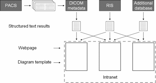
In a second step the modality parameters are imported in the DICOM metadata repository database that currently contains over 18 million image headers from the same amount of images. The structure of the database conforms to the DICOM standard and contains over 200 of the most interesting DICOM header parameters. New parameters can be inserted on demand.
The database contains no images and the patients are not identified.
As the database previously described contains most of the administrative and technical data that the PACS database contains, there is no need to retrieve this kind of information from the PACS database. Moreover the database can be used for general statistical purposes without any risk to interfere with the clinical workflow. The database can be used for statistical analysis outside the radiology department as the patient identity is unknown and the images are removed. The latter also provides the possibility to maintain the metadata repository on a standard computer.
Extracting the data from the metadata repository and the RIS (Radiology Information System) database
The RIS database and the metadata repository database is accessed using SQL (Structured Query Language). The queries are scheduled to produce data summaries at desired frequencies. The output is used as input for web graphs but can also be accessed as data files and processed in spreadsheets on demand.
Presenting the data
The structured data files are used by a graph program (Easy Charts, Oslo, Norway) to generate convenient graph formats.
The graphs are embedded in standard .asp (Active server pages) pages with the use of templates for each type of graph.
The result is a fully automated workflow that extracts new DICOM header parameters every night, imports the parameters in a separate database and extracts and displays the parameters on the intranet.
The skills needed to implement the workflow are limited to basic programming. To produce graphs, a knowledge of SQL (Structured Query Language) is necessary. The final web pages with the graphs are made by the use of standard HTML (Hypertext Markup Language). The workflow is built on standard components with low cost. The up-time has been excellent since the start in November 2004, no unexpected events has occurred.
The use of DICOM header metadata in dose optimization
Exposure index is a valuable parameter in the DICOM header as it describes the dose to the detector Citation[4]. When making timelines showing the exposure index for the CR image plate detectors in use for different examinations it became clear that the dose levels in the institutions were far from optimized.
A pilot project was initiated with the objective to normalize the patient doses in one examination room with the help of the meta data in the database. The information that was used covered, besides exposure index, all administrative data needed to isolate dose levels in specific projections. When adjusting exposure levels the image processing was monitored with the help of the exposure index value. The desired dose level was decided with the support of an experienced radiologist and the recommendations of the CR manufacturer, Fuji Film.
The daily mean values of the exposure index were monitored as the set value for the automatic exposure control for the projections were changed (). The subsequent change in daily mean values of the exposure index was compared with the dose reduction as measured with a 10 cm perspex phantom and the manually registered Dose Area Product where available ().
Figure 2. Follow-up graph. The effect on exposure index (S-value) after dose reduction on lumbary spine anterior projection.
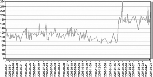
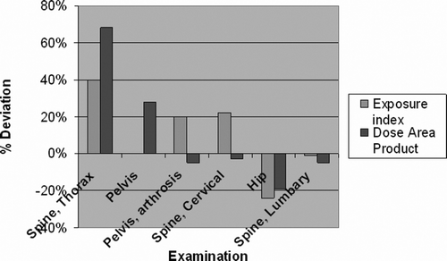
Figure 3. Deviation from expected change in exposure determined by reference method (perspex exposures with AEC) after dose normalisation. Deviation calculated as difference in mean one month before and one month after the exposure reduction.
Examinations from cervical spine down to hip and pelvis was investigated and normalized.
In a follow-up project all examination rooms in the same institution was normalized using the same principle.
For the particular detector in use the only result parameter in use was the exposure index.
Results
When monitoring the change in dose to the detector the exposure index is clearly elevated indicating a reduced dose, the actual exposure index being inversely proportional to the detector dose (). The absolute dose reductions were in the range of 10–50% for the examinations involved.
The deviation from the expected change after dose reduction was calculated as the difference between the exposure one month before and one month after the change in exposure. The Dose area product and the exposure index indicates large deviations from the exposure change measured with AEC (Automatic Exposure Control) on a 10 cm, 24×24 cm perspex slab.
Discussion
The DICOM meta data repository has been helpful in creating an awareness that the exposure levels in our county is far from normalised. The possibility to collect and compare data from a centralised point of view has enhanced the possibilities to detect exposure differences. When comparing the indicated exposure changes obtained by different methods it seems that the conventional method to assess the change with a perspex phantom might be inferior as it deviates significantly from the measurements made on patient images using Dose Area Product and exposure index although further investigations are needed.
When making a large scale normalisation of patient exposures, many examination rooms might need adjustment. The possibility to selectively monitor the projections in any examination room offers a safe and effective way to ensure that no incorrect adjustments are made.
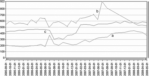
The trend graphs for exposure index have also shown promising tendencies to warn for technical errors. shows some examples of typical errors and tendencies.
Figure 4. Trend showing monthly mean values for exposure index (S) for Thorax anterior projection for different examination rooms. The exposure index is inversely proportional to the dose to the detector. a. Successive change in exposure index due to dose adjustments. b. X-ray generator malfunction with resulting decrease in tube output. c. Manual error in CR reader calibration.
CR image plate detectors have less informative meta data than flat panel direct digital detectors. The reason for this being that the detector in the CR case is not connected to the rest of the imaging equipment. The image file thus only receive information on what can be obtained from the image readout equipment, typically image processing and pixel value distribution, the latter being the basis for the calculation of the exposure index. The flat panel direct digital detectors in use are well integrated in the imaging equipment and the imaging process is well described in the image file. The data obtained from this type of imaging systems have a large potential as the troubleshooting possibility as well as the dosimetric aspects of the images are described in larger detail compared to CR image plates. Current work is oriented towards the use of DICOM meta data in dose optimization of flat panel direct digital systems as well as CT systems.
Future developments
The application currently in use needs the header data in an extracted text file for further processing. This means that a filter program needs to access the actual image files on the image server. However technically simple, this is not allowed by most PACS vendors. In this project the extraction software was developed in close cooperation with the vendor (Agfa Healthcare) and carefully validated before it was installed.
The data extraction needs to be made in a way that ensures vendor independence. In order to achieve this a DICOM client version is under construction.
The new implementation of the data retrieval system appears to the outside world (PACS, modalities) as any dicom aware workstation. Except for the need to set up a new dicom node in PACS no action needs to be taken at the server side. It is also possible to associate this application directly to one or more modalities. For example, if only data from the CT scanners is desired it is possible to configure the application to request new images directly from the scanners with no PACS involved.
With the new methodology, the data mining part is a special implementation of an ordinary workstation query. The header data from all received dicom objects are extracted and formatted in the same way as the old data extraction tool exports its data.
References
- National Electrical Manufacturers Association (NEMA), Digital Imaging and Communications in Medicine (DICOM), 2008
- Riddle WR. Extracting data from a DICOM file. Med Phys 2005; 32: 1537–41
- Vano E. A survey of patient dose and clinical factors in a full-field digital mammography system. Rad Prot Dosim 2005; 114: 375–9
- Warren-Forward H. An assessment of exposure indices in computed radiography for the posterior-anterior chest and the lumbary spine. Br J Rad 2007; 80: 26–31