Abstract
Introduction. Interfractional set-up errors were assessed from daily portal images (PI) registration for head and neck cancer patients. We aimed to evaluate whether a daily PI is worthwhile and we derived the Planning Target Volume (PTV) margins from the estimation of systematic and random errors. Material and methods. Twenty patients were treated in supine position with a fixed 5-point mask immobilisation system and head-and-knee supports. DRRs (Digitally Reconstructed Radiograph) were obtained from the planning CT-scan and considered the reference images to be compared with two orthogonal PI by matching bone anatomy landmarks. A total of 567 PI were done. For the set-up errors analysis, we determined the systematic, random, and overall standard deviations (SD), as well as the overall means in three directions (cranio caudal CC, medio lateral ML and anterior posterior AP). PTV-margins were calculated according to three methods. Differences of SD regarding the overall displacements among portals performed every day and each 2, 3, or 4 days were tested. Results. The systematic set-up errors were less than 1 mm in the three directions whereas the random set-up errors were around 2 mm. PTV margins varied from 3 to 4 mm in the 3 directions. Corrections were significant in the CC direction only, in which the set-up error increased significantly when the scenario of one PI every 3 fractions was adopted. Conclusions. It is of practical importance to apply on-line protocols with contouring of the bony landmarks on the PI in order to decrease the systematic mean error in this patient group. This study suggested that a PI in AP and ML directions once a week and every two days in the CC direction would be adequate to overcome the problem of set-up errors.
The efficacy of the radiotherapy treatment depends partially on patient's set-up at each fraction, especially when the head and neck region is treated, and where safety margins are narrow due to the close proximity of organs at risk such as salivary glands, spinal cord and brain stem. Furthermore, optimized intensity modulated radiation therapy (IMRT) plans usually have high dose gradients between tumor volume and adjacent normal tissue. Set-up uncertainties in patient positioning can produce undertreatment of some part of the tumor, increasing the possibility of local tumor recurrence, while some non-target tissues may be over-irradiated. Geometric uncertainties are more important in IMRT planning as compared with conventional treatments and it is worthwhile to quantify, and if possible to reduce, patient set-up errors during treatment.
The main issue is to reproduce the patient position in each treatment fraction as established at the time of the planning CT-scan. Different immobilization and reliable alignment systems have been used to reduce the patient setup error Citation[1–6]. However, patient positioning is based on the outer surface of the body and therefore effects of patient-dependent factors, such as weight loss and cooperation, are not taken into account.
Portal imaging (PI) allows for easily repeated imaging and image processing, rapid quantitative assessment of treatment set-up errors, and automated verification of the field shape. Several publications on PI guided set-up corrections for conformal and IMRT have been reported Citation[7–15], but they have included small patient groups or did not use daily PI, making difficult to draw conclusions on improving the positioning of patients and the needed frequency of PI verification.
In this study, we performed interfractional set-up verification for each day of treatment in 20 patients. We compared the Digital Reconstructed Radiography (DRR) and the PI, to ensure a good patient positioning at the time of irradiation by using internal anatomic landmarks. The aim of this study was to analyze the set-up accuracy and the efficacy of the daily portal imaging on-line protocol for the positioning of patients treated with IMRT.
Patients and methods
Patients
Twenty head and neck cancer patients treated with IMRT using dynamic multileaf collimation were included in this study. Doses ranged from 46 to 72 Gy delivered in 23–36 fractions over 5–7 weeks. Patient and tumor characteristics are listed in . Bilateral cervical lymph nodes and primary target volumes were irradiated. Target volume was delineated according to the CT-scan planning images. Patients were treated in supine position on a Varian/Clinac 2300 C/D. A fixed 5-point mask system was used for immobilization of the head, neck and shoulders (MCP Posicast PRL on a Posifix carbon plate). The head support was adapted to the neck position of the patient.
Table I. Characteristics of 20 head and neck cancer patients.
Generation of DRR and portal imaging
DRRs were generated from the radiotherapy planning Computed Tomography scan with a slice thickness of 3 mm. The virtual simulation software was the VARIAN/Somavision and a mixed bone/soft tissue contrast was adjusted to approach the PI quality of the linac. Anterior and lateral DRRs were done with a 10×10 cm2 field superposed center on the treatment isocenter. Daily portal images were obtained with a 6 MV photon energy beam on a 256×256 ionisation chamber arrays (Varian). Anterior and lateral PI were generated with double exposures (a small field of 10×10 cm2 and a second field of 20×20 cm2) of 2–3 monitor units.
Before the first treatment session, patient positioning was checked in a conventional simulator and the planned isocenter projection marked on the patient mask. At the treatment unit, patients were positioned by aligning lasers on the marked isocenter. If the PI were incorrect (tolerance level was 2 mm), the patient position was corrected within the mask and the PI was performed to recheck the positioning. The additional table parameters were unchanged and kept constant every day. A total of 567 PI were completed.
Set-up error evaluation
The portal vision software (Varian, Portal Vision Version 6.1) allowed detection and quantification of the set-up error by comparison and superposition with the portal image and the DRR. The superposition was performed by achieving the best possible matching of the outlines of the 10×10 cm2 fields (the so called “split” method). The anatomic landmarks of interest were the superior orbital ridges, the nasal septum and the anterior border of the mandible which were easily identifiable on both images. The considered daily set-up errors were the displacement measured between the landmarks of the DRR and the ones of the daily PI. The cranio-caudal displacement (CC) was measured according to the superior orbital ridges, and the medio-lateral (ML) displacement was measured according to the nasal septum; both on the anterior portal imaging. The anterior border of the mandibule determined the anterior-posterior (AP) displacement on the lateral imaging.
If there were more than one PI in a day, the first and the last portal of the day were used. Data were analysed taking into account negativity and positivity of each displacement. Positive×values indicated a lateral movement to the right, positive y values represented an anterior movement and positive z values represented a cranial movement. The whole data were collected by only one observer (BP).
Statistical methods
A statistical analysis was performed on the measured displacements or set-up errors. For the set-up errors, random (day-to-day variation, σ), systematic (the variation of the mean displacement of patients, Σ), and overall standard deviations (total variation around the overall mean, SD), as well as the overall mean displacement, M) were determined according to Remeijer et al. method Citation[16].
PTV-margins were calculated according to the three methods proposed by Stroom et al. Citation[17], Van Herk et al. Citation[18] and ICRU-62 Citation[19]. These methods were defined using measured distributions of geometrical uncertainties for groups of previously treated patients, which allow attributing a different weight to systematic and random errors. ICRU-62 mentions that systematic and random uncertainties should, in an “ideal approach”, be added in quadrature to obtain one SD, i.e , which should then be used for margin calculation. Margin equal to 1.96* SDtot would then include 95% of the CTV. ICRU methods provide standard aproach to radiation oncology community and they suggested that random and systematic uncertainties have equal effects on a patient dose distribution. However, Stroom and Van Herk formules distinguished systematic errors from random errors as their effects on dose distribution are quite different.
Stroom et al. Citation[17] proposed a CTV-to-PTV margin recipe that properly accounts for the different consequences of systematic and random errors. They found that a margin equal to 2Σtot+0.7σtot ensured an adequate CTV coverage. This recipe implies that the effect of systematic errors is about three times more important than the effect of random errors. Van Herk et al. Citation[18] using another criterion for margins prescription (on average more than 99% of the CTV should at least receive 95% of the dose as compared with 90% of the patients receiving the minimum dose of at least 95%) than that of Stroom et al., resulted in margins equal to 2.5Σtot+0.7σtot.
Differences of SD of the overall displacements between portals every day and for those each 2, 3, 4 or 5 days were tested using the Levene test for homogeneity of SDs Citation[20]. A p-value of <0.05 was considered statistically significant.
Results
A total of 567 anterior and lateral portal images for the 20 patients were analyzed in this study. Of the 567 images analyzed, 462 first lateral portal images (∼80%) and 526 (∼90%) in the anterior direction were taken without an adjustment of the patient position.
We evaluated set-up errors separately in the three dimensions. gives the overall mean, the overall SD and the random SD of the treated patient population before correction (first PI) and after correction (last PI). Distributions of the displacements are shown in . The largest displacement was in the cranial/caudal direction. The SD of the systematic and random displacements in CC direction was 1.20 and 2.26 mm, after correction they were reduced to 0.95 and 1.93 respectively. The overall mean values after corrections showed a displacement of 1.2 in the anterior-posterior and media-lateral direction and of 1.75 in the cranio-caudal direction.
Figure 1. Distribution of displacements in each direction for the first portal images and after last portal images: (a) cranio-caudal, (b) medio-lateral, (c) anterior-posterior.
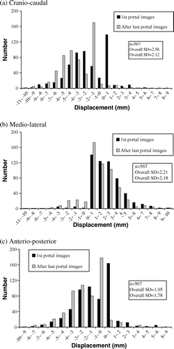
Table II. Set-up errors in CC, ML and AP directions and PTV margins for three models Citation[18–20] and in the three spatial directions
By combining the 2 SDs according to the three different models Citation[17–19], PTV margins were calculated and are shown in . After corrections, it varied from 2.82 to 4.22 mm in the three directions.
Finally, SDs of overall displacements are shown in for five different scenarios: one PI was done at each fraction and every 2, 3, 4 or 5 days. The overall displacement SD of each scenario was compared with the SD of the “a PI every day” scenario. In the CC direction, the SD of the “a PI every day” scenario was statistically lower than in the 3, 4 and 5-day scenarios but not than that of the 2-day scenario. In the other directions, the SD was not significantly lower with the “a PI every day” scenario than with the other scenarios.
Table III. Overall SDs of the measurements for 5 scenarios: daily portal, every 2, 3, 4 or 5 days. P-values for Levene test for homogeneity of SDs
Discussion
The treatment effects of patient set-up errors are more pronounced in IMRT planning because of the high dose gradients obtained to spare organs at risk that are adjacent to the target volume. Xing et al. Citation[21] observed that 3 mm error of the couch location in the AP direction resulted in a 38% decrease of the minimal target dose or in a 41% increase of the minimal spinal cord dose. Therefore, it is crucial to quantify, and then reduce, patient set-up errors during radiation treatment. The purpose of the current study was to analyze the influence of daily PI for reducing set-up errors and subsequently to decide on the appropriate PTV margins definition for the head and neck IMRT treatments in our department. We applied an on-line protocol for 20 patients with a majority of oro- or nasopharynx tumors. Security on daily patient's positioning is usually based on the immobilization mask, that it may be different with the position of the internal anatomy. Therefore, we used orbital ridge, nasal septum and mandible as internal bone landmarks for calculating set-up errors.
Two classes of set-up uncertainties are identified; systematic and random. Systematic error is the deviation between the planned patient position and the average patient position over the whole course of radiation therapy, and machine related errors and target delineation uncertainties are part of this systematic errors. The random error is the day to day deviation from the average target position introduced with internal motion, and it reflects the patient related or mask related factors.
As in other series of the literature, we analyzed errors according to random and systematic set-up errors. In our study, we found that the systematic errors in CC, ML and AP were respectively 1.20, 0.89 and 0.93 without corrections, and 0.95, 0.80 and 0.84 after corrections. We observed that the overall mean values were 1.2 to 1.8 mm after corrections in whatever the considered direction. These systematic errors could be due to (i) the precision on the lasers alignment either of the simulator or of the treatment unit (tolerance level of +/− 2 mm) or (ii) a systematic error of the observer, or (iii) or of the set-up.
There are several publications addressing this issue for conformal RT. Hurkmans et al reviewed some of these publications for the head and neck region treated with conformal RT Citation[22]. In these studies, standard deviations of the systematic and random set-up errors varied respectively between 1.6–4.6 mm and 1.1–2.5 mm. Data for set up verification by PI for head and neck IMRT is very limited. Suzuki et al. published a study Citation[23] in which the mean of the systematic set-up errors for all directions ranged within 1 mm, the average of the individual random set-up errors ranged from 0.7 to 1.6 mm. However, they made verifications of the isocenter every day for the first week of the treatment and weekly thereafter.
It is generally accepted that systematic set-up errors influence more the physical dose distribution than the random set-up errors Citation[24]. In the majority of the studies, systematic errors were usually larger than random errors. However, in our study while the systematic set-up errors after correction were less than 1 mm in the three directions, we found that the random set-up errors were around 2 mm. It is difficult to determine the inter-fractional random set-up errors by weekly PI. It may be one of the reasons why our results are different from other studies where set-up error evalutions were made weekly or only in the first few fractions of the treatment.
When we tried to define how many PI should be done per week, we found that the overall SD displacement increased significantly in the CC directions when the PI was done every 3 days or less, but that PI could be done once a week without increasing overall SD displacement in ML and AP directions. Therefore, in practice, PI should be done at least every 2 days in the CC direction. In the other two directions, a daily PI seems not so useful and could be performed once a week.
The most common approach to overcome uncertainties in patient set-up and organ motions is to add a margin to the clinical target volume (CTV) to create a PTV according to the ICRU 50 Citation[25]. However, improving the patient positioning and performing regular position corrections would result in smaller PTV margins, and consequently in a lower normal tissue complication probability. Therefore, we calculated PTV margins without or after correction according to the ICRU, Stroom and Van Herk models Citation[18], Citation[19].
PTV margins were between 3 to 5 mm without correction in the three directions, after correction it was estimated between 2 to 4 mm. Based on these results we can conclude that a 5 mm PTV margin can be enough for adequate dose distribution in IMRT, and that this margin could be reduced to 4 mm if daily verifications are made. However, the set-up error is only one of the problems from several uncertainty reasons and we can only hypothesize at the present time that the use of new image-guided-radiotherapy modalities would allow to decrease the PTV margin.
Acknowledgements
Declaration of interest: The authors report no conflicts of interest. The authors alone are responsible for the content and writing of the paper.
Related Research Data
References
- Willner J, Hadinger U, Neumann M, Schwab FJ, Bratengeier K, Flentje M. Three dimensional variability in patient positioning using bite block immobilization in 3D conformal radiation treatment for ENT-tumors. Radiother Oncol 1997; 43: 315–21
- Gilbeau L, Octave-Prignot M, Loncol T, Renard L, Scalliet P, Grégoire V. Comparison of setup accuracy of three different thermoplastic masks for the treatment of brain and head and neck tumors. Radiother Oncol 2001; 58: 155–62
- Rabinowitz I, Broomberg J, Goitein M, McCarthay K, Leong J. Accuracy of radiation field alignment in clinical practice. Int J Radiat Oncol Biol Phys 1985; 11: 1857–67
- Kortmann RD, Hess CF, Jany R, Meisner C, Bamberg M. Reproducibility of field alignment in difficult patient positioning. Int J Radiat Oncol Biol Phys 1994; 29: 869–72
- Kumar S, Burke K, Nalder C, Jarett P, Mubata C, A'hern R, et al. Treatment accuracy of fractionated stereotactic radiotherapy. Radiother Oncol 2005; 74: 53–9
- Hess CF, Kortmann RD, Jany R, Hamberger A, Bamberg M. Accuracy of field alignment in radiotherapy of head and neck cancer utilizing individualized face mask immobilization--a retrospective analysis of clinical practice. Radiother Oncol 1995; 34: 69–72
- Bel A, Keus R, Vijlbrief RE, Lebesque JV. Setup deviations in wedged pair irradiation of parotid gland and tonsillar tumors, measured with an electronic portal imaging device. Radiother Oncol 1995; 37: 153–9
- Bel A, Van Herk M, Bartelink H, Lebesque JV. A verification procedure to improve patient set-up accuracy using portal images. Radiother Oncol 1993; 29: 253–60
- Bijhold J, Lebesque JV, Hart AA, Vijlbrief RE. Maximizing setup accuracy using portal images as applied to a conformal boost technique for prostatic cancer. Radiother Oncol 1992; 24: 261–71
- Creutzberg CL, Althof VG, Huizenga H, Visser AG, Levendag PC. Quality assurance using portal imaging: The accuracy of patient positioning in irradiation of breast cancer. Int J Radiat Oncol Biol Phys 1993; 25: 529–39
- Dong L, Boyer AL. An image correlation procedure for digitally reconstructed radiographs and electronic portal images. Int J Radiat Oncol Biol Phys 1995; 33: 1053–60
- Hong TS, Tomé WA, Chappell RJ, Chinnaiyan P, Mehta MP, Harari PM. The impact of daily setup variations on head-and-neck intensity-modulated radiation therapy. Int J Radiat Oncol Biol Phys 2005; 61: 779–88
- Asselen BV, Dehnad H, Raaijimakers CP, Roesink JM, Lagendjik JJ, Terhaard CH. Implanted gold markers for position verification during irradiation of head-and-neck cancers: A feasibility study. Int J Radiat Oncol Biol Phys 2004; 59: 1011–7
- Gildersleve J, Dearnaley DP, Evans PM, Swindell W. Reproducibility of patient positioning during routine radiotherapy, as assessed by an integrated megavoltage imaging system. Radiother Oncol 1995; 35: 151–60
- Yan D, Wong J, Vicini F, Michalski J, Pan C, Frazier A, et al. Adaptive modification of treatment planning to minimize the deleterious effects of treatment setup errors. Int J Radiat Oncol Biol Phys 1997; 38: 197–206
- Remeijer P, Rasch C, Lebesque JV, Van Herk M. A general methodology for three-dimensional analysis of variation in target volume delineation. Med Phys 1999; 26: 931–40
- Levene H. Robust tests for the equality of variance. Contributions to probability and statistics, I Olkin. Stanford University Press, Palo Alto, CA 1960; 278–292
- Stroom JC, Heijmen BJM. Geometrical uncertainties, radiotherapy planning margins, and the ICRU-62 report. Radiother Oncol 2002; 64: 75–83
- Van Herk M, Remeijer P, Rasch C, Lebesque JV. The probability of correct target dosage: Dose-population histograms for deriving treatment margins in radiotherapy. Int J Radiat Oncol Biol Phys 2000;47:1121–35.
- ICRU Report 62. Prescribing, recording, and reporting photon beam therapy. (Supplement to ICRU Report 50). Bethesda, MD; 1999.
- Xing L, Lin Z, Donaldson SS, Le QT, Tate D, Goffinet DR, Wolden S, et al. Dosimetric effects of patients displacement and collimator and gantry angle misalignment on intensity modulated radiation therapy. Radiother Oncol 2000; 56: 97–108
- Hurkmans CW, Remeijer P, Lebesque JV, Mijnheer BJ. Set-up verification using portal imaging; review of current clinical practice. Radiother Oncol 2001; 58: 105–20
- Suzuki M, Nishimura Y, Nakamatsu K, Okumura M, Hashiba H, Koike R, et al. Analysis of interfractional set-up errors and intrafractional organ motions during IMRT for head and neck tumors to define an appropriate planning target volume (PTV)- and planning organs at risk volume (PRV)-margins. Radiother Oncol 2006; 78: 283–90
- Manning MA, Wu Q, Cardinale RM, Mohan R, Lauve AD, Kavanagh BD, et al. The effect of setup uncertainty on normal tissue sparing with IMRT for head-and-neck cancer. Int J Radiat Biol Phys 2001; 51: 1400–9
- ICRU Report 50, Prescribing, recording, and reporting photon beam therapy. Bethesda, MD; 1993.