Abstract
Background. The aim of the study was to investigate the results of treatment of malignant parotid gland tumours at a single centre during a 56 year period, focusing on tumour control and survival. Patients and methods. At Uppsala University Hospital, Sweden, 144 patients (73 male and 71 female) with parotid cancer were treated between 1948 and 2004. The mean and median ages were 62 and 65 years, respectively (range 16–89 years). Surgery was the primary treatment in 113 (78%) patients followed by radiotherapy in 81. Postoperative radiotherapy in doses of 64–66 Gy, where the intention was curative and delivered with either split course or not, was administered to a majority of patients after 1970. The split-course mode was practised between 1970 and 1989. The median follow-up time was 8.3 years for patients still alive. There were 57 (40%) relapses, of which 40 were local recurrences with 26 inside the treatment volume.Results. The overall 5-year survival was 53%. The majority of tumour-related deaths appeared in the first 3–5 years after diagnosis. Age, co-morbidity, the presence of lymph node metastases, adenoid cystic carcinoma and extent of disease were important for outcome; gender, however, was not. We found no difference in the survival between patients following split course therapy versus continuous fractionation. No difference could be seen in the survival of patients treated in the 1970s versus the 1990s. Conclusions. Age, nodal engagement, a higher T-stage, adenoid cystic carcinoma histopathology, facial palsy and intercurrent disease worsen the outcome of patients, whereas gender does not. Treatment principles at our hospital have been surgery followed by radiotherapy since the early 1970s even though a split course technique was practised during a part of this period. Survival has not improved markedly. Thus, there is scope for improvement for this group of patients.
Malignant tumours of the parotid gland are unusual. They represent about 0.5% of all cancers and less than 5% of tumours in the head and neck region Citation[1], Citation[2]. In Sweden, approximately 0.6–0.8/100.000 persons are diagnosed each year, resulting in 80–90 new patients Citation[3]. Most of them are older than 60 years at the time of diagnosis; however, these tumours can also affect younger individuals Citation[4], Citation[5]. There is no proven gender predominance Citation[6]. Mucoepidermoid carcinoma is the most common tumour followed by adenocarcinoma, mixed type malignant tumour, adenoid cystic carcinoma, acinic cell carcinoma, undifferentiated and squamous cell carcinomas Citation[1], Citation[2].
The etiology of parotid gland carcinoma is obscure. It has been associated with exposure to ionizing radiation Citation[7], familial predisposition and inhalation of wood dust Citation[1]. However, there is no apparent association with any etiological factors in most cases Citation[2].
The treatment of choice for malignancies of the parotid gland, as in other salivary gland tumours, is surgery with or without adjuvant radiotherapy Citation[1], Citation[2], Citation[8]. The aim of the surgery is to remove the tumour radically without compromising the facial nerve. In cases where the facial nerve is surrounded by tumour, it has to be sacrificed. Radiotherapy has been used as a complement to surgery either preoperatively or, in most cases, postoperatively, for tumours with a high malignancy grade and/or advanced stage (III–IV) Citation[6], Citation[9], Citation[10]. For inoperable patients, radiotherapy has been the only treatment given. 3-D conformal radiotherapy techniques have been used in the Scandinavian countries since the early 1970s. Chemotherapy has not been used up-front as a treatment of choice.
Clinical stage, facial nerve involvement and nodal status have a significant impact on treatment outcome Citation[8], Citation[10–13].
The aim of the present study was to investigate the clinical outcome for primary parotid gland carcinoma in material from one centre during a time span of 56 years (1948–2004) focusing on local control and survival. This long follow-up period implies that some development in treatment should have occurred. The study was approved by the Ethical Board of the Medical Faculty of the University in Uppsala, Sweden.
Materials and methods
Patients
At the Departments of Oncology and Otorhinolaryngology (ORL) at the University Hospital, Uppsala, Sweden, 144 patients with cancer of the parotid gland, were treated between 1948 and 2004. The median age of this group was 65 years (range 16–89). Seventy-three patients were men (51%) and 71 women (49%) ().
Table I. Patient characteristics. The age, gender, T-stage, N-stage, M-stage and treatment are shown.
The patient files were retrieved from the ORL and/or the Oncology Departments. Information on the surgery, radiotherapy and the subsequent follow-up were extracted from the available records. Concurrent illnesses were noted in some records. Most patients were monitored at the Oncology Department and/or at an ORL Department either at the University Hospital, Uppsala, or at a local county hospital. Records for follow-up or correspondence provided information on recurrence, time to recurrence, treatment at recurrence and cause of death. Twelve (8%) patients were lost to follow-up. Causes of death (see ) were extracted from the Swedish Cancer Registry at The Board of Health and Welfare of Sweden. We were, therefore, able to find the cause of death for the patients by means of investigating these records. It was possible for us to state the cause of death even though the patients were lost to follow-up in the medical records.
Table II. Histopathology according to original diagnosis for all studied patients. The most common diagnosis was mucoepidermoid carcinoma, followed by adenoid cystic carcinoma and adenocarcinomas.
Symptoms at diagnosis
One-hundred-and-nineteen patients (83%) presented with a swelling, which led the patient to seek medical advice. In a total of 20 cases (14%), facial palsy was present at the time of diagnosis, and, for 12 of these patients, a lump was also present. The median time of duration of symptoms was 6 months (range 1 week to 32 years) up to diagnosis.
The most frequent tumours were the mucoepidermoid carcinomas (25%), adenoid cystic carcinomas (18%), adenocarcinomas (17%) and acinic cell carcinomas (15%) (see ).
Of the 144 patients, 81 patients (56%) had surgery combined with radiotherapy (RT), an additional 32 patients (22%) underwent surgery alone and 26 patients (18%) received only RT. Five patients (3%) did not receive any treatment due to their poor medical condition and/or advanced age. In 34 patients (24%), RT was administered postoperatively because of non-radical surgery.
TNM-staging did not exist in the medical charts at all. The TNM-staging was performed retrospectively in all cases according to the 6th edition of the Union Internationale Contre le Cancer, UICC Citation[14].
The original histopathological diagnosis was used. Due to the large time span and difficulties in finding all the specimens, no pathological reviewing of the material was performed.
Surgery
For 113 patients (78%), radical surgery was the primary treatment. Sixty-eight (47%) of these patients underwent primary surgery and received radiotherapy postoperatively. In 32 of these cases, the patients did not receive any other primary treatment than surgery. Thirteen had surgery after radiotherapy. Thirty-one (22%) patients did not undergo surgery due to advanced disease or poor medical condition.
Surgery was performed whenever the tumour was deemed operable. The patients received postoperative radiotherapy if the margins were positive after surgery, if there was residual disease or if the tumour was of a more aggressive type, i.e. adenoid cystic carcinoma. Neck dissection was performed when the tumour in the lymph nodes was >N1 or when there was a residual lump that had not disappeared with radiotherapy.
The surgery consisted of either total or partial parotidectomy. Neck dissection was performed in five cases according to records. For the remaining patients, surgery was not performed because of the extent of the disease.
Radiotherapy
Eighty-one patients were treated with RT as part of their primary treatment, 34 of those because of non-radical surgery, meaning that the surgery aimed for cure, but the postoperative margins were positive and radiotherapy was added to enable cure. Primary radiotherapy was the treatment of choice when the tumour was considered unsuitable for surgery because of the extent of the tumour. An additional 26 patients received RT alone without surgery. Before 1970, radiotherapy was administered once daily with a 100–180 kV Roentgen- or Cobalt source and a multi-field technique. Between 1948 and 1969 all but one patient had postoperative radiotherapy. Before 1970, the treatment was given with one daily fraction and according to the records, no pauses were granted during the treatment period. After 1970, the linear accelerators came into use and most patients received their treatment with these accelerators. During the 1980s, more treatments were computer planned with increased accuracy and precision of the treatment. The treatment has been delivered with 3D conformal radiotherapy since the arrival of the linear accelerators in the early 1970s according to our local standard.
The common standard at our department since the arrival of the linear accelerators was treatment schedules with daily fractions of 2 Gy with a weekly dose of 10 Gy to a total dose of 64–66 Gy. The median area of the treatment fields was 140 cm2 (range 48–315). This was based on 52 cases in whom the surface area could be defined. The treatment volumes could not be estimated.
Of the 107 patients who received radiotherapy, 45 received their treatment with a split-course procedure, the treatment was delivered with a planned pause of 14 days after 4 weeks of RT corresponding to approximately 40 Gy.
The split course was standard treatment between the years 1970 and 1989. The split course was introduced since it was observed that the patients suffered considerable acute reactions to the skin and it was therefore considered inappropriate to give the entire radiotherapy in one series. Two patients had a pause when surgery was performed. These two patients received additional radiotherapy after surgery.
Palliative doses never exceeded 50 Gy and ranged from 10 to 46 Gy.
Five patients received no active therapy but were given best supportive care (BSC), ().
Statistical methods
Overall, cancer-specific and relapse-free survival were analysed using the Kaplan-Meier method. Patients who were lost to follow-up were analysed to the last known date of contact. If known, the cause of death was given. Differences in survival between groups were calculated with the log rank test (Statistica Software release 8), χ2 test and a multivariate analyses was performed. The survival was calculated for 136 of 144 patients. One patient was diagnosed and operated abroad and the exact date of diagnosis could not be found. For two other patients, it was not possible to find the date of diagnosis.
Results
The clinical characteristics of the patients are presented in . The patients in our study were diagnosed in different time periods. Sixteen patients were diagnosed before 1970, 55 patients were diagnosed between 1970 and 1979, 42 patients were diagnosed 1980–1989 and 28 patients were diagnosed after 1989. For two patients, who were diagnosed with cancer of the parotid gland, it was not possible from the medical files to conclude when these diagnoses were established.
Data retrieved from the patient files were sufficient for TNM-staging in retrospect for 97 cases (67%) for T-staging. The N-staging was done with N + or N0 in 115 cases (80%). Eight patients (6%) had distant metastasis at diagnosis and were consequently staged as M1. T- and N-stage could not be found in 47 and 29 cases respectively.
At our department, surgery followed by radiotherapy has been the treatment of choice for patients with adenoid cystic carcinomas, and for patients with any other tumour types, when the margins were not radical. In cases where there was evidence of infiltration into the nerves or advanced stage, postoperative radiotherapy was also delivered.
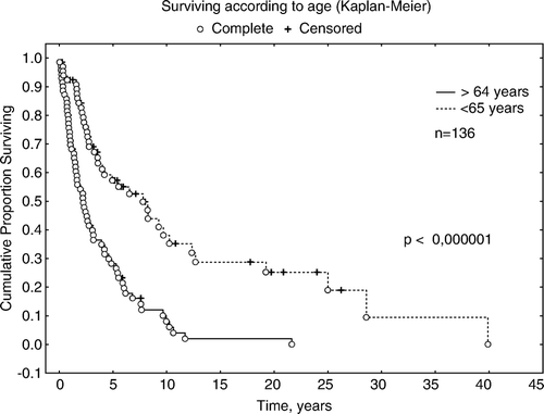
The overall five-year survival was 53% (). The five-year survival for patients younger than 65 years was 58% while the 5-year survival for patients 65 years and older was 23%, p < 0.00001 ().
Figure 1. Survival in the whole group, overall survival. Information was available for 139 patients. Median survival was 6.1 years and mean survival was 10.5 years.
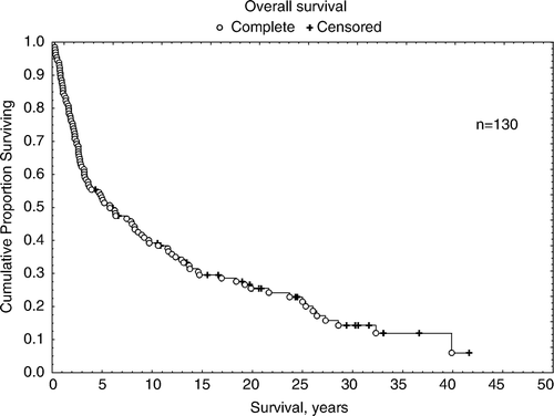
Figure 2. Survival and age. Groups younger and older than the median of 65 years. Log-rank test p < 0.00001.
Histopathology was diverse in this relatively small group of patients. The best 5-year survival of 70% was reported in patients with adenoid cystic cancer. Patients with mucoepidermoid carcinoma had the poorest prognosis with a 5-year survival of 30%. There were no significant differences (p = 0.198).
Twenty (14%) patients had facial palsy at diagnosis and these patients had a poorer prognosis than patients without facial palsy. The 5-year survival in this group was 18%, compared with patients without facial palsy for whom the 5-year survival was 55%. This was a significant difference in survival (p = 0.017). Patients with a more advanced stage had a poorer prognosis than those with earlier stages at diagnosis. Stage I disease had a 5-year survival of 59% while stage II had one of 48%, stage III 37% and stage IV 22% respectively. The differences are significant (p = 0.0094). Data are not shown. Stage and survival for the described groups was only performed for patients where staging could be achieved.
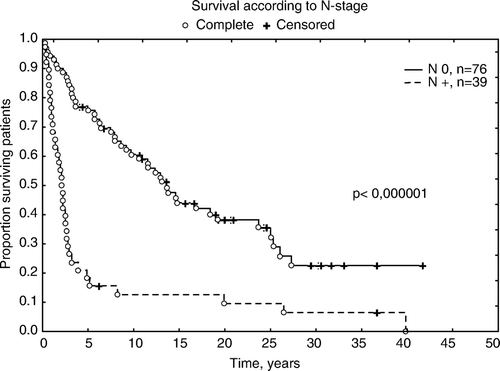
Survival according to N-stage showed a 5-year survival for N0-stage of 75% whereas N + had a 5-year survival of 15% ().
Figure 3. Survival depending on N-stage. N0 = 76, N + = 39. It was possible to define the N-stage in 115 cases. Since the information was not very detailed we grouped patients with no known lymph node metastases (N = 0) or with known lymph node metastases present (N + ) at diagnosis. The outcome for patients with N + disease was worse, with a 5-year survival of 15% while N0 disease had a 5-year survival of 78%. p < 0.00001.
Fifty-two (36%) patients died of parotid gland carcinoma, 22 (15%) died from other cancers while 44 (30%) died from other causes, for instance heart disease, pneumonia and other infectious diseases. Twenty-six patients (18%) were still alive at closing date of the study (February 1, 2007).
We compared the outcome for patients treated in the 1970s with that of patients treated in 1990s. The median radiation dose to the target was 59.9 Gy to patients in the 1970s and 62.2 Gy to patients in the 1990s. There was no significant difference in survival between the two periods (p = 0.475), data are not shown.
Of the 139 patients treated, nine progressed locally in direct connection with treatment or developed distant metastases within a few weeks of treatment. One-hundred and twenty-eight patients (89%) achieved local control after primary treatment but none of the eight patients that presented with distant metastases at diagnosis achieved disease control.
For a total of 102 patients, information concerning recurrence or not could be detected. Fifty-seven patients (40%) suffered a relapse. Forty patients (28%) reported local recurrences. In 26 (18%) of these cases, the recurrences occurred within the treatment volume. The radiation doses in these patients were not lower within the treated volumes than in others. The median and mean times to recurrence were 1.4 years and 3.2 years respectively. Patients treated according to the split-course schedule did not exhibit more local recurrences than the patients with continuous radiation treatment. Data are not shown.
Nineteen patients received radiotherapy with a palliative intent. Twenty-five (17.4%) patients received palliative chemotherapy after recurrence.
Overall survival was 53% in the whole group as concluded at the end of the study (February 1, 2007). Younger patients (<65 years) had better survival and prognosis than older patients. Survival was, in all aspects, better in the younger age group, whether defined as cancer specific or not. In the age-group younger then 65 years, the 5-year survival was 74% while it was 37% in the group 65 years and older. The patients in the older age group had more intercurrent diseases, which both hampered their treatment and caused more morbidity. The difference in survival was significant (p < 0.000001) (see ).
There was no difference in survival between genders (p = 0.49). Data are not shown.
All in all, 103 (72%) patients had died when this analysis was performed. Fifty-two patients (36%) died from cancer of the parotid gland, 10 (7%) died of other cancers, 22 (14%) died from cardiovascular or pulmonary disease and the remaining 34 patients (24%) died of other causes.
Twenty-six (18%) patients were alive at follow-up on February 1, 2007. Twelve (8.0%) patients, including one from abroad, were lost to follow-up. However, reported cause of death and time to death were extracted from the Death Registry of Sweden and the information was available for all deceased patients with the exception of three lost to follow-up.
It has not been possible to perform an adequate analysis of the median-relapse free, cancer- specific or overall survival due to a lack of data in a number of cases as regards the presence or absence of viable cancer at the time of death.
Discussion
The relatively low frequency of malignant parotid tumours presents many problems; prospective studies would demand collaboration from several centres for many years in order to recruit enough patients to attain an acceptable level of significance. The studies presented in the literature are mainly retrospective in nature. Due to the variety of histopathology, it is difficult to achieve a complete survey of the disease. In order to obtain homogenous material, the group most often studied is the largest group, mucoepidermoid cancer Citation[12], Citation[15], Citation[16].
In the 1960s and 1970s, a series of investigations concerning the parotid gland was performed by Eneroth et al. They investigated the effects of radiotherapy on salivary glands and showed that radiotherapy had a diminishing effect on function and size of the parotid gland after radiotherapy Citation[17].
There seems to be agreement in literature that parotid gland tumours should be treated with surgery with or without radiotherapy Citation[1], Citation[9], Citation[10], Citation[18], Citation[19]. In previous years, many centres used single modality treatment Citation[1], Citation[2], but since the beginning of the 1990s there has been a trend towards the addition of postoperative radiotherapy with improved outcome as a result Citation[20].
Östman et al Citation[21], however, found in their comparison of all salivary gland tumours in Sweden during three time-periods no difference in survival, which is in accordance with our results. This may be surprising but, when we scrutinized the material, we realised that the patient cohort is basically the same and the treatment modalities have not changed markedly since the 1970s. In our material, which before 1970 mainly comes from the Department of Oncology, might be bias, treatment was surgery with postoperative radiotherapy in patients with advanced disease and aggressive histopathology.
Since the linear accelerators came into use in the 1970s, surgery in combination with postoperative adjuvant radiotherapy has been implemented.
The 5-year survival rate in our material was 53%. This is in the lower range but comparable to the findings of others showing survival rates of 50–81% Citation[6], Citation[9], Citation[11], Citation[13], Citation[18], Citation[19]. In our study the median age was 65 years, which is higher than in most comparable studies, where the median age ranged between 50–61 years Citation[9], Citation[10], Citation[18–20] except in a study by Kohl in which the median age was the same as in our study. The discrepancy to other studies may also be a consequence of selection in these studies.
As in other studies, gender had no impact on survival Citation[8], Citation[12]. In several reviews Citation[8–10], Citation[20], it has been reported that tumour size, malignancy grade and extent of the tumour are major determinants predicting survival, which we also observed. Presence of lymph node metastases implies a larger risk of recurrence and worsens the prognosis in our study, as confirmed by others Citation[6], Citation[8], Citation[10], Citation[11], Citation[16]. It is consistent with our findings that patients with more advanced disease have a much poorer outcome in terms of overall survival and recurrence rate. Nerve infiltration and facial palsy was associated with poorer outcome and poorer survival in our study as well as in a study by Zbären Citation[6].
The split-course method was used in our material between 1972 and 1989. This procedure did not affect the outcome, which was shown in our study in the comparison of treatment given during the 1970s when the split-course procedure was used compared with the 1990s when it was not. We wanted to estimate the treatment volume but found that this was not possible. The figures are therefore given for treatment surface area. They give, however, a fair estimate of the volumes treated in the material.
It was at an early stage of the study obvious to us that a retrospective analysis of the material would be problematic to achieve because of difficulties in finding the histopathological material due to the large time span. No such retrospective histopathological analysis has therefore been performed. The distribution of histopathology seems, however, to be in accordance with the findings of other authors. The data from the medical records have been sparse in some cases. Despite this, we have only lost 12 (8%) of 144 patients to follow-up. In all but three cases, we found the registered cause of death.
In our study, postoperative radiotherapy has been performed in cases with nerve involvement and non-radical resection as well as in cases of adenoid cystic carcinoma and advanced stage disease. Postoperative radiotherapy has been used as standard treatment since the 1970s in our material and is recommended in several recently published studies Citation[8], Citation[12], Citation[13]. However, the role of radiotherapy has been debated by several authors through the years but there now seems to be agreement that non-radical surgery, inoperable tumour due to tumour extension and more advanced stages require adjuvant radiotherapy. This also seems to be true for certain histopathological types (adenoid cystic carcinoma) Citation[8], Citation[9], Citation[13], Citation[20].
Thus, our conclusions are that age, nodal engagement, higher stages, adenoid cystic carcinoma, facial palsy and intercurrent disease worsens the outcome of patients, while gender does not. We are not content with the finding that the addition of radiotherapy treatment, which has changed from split to continued radiotherapy for an uninterrupted period of 6–6.5 weeks, has not made any real difference in outcome concerning local control and/or survival since the 1970s. In our study, we could not demonstrate any improvement in outcome, mainly because the treatment did not change markedly since the 1970s in spite of the use of split course technique during a period. The adjuvant radiotherapy prolongs the time of local control and survival in patients with non-radical surgery, nerve involvement, adenoid cystic carcinoma and high stage disease.
We believe that new treatment methods must be developed to improve outcome in this ageing group of patients. Hyperfractionated radiotherapy has been discussed by Wang et al. in a study from 1991 as a means of improve the outcome in unresectable cases Citation[22].
No large prospective studies have been performed but should be recommended.
New treatment modalities are needed to improve the outcome for patients with parotid cancer.
The current rapid development in molecular biology will hopefully produce new tools for diagnostics and treatment with high specificity to target sensitive phases in cycling tumour cells. A combination of surgery, external radiotherapy and substances specifically addressing remaining tumour cells may be envisioned.
Conclusion
Diagnosis before or after 1980 showed no significant difference in survival, no significant difference N + versus N0, no significant difference advanced disease versus not advanced disease, no significant difference according to age and not more significant numbers of facial palsies.
Overall survival is significantly influenced by age, facial palsy, N + and advanced stage but not influenced by gender, histopathological diagnosis, time period of diagnosis or split course radiotherapy in a multivariate analysis.
There is no difference in outcome according to split course radiotherapy; the use of split course radiotherapy is similar between the groups when it comes to appearance of facial palsy, N + and age.
Acknowledgements
Declaration of interest: The authors report no conflicts of interest. The authors alone are responsible for the content and writing of the paper.
References
- DeVita VT, Jr, Hellman S, Rosenberg SA. Cancer, principles & practice of oncology5th ed. Lippincott-Raven, Philadelphia-New York 1997
- McGurk M, Renehan A. Controversies in the management of salivary gland disease. Oxford University Press, Oxford 2001
- http://www.socialstyrelsen.se/en/Statistics/, 2008.
- Sato T, Kamata SE, Kawabata K, Nigauri T, Mitani H, Beppu T, et al. Acinic cell carcinoma of the parotid gland in a child. Pediat Surg Int 2005; 21: 377–80
- Claros, P, Dominte, G, Claros, A, Castillo, M, Cardesa, A, Claros. A parotid gland mucoepidermoid carcinoma in a 4-year-old child. Int J Pediat Otorhinolaryngol 2002;63:67–72.
- Zbären P, Schüpbach J, Nyuens M, Stauffer E, Greiner R, Häusler R. Carcinoma of the parotid gland. Am J Surg 2003; 186: 57–62
- Saku T, Hayashi Y, Takahara O, Matsuura H, Tokunaga M, Tokuoka S, et al. Salivary gland tumors among atomic bomb survivors, 1950–1987. Cancer 1997; 79: 1465–75
- Koul R, Dubey A, Butler J, Cooke AL, Abdoh A, Nason R. Prognostic factors depicting disease-specific survival in parotid-gland tumors. Int J Radiation Oncology, Biol. Phys 2007; 68: 714–8
- Tullio A, Marchetti C, Sesenna E, Brusati R, Cocchi R, Eusebi V. Treatment of carcinoma of the parotid gland: The results of a multicenter study. J Oral Maxillofac Surg 2001; 59: 263–70
- Bron LP, Traynor SJ, McNeil EB, O′Brien CJ. Primary and metastatic cancer of the parotid: Comparison of clinical behaviour in 232 cases. Laryngoscope 2003; 113: 1070–5
- Bhattacharyya N, Fried MP. Determinants of survival in parotid gland carcinoma: A population-based study. Am J Otolaryngol 2005; 26: 39–44
- Pires FR, de Almeida OP, de Araujo VC, Kowalski LP. Prognostic factors in head and neck mucoepidermoid carcinoma. Arch Otolaryngol Head Neck Surg 2004; 130: 174–80
- Harbo G, Bundgaard T, Pedersen D, Sogaard H, Overgaard J. Prognostic indicators for malignant tumours of the parotid gland. Clin Otolaryngol 2002; 27: 512–6
- Sobin, LH, Ch. Wittekind. TNM, Classification of malignant tumours, UICC, 6th ed. New York; 2002
- Kokemueller H, Swennen G, Brueggemann N, Brachvogel P, Eckardt A, Hausamen JE. Epithelial malignancies of the salivary glands: Clinical experience of a single institution–a review. Int J Oral Maxillofac Surg 2004; 33: 423–32
- Boahene DK, Olsen KD, Lewis JE, Pinheiro AD, Pankratz VS, Bagniewski SM. Mucoepidermoid carcinoma of the parotid gland: The Mayo clinic experience. Arch Otolaryngol Head Neck Surg 2004; 130: 849–56
- Eneroth CM, Henrikson CO, Jakobsson PÅ. The effect of irradiation in high doses on parotid glands. Acta Otolaryngol 1971; 71: 349–56
- Wahlberg P, Anderson H, Biörklund A, Möller T, Perfekt R. Carcinoma of the parotid and submandibular glands – a study of survival in 2465 patients. Oral Oncol 2002; 38: 706–13
- Luukka H, Klemi P, Leivo I, Koivunen P, Laranne J, Mäkite A, et al. Salivary gland cancer in Finland 1991–96. An evaluation of 237 cases. Acta Otolaryngol 2005; 125: 207–14
- Garden AS, El-Naggar AK, Morrison WH, Callender DL, Ang KK, Peters L J. Postoperative radiotherapy for malignant tumors of the parotid gland. Int J Radiat Biol Phys 1997; 37: 79–85
- Östman J, Anneroth G, Gustafsson H, Tavelin B. Malignant salivary gland tumours in Sweden 1960–1989–an epidemiological study. Oral Oncol 1997; 33: 169–76
- Wang CC, Goodman M. Photon irradiation of unresectable carcinomas of salivary glands. Int J Radiat Oncol Biol Phys 1991; 21: 569–76