Adhesive capsulitis, also known as frozen shoulder, is characterized by progressive shoulder pain associated with functional restriction of passive and active motion due to inflammation of the synovial lining and capsule (Citation1, Citation2). It has an estimated incidence of 2–5%, with a predominance in middle-aged women and in the non-dominant arm (Citation2). Several conditions have been identified as risk factors, including prolonged immobilization, diabetes mellitus, dyslipidaemia, thyroid disorders, chronic obstructive pulmonary disease, and cerebrovascular diseases (Citation1, Citation3).
We report a case of adhesive capsulitis secondary to a follicular lymphoma in a woman with no cardiovascular risk factors.
A right-handed 61-year-old woman, with a history of ulcerative colitis in remission under mesalazine treatment, presented to our rheumatology consultation with left shoulder pain progressively worsening over 9 months, associated with significant functional limitation in the past 2 months. The patient denied precipitating trauma, previous infection, or history of corticosteroid therapy, as well as other joint complaints or constitutional symptoms. Physical examination revealed pain and marked restriction of active and passive motion of the left glenohumeral joint in all planes (abduction 45°, flexion 60°, extension 5°, external rotation 10°, internal rotation up to the left iliac crest), without abnormal findings in other joints. No skin changes or muscle atrophy were observed. The patient showed good general condition, without palpable lymphadenopathy or organomegaly.
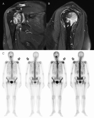
X-ray of the shoulders revealed no alterations. Magnetic resonance imaging (MRI) of the left shoulder demonstrated bone marrow hypersignal of the humeral head with extension to the glenoid cavity and acromioclavicular region (arrow), as well as thickening and hyperintensity of the inferior glenohumeral ligament (asterisk) (). Bone scintigraphy showed early increased radiotracer uptake in the left glenohumeral region and, later, in a heterogeneous manner in the cranial vault, proximal half of the left femur, and proximal third of the right femur, as well as mildly increased radiotracer uptake in other sites (), ruling out algoneurodystrophy or osteonecrosis. Laboratory tests were normal, including complete blood count (haemoglobin 13.4 g/dL, leucocytes 6.55 × 10⁹/L, neutrophils 4.85 × 10⁹/L, lymphocytes 1.01 × 10⁹/L, and platelets 157 × 109/L), inflammatory markers (erythrocyte sedimentation rate 12 mm/h, C-reactive protein 0.5 mg/L), serum protein electrophoresis, and peripheral blood immunophenotyping. The autoimmune panel showed borderline anti-nuclear antibodies (titre 1:100, granular and nucleolar patterns), and the other results were negative for anti-double-stranded DNA, anti-nucleosome, anti-phospholipid, anti-neutrophil cytoplasmic and anti-thyroid antibodies, extractable nuclear antigen screening, rheumatoid factor, and anti-citrullinated protein antibody.
Figure 1. (A, B) Coronal and sagittal T2-weighted magnetic resonance imaging (MRI) of the left shoulder and (C) bone scintigraphy of a 61-year-old female presenting with a 9 month history of frozen shoulder. (A, B) MRI demonstrates bone marrow hypersignal of the humeral head with extension to the glenoid cavity and acromioclavicular region (arrow), as well as thickening and hyperintensity of the inferior glenohumeral ligament. (C) Bone scintigraphy shows early increased radiotracer uptake in the left glenohumeral region and later in other locations.

Given the non-specific findings with possibility of malignancy, computed tomography (CT)-guided biopsy of the left humeral head lesion was performed after 14 months of symptoms. Anatomopathological analysis and immunophenotyping were in favour of follicular lymphoma. Complementary investigations with bone marrow aspirate and biopsy, CT of the thorax, abdomen, and pelvis, and a positron emission tomography scan led to a diagnosis of follicular lymphoma (International Prognostic Index score 3 and Ann Arbor stage IV-A).
After diagnostic confirmation, ultrasound-guided shoulder hydrodistension was performed, with significant symptomatic relief and full recovery of shoulder mobility. Partial tumour response was achieved after six cycles of chemotherapy at 3 week intervals, which included three cycles of R-CHOP (rituximab, cyclophosphamide, doxorubicin, vincristine, and prednisolone) followed by a switch to three cycles of CHOP (cyclophosphamide, doxorubicin, vincristine, and prednisolone) owing to the intercurrence of severe acute respiratory syndrome coronavirus-2 (SARS-CoV-2) infection. It was proposed that the patient undergo maintenance therapy with rituximab every 8 weeks for 2 years, which is currently pending initiation.
Although rare, adhesive capsulitis can be a manifestation of solid and haematological malignancies (Citation4), as well as masking shoulder tumours (Citation5). The diagnosis is essentially clinical, but imaging tests are necessary to exclude differential diagnoses, namely glenohumeral osteoarthritis, osteonecrosis, fractures of the proximal humerus, rotator cuff tendinopathy, or even local tumours (Citation3, Citation6).
We encountered a case of an isolated musculoskeletal manifestation that culminated in the diagnosis of an advanced haematological malignancy. This case highlights the need to consider malignancies as un underlying cause of adhesive capsulitis, particularly if there is an atypical presentation, such as the presence of structural bone changes besides thickening of the inferior glenohumeral ligament.
Misdiagnosis as frozen shoulder or waiting for the failure of conservative treatment to expand the evaluation can cause a significant delay in achieving an accurate diagnosis of malignant tumours, and therefore negatively impact the prognosis (Citation5). On the other hand, the benefits of screening for malignancy among patients with frozen shoulder need to be weighed against the risks, such as cost and associated anxiety (Citation1), especially since malignant tumours in and around the shoulder are uncommon (Citation5).
Our patient did not present clear risk factors for adhesive capsulitis, prompting us to undertake further investigation. In conclusion, we consider that a high degree of suspicion of malignancy is crucial and may make the difference to the patient’s prognosis.
Ethics approval
Written informed consent was obtained from the patient for the publication of this case report and the accompanying images.
Disclosure statement
No potential conflict of interest was reported by the authors.
References
- Pedersen AB, Horváth-Puhó E, Ehrenstein V, Rørth M, Sørensen HT. Frozen shoulder and risk of cancer: a population-based cohort study. Br J Cancer 2017;117:144–7.
- Cogan CJ, Cevallos N, Freshman RD, Lansdown D, Feeley BT, Zhang AL. Evaluating utilization trends in adhesive capsulitis of the shoulder: a retrospective cohort analysis of a large database. Orthop J Sports Med 2022;10:23259671211069577.
- Jacob L, Gyasi RM, Koyanagi A, Haro JM, Smith L, Kostev K. Prevalence of and risk factors for adhesive capsulitis of the shoulder in older adults from Germany. J Clin Med 2023;12:669.
- Gheita TA, Ezzat Y, Sayed S, El-Mardenly G, Hammam W. Musculoskeletal manifestations in patients with malignant disease. Clin Rheumatol 2010;29:181–8.
- Sano H, Hatori M, Mineta M, Hosaka M, Itoi E. Tumors masked as frozen shoulders: a retrospective analysis. J Shoulder Elbow Surg 2010;19:262–6.
- Robinson CM, Seah KT, Chee YH, Hindle P, Murray IR. Frozen shoulder. J Bone Joint Surg Br 2012;94:1–9.
