
Abstract
Seven new species of antipatharian corals are described from the territorial waters of New Zealand and adjacent regions. Differential diagnoses are given and comparisons are made to related nominal species. Described as new are: Antipathes leptocrada; Antipathes craticulata; Asteriopathes octocrada; Phanopathes zealandica; Tetrapathes latispina; Parantipathes dodecasticha; and Parantipathes robusta.
http://zoobank.org/urn:lsid:zoobank.org:pub:DCF9A2BD-63DE-4240-AA4E-0B8BE7E095F1
Introduction
The antipatharian coral fauna of New Zealand has, until recently, been relatively unstudied. The first species from the area, Antipathes fruticosa, was described by Gray (Citation1857) in 1857. Thirty-two years later a second species, Antipathes strigosa, was included in the report on the Challenger Expedition by Brook (Citation1889). In 1911, seven species were collected at a single station off North Cape during the British Antarctic (Terra Nova) Expedition, and five of these were later described as new to science (Totton Citation1923). Very little was published on black corals in New Zealand over the next 50 years; however, interest in the group increased when it was discovered that one particular species was present in the fiords of the South Island (Grange et al. Citation1981). That species, given the name Antipathes fiordensis (Grange Citation1990; later re-assigned to the genus Antipathella, see Opresko Citation2001), has been the subject of numerous ecological and biological studies (Grange Citation1985, Citation1991; Grange & Singleton Citation1988; Grange & Goldberg Citation1993; Goldberg Citation1991; Goldberg et al. Citation1990; Miller Citation1996, Citation1997, Citation1998; Parker et al. Citation1997). Beginning in the mid-1960s, the New Zealand Oceanographic Institute (now the National Institute of Water and Atmospheric Research [NIWA]) began a series of exploratory research cruises during which a large number of mainly deep-water antipatharian corals were collected. The specimens were deposited at the NIWA facility at Greta Point and were largely unstudied until the late 1990s when, during an initial visit to NIWA, I examined the collection and found that it contained as many as 58 species of antipatharians. The species were provisionally identified and listed in the New Zealand Inventory of Biodiversity (Cairns et al. Citation2009). The NIWA collection forms the basis for an ongoing monographic revision of the New Zealand fauna. During the course of this study, many new taxa have been identified, and seven of these new species are the subject of this article. As a corollary to the taxonomic studies, a field guide to the most commonly occurring genera of black corals in the New Zealand region was recently published (Opresko et al. Citation2014).
Materials and methods
This study is based on specimens deposited in the NIWA Invertebrate Collection (NIC) over the past 40 plus years. The material was collected as a result of various trawling survey programmes and cruises sponsored by NIWA and its predecessor the New Zealand Oceanographic Institute (NZOI), including: Challenger Centenary Cruise (1974), Norfolk Rise Biology (1975), Norfolk Ridge II (1977), Southern Benthomass (1981), Cook Island Expedition (1981), Bay of Plenty Geology (1989), Kermadec Volcanics NZAPLUME-1 (1999), Bay of Plenty and southern Kermadec Arc Seamounts (2000), Kermadec Ridge Volcanics (2002), Kermadec Volcanics NZAPLUME-III (2004), Bay of Plenty and Hikurangi Plateau Seamounts (2004).
Specimens were studied at NIWA and at the US National Museum of Natural History (USNMNH), Smithsonian Institution, Washington, DC. Subsamples of some specimens have been deposited at the USNMNH (museum catalogue numbers are designated with the prefix ‘USNM’). Specimens from the collections of the Natural History Museum, London are indicated with the prefix ‘BMNH’ before the catalogue number; those from the collections of the South Australian Museum are indicated with the prefix ‘SAM’ before the catalogue number.
For detailed study of the microscopic skeletal elements of the corals a Zeiss EVO MA 15 scanning electron microscope (SEM) housed at the USNMNH was utilised. The samples were sputter-coated with a 30–40 nm layer of 60% gold: 40% palladium. Measurements of the microscopic skeletal features were made directly using a dissecting microscope or from the photographs taken with the SEM. SEM stub numbers are from a series established by the author at the USNMNH. All SEM stubs are deposited at the USNMNH; however, copies of the SEM photos are to be kept in the collections database at NIWA.
The holotypes and paratypes of all new species are deposited in the NIC at Greta Point, with selected schizoholotypes and schizoparatypes in the collections of the USNMNH.
Taxonomic account of new species
Phylum Cnidaria
Class Anthozoa
Order Antipatharia
Family Antipathidae Ehrenberg, Citation1834
Genus Antipathes Pallas, Citation1766
Antipathes leptocrada n. sp.
(–)
Figure 1 Distribution of species. The type locality of Antipathes craticulata n.sp. is in the Cook Islands at 20.12°S and 157.35°W.
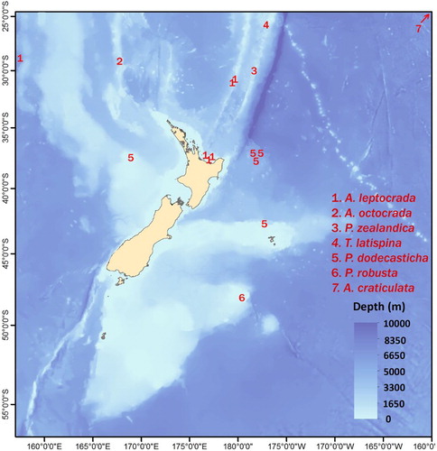
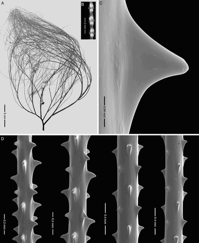
Figure 2 Antipathes leptocrada n. sp., holotype NIWA 19778 (schizoholotype, USNM 1202915/SEM stub 371). A, Corallum; B, polyps; C, single spine; D, spines on branchlets.
Material examined
Holotype: NIWA 19778 (schizoholotype USNM 1202915/SEM stub 371), Stn P925, 11 Dec 1979, west of the Lord Howe Seamount Chain, 27.99°S, 155.63°E, 420 m. Paratypes: NIWA 16050 (schizoparatype USNM 1174710/SEM stub 335), Stn TAN0413/74, 12 Nov 2004, Bay of Plenty, 37.47°S, 177.22°E, 200–175 m; NIWA 16058 (schizoparatype USNM 1174715/SEM stub 289), Stn TAN0413/7, 8 Nov 2004, 36.83°S, 177.47°E, 1443 m; NIWA 19779 (schizoparatype USNM 1202916), Stn X121, 23 Nov 1989, 37.41°S, 177.20°E, 340 m; NIWA 19880 (schizoparatype USNM 1202934/SEM stub 323), Stn K863, 31 July 1974, 30.54°S, 178.54°W, 200 m; NIWA 19883 (schizoparatype USNM 1202935/SEM stub 340), Stn K840, 28 July 1974, 30.29°S, 178.42°W, 398 m (see for collecting sites).
Description
Species forming upright, branched, bushy corallum with thin, flexible, elongate branchlets and narrow distal branch angles. Holotype (NIWA 19778, A) c. 29 cm tall (as preserved), 20 cm wide, with a basal stem diameter of 3 mm. Holdfast absent. At least five orders of branches. Narrow distal branch angles, mostly 30–50°. End-branchlets 24 cm or more in length, with basal diameter of 0.3 mm. End-branchlets 0.13 mm in diameter at a distance of 3 cm from tip. Branchlets uniserial over short distances or irregularly bilateral. Spines small, smooth, slightly compressed laterally, rarely bifid, subequal around circumference of axis; distal edge often hooked downward at apex (i.e. towards proximal end of spine); proximal edge concave, then flared out along axis (C–D). On axis 0.15 mm in diameter, polypar spines about 0.08 mm tall, abpolypar spines 0.07 mm. Spines in axial rows, five of which visible in lateral view (includes only rows in which the base of the spines can be seen) and 0.36–0.53 mm apart in each row. Distal edge of polypar spines up to 0.1 mm in length; proximal edge up to 0.16 mm long. On branchlet 0.26 mm in diameter, polypar spines 0.064–0.07 mm tall, abpolypar spines 0.058–0.064 mm; mutual distance 0.47–0.54 mm; five rows of spines visible in lateral view. Polyps 1 to 1.5 mm in transverse diameter, with 6 to 6.5 polyps per centimetre (B).
Paratypes similar to holotype except in density of branching; some colonies having only a few orders of branches. Longest end-branchlets 12–26 cm long. Spines typically 0.05–0.07 mm tall on branchlets 0.2 to 0.3 mm in diameter. Four to six rows of spines visible (lateral view); with spines 0.35–0.43 mm apart within each row. Polyps 1–1.5 mm in transverse diameter, with six to eight polyps per centimetre.
Etymology
From the Greek, leptos (thin) and crada (branch), in reference to the very thin flexible branches characteristic of the species.
Discussion
The species bears a general resemblance to Antipathes densa Silberfeld, 1909 and Antipathes furcata Gray, 1857, but with longer, thinner, more flexible branchlets and smaller spines with a distinctively different shape. Silberfeld (Citation1909a,Citationb) reported that in A. densa (A) the branches reach, at most, 16 cm in length; the diameter of the smallest branches amounted to 0.178 mm; the spines on the end-branches ranged in size from 0.135 mm to c. 0.178 mm (maximum 0.2 mm), and the spines on branches 0.64 mm in diameter were 0.157 mm tall. The spines were described as standing out at right angles to the axis with the proximal side sloping obliquely to form a narrow angle with the axis; the distal edge, in some cases concave so that the tip is directed upward. The type specimens of A. densa are missing; therefore, Silberfeld's description cannot be verified; however, there appear to be sufficient differences in the reported length of the end-branchlets and size and shape of the spines to adequately differentiate A. leptocrada from this species.
Figure 3 Species related to Antipathes leptocrada n. sp. A, Antipathes densa Silberfeld, digital scan of published image of syntype (Silberfeld Citation1909b, plate 1, , scale not provided; reprinted with kind permission from Bayerischen Akademie der Wissenschaften); B, Antipathes furcata Gray, holotype, BMNH 1843.2.6 or 1843.2.8; C, A. furcata, schizoholotype, spines on branchlet, USNM 99596/SEM stub 250.
In A. furcata, the branches are straight and directed vertically and most reach the top of the corallum (B). According to Schultze (Citation1902), the end-branchlets in the type are not more than 9 cm long. The spines are triangular, compressed, and up to about 0.07 mm tall (C). The long axis of the spines is placed at right angles to the skeletal axis, and the distal and proximal edges are subequal in length. Although the size of the spines is similar to that in A. leptocrada, the shape of the spines is distinctly different, and the two species can be further differentiated by the length of the end branchlets (26 cm vs. 9 cm).
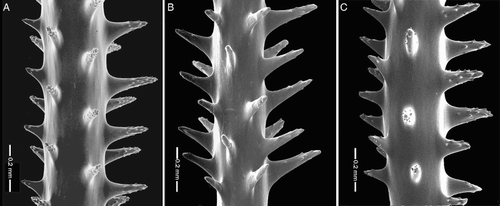
Antipathes craticulata n. sp.
(, , A)
Material examined
Holotype: NIWA 19800 (schizoholotype USNM 1202927/SEM 338), Stn T178, 14 Sept 1981, 20.12°S, 157.35°W, north of Ma'uke Island, Cook Islands, 348 m.
Description
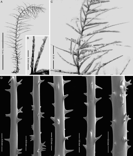
Species forming a small, flabellate corallum with short curved branchlets. Holotype (A) approximately 8.5 cm tall and 9.5 cm wide; basal stem diameter 1.1 mm. Corallum with overlapping and coalescing branchlets. Stem only slightly thicker than branches. Branching starts 1 mm above base. Few distinct primary branches. Up to 15 orders of branches. Branches and branchlets uniserial over short distances (two or three in a row) or very irregularly bilateral; three to five branches and/or branchlets per centimetre. End-branchlets (A) short, mostly 5–10 mm long (maximum 13 mm) and 0.54–0.70 mm in diameter at their midpoint (0.18–0.24 mm excluding spines). Branches and branchlets often originating on convex outer side of lower order branches. Distal angle of insertion of branches and branchlets mostly 60° to 90°; many ramifications curved toward distal end of branch from which they arise.
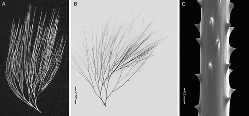
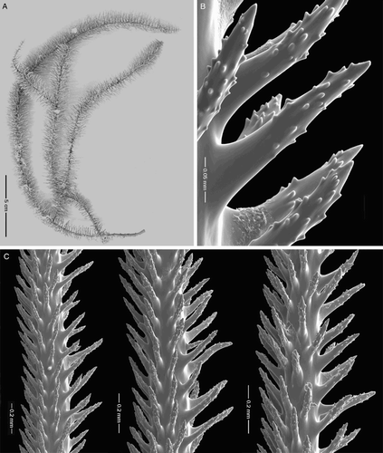
Figure 4 Antipathes craticulata n. sp., holotype NIWA 19800 (schizoholotype USNM 1202927/SEM stub 338). A, Colony; B, close-up view of spines; C, spines on branchlets.
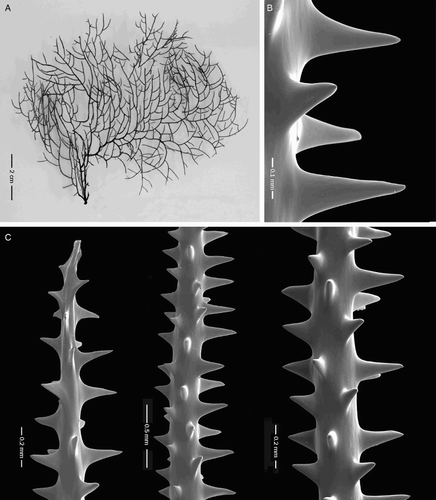
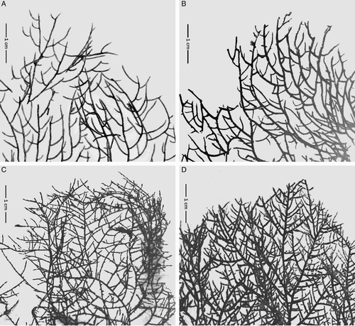
Figure 5 Branching in Antipathes craticulata and related species. A, Antipathes craticulata n. sp., NIWA 19800; B, Tylopathes dubia Brook, BMNH 83.8.29.5; C, T. elegans Brook, BMNH 83.9.13.14; D, T. hypnoides Brook, BMNH 86.2.8.2. Note: Brook's three species were not included in the genus Tylopathes as re-defined by Opresko (2006), and most likely should instead be referred to the genus Antipathes.
Spines (B–C) strong, conical, slightly compressed laterally; unequal in size around circumference of axis. Spines smooth and without apical lobes or bifurcations. On branchlets 0.2–0.3 mm in diameter, polypar spines mostly 0.22–0.27 mm tall (up to 0.30 mm) and 0.11–0.13 wide near base; abpolypar spines 0.13–0.18 mm tall. Spines on branchlets arranged in longitudinal rows, four to six seen in lateral view. Within each row, spines 0.22–0.45 mm apart (3–5 per millimetre). Spines on stem and largest branches slightly smaller than those on branchlets, 0.18 to 0.20 mm tall.
Polyps in a single row, mostly on lateral sides of branches and branchlets. Polyps 0.6–0.85 mm in transverse diameter (from proximal side of proximal lateral tentacles to distal side of distal lateral tentacles); interpolypar space between adjacent polyps mostly 0.6–0.7 mm; about eight polyps per centimetre.
Etymology
The species name is derived from the Latin craticulus (latticed), in reference to the lattice-like appearance of the corallum.
Discussion
In superficial appearance this new species resembles a small colony of Phanopathes or Acanthopathes of the family Aphanipathidae (see Opresko Citation2004). The spines of aphanipathids typically have tubercles whereas no tubercles are present on the spines of the type specimen of A. craticulata. Nevertheless, it is possible that tubercles could have been secondarily lost in this species. DNA sequencing would be needed to determine the true affinities of this species; however, based on spine morphology alone, it is here referred to Antipathes.
In 1914 van Pesch identified a specimen in the Siboga collection (Sta. 305, Solor Strait, 113 m) as Antipathes plana Cooper (Citation1909). Van Pesch's species description is as follows: ‘COLONY: Short stem, branched in a plane; fan-shaped; network through frequent fusions. Branches often inserted at right angles, and afterwards curved upward.—Ultimate branches a few mm in length; are inserted on the anterior side of the colony. SPINES: Smooth; distal side at right angles with the axis. Length 100–200 μ; four longitudinal rows, with a mutual distance between the spines of 210 μ. POLYPS: Tentacles rounded, knob-shaped, radiate. Oral cone dome-shaped with round mouth. Diameter of oral cone 180 μ; interpolypar distance 0.75–1.1 mm.’ The spines of van Pesch's specimen (van Pesch Citation1914, p. 49, fig. 23) resemble those of the type of A. craticulata, especially in regard to the relative large size of the abpolypar spines. The NIWA specimen, however, differs from the Siboga specimen in that the spines are larger (0.22–0.30 mm vs. 0.1–0.2 mm).
Three species described by Brook (Citation1889), Tylopathes dubia, T. elegans and T. hypnoides, resemble A. craticulata in having a flabellate corallum with short, straight or slightly curved terminal branches. In terms of the density of the branching, A. craticulata, with 3–5 branchlets per centimetre (A) is more similar to A. dubia (4–6 branchlets per centimetre; B) than to T. elegans (8–10 branchlets per centimetre, C) or T. hypnoides (12–14 branchlets per centimetre, D). Antipathes craticulata and T. dubia differ, however, in the size and shape of the spines. Examination of a subsample of the type of A. dubia (USNM 100362) revealed that the spines on the branchlets are only 0.11–0.13 mm tall, considerably smaller than the spines in A. craticulata (0.22–0.33 mm). The spines in T. elegans and T. hypnoides are about 0.1 mm and therefore also smaller than those of A. craticulata.
Note: the genus Tylopathes, with the type species Tylopathes crispa Brook, was assigned to the newly established family Stylopathidae by Opresko (Citation2006) in 2006. At that time the three species described by Brook, T. dubia, T. elegans and T. hypnoides, were not considered to be congeneric with T. crispa due to differences in the morphology of the spines. The true affiliation of these three species is yet to be determined, although based on morphology of the spines, it is most likely that all three belong in the genus Antipathes.
Family Aphanipathidae Opresko, 2004
Genus Asteriopathes Opresko, 2004
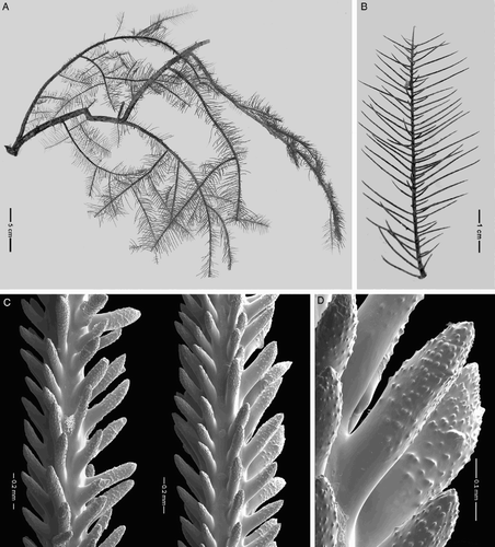
Asteriopathes octocrada n.sp.
(, , A)
Material examined
Holotype: NIWA 19798 (schizoholotype USNM 1202925/SEM stub 317), Stn I85, 22 July 1975, 29.13°S, 168.25°E, 290 m (see for type locality).
Description
Colony (A) very sparsely branched and pinnulate; stem and branches bottlebrush in appearance; pinnules simple, arranged bilaterally and in alternating groups, each consisting of up to eight pinnules. Pinnules in each group originating at nearly the same level on the axis. Pinnules mostly less than 1 cm long (maximum 1.2 cm). Pinnules 0.17–0.19 mm in diameter excluding spines and 0.47–0.56 mm in diameter including spines. Five groups of pinnules per centimetre on each side of axis; up to 80 pinnules per centimetre for both sides. Spines (B–C) tall, acicular, and covered with small tubercles over about two-thirds of surface. Polypar spines up to 0.26 mm in height and about 0.07 mm in width at the base (in distal-proximal direction); inclined distally (distal angle about 45°); usually straight, but sometimes curved distally or proximally. Some polypar spines in interpolypar areas taller than others. Abpolypar spines up to 0.2 mm tall and 0.06 mm wide at base; inclined distally (distal angle about 30°); straight or curved distally. Polypar spines 0.16 mm apart with 6–7 per millimetre; abpolypar spines about 0.1 mm apart with up to 10 per millimetre. Approximately 15 to 20 tubercles per spine seen in lateral view (including those seen in profile on the edge of the spine). Spines arranged in irregular axial rows, usually six or seven (rarely eight) seen in lateral view. Polyps not preserved.
Figure 6 Asteriopathes octocrada n. sp., holotype NIWA 19798 (schizoholotype USNM 1202925/SEM stub 317). A, Colony; B, close-up view of spines; C, spines on pinnules.
Etymology
The species name is derived from the Greek octo (eight) and crada (branch), in reference to the occurrence of the pinnules in groups of eight.
Discussion
Asteriopathes octocrada n. sp. resembles A. arachniformis Opresko (Citation2004) in having very short pinnules usually less than 1.2 cm long. It differs from that species in having more pinnules per group (8 vs. 5–6), in having larger, thicker polypar spines (up to 0.26 mm in height vs. 0.14–0.22 mm), and in having slightly more tubercles per spine (15–20 vs. 5–15) (A–B). The spines of A. octocrada (A) resemble those of A. colini Opresko (C), but the two species differ in number of pinnules per group (8 vs. 4–6) and in length of pinnules (up to 1.2 cm vs. 2.5 cm).
Figure 7 Spines of Asteriopathes octocrada n.sp. and related species. A, Asteriopathes octocrada, holotype USNM 1202925/SEM stub 317; B, A. arachniformis Opresko, holotype, USNM 1007096/SEM stub 142; C, A. colini Opresko, holotype, USNM 1007092/SEM stub 145.
Genus Phanopathes Opresko, 2004
Phanopathes zealandica n. sp.
(, )
Material examined
Holotype: NIWA 19807 (schizoholotype USNM 1202929/SEM stub 320), TAN0205/75, 23 April 2002, Kermadec Ridge, 30.02–30.03°S, 178.71–178.72°W, 240–158 m (see for type locality).
Description
Small, flabellate colony. Holotype (A) about 8 cm high and equally as wide. Basal diameter of stem 0.9 mm. Larger branches up to about 6 cm in length. Most smaller branchlets slightly curved; distal branch angles (angle formed by distal side of a branch and lower order branch from which it arises) usually quite wide (60° or more), with branches curved distally. Branching uniserial over short distances (generally not more than three branchlets in a row) or very irregularly bilateral. Branches and branchlets generally ascending; overlapping in places and occasionally fused. Unbranched end-branchlets mostly 1.3 cm or less in length (rarely up to about 2.5 cm), and spaced at varying distances apart (2 mm to 2 cm), mostly with 3–6 branches/branchlets per 2 cm. Skeletal spines (B–C) on branchlets dimorphic with polypar spines larger than abpolypar spines. Polypar spines on branchlets tall, conical to acicular (up to 0.34 mm on axis 0.3 mm in diameter); abpolypar spines mostly 0.14–0.15 mm. Polypar spines with tubercles on upper half of surface; usually four to six tubercles (maximum eight) visible in lateral view including those seen in profile on edge of spine (B). Abpolypar spines usually with fewer tubercles than polypar spines, and in some cases lacking tubercles. Abpolypar spines more distally inclined than polypar spines which project out nearly at right angles to axis. Spines on branchlets spaced 0.37–0.5 mm apart (about three per millimetre) and arranged in five or six longitudinal rows (complete spines including base) as seen in one lateral view. Polyps poorly preserved; about 0.6 to 0.7 mm in transverse diameter.
Figure 8 Phanopathes zealandica n. sp., holotype, NIWA 19807 (schizoholotype, USNM 1202929/SEM stub 320). A, Colony; B, single spine; C, spines on branchlets.
Etymology
The species name zealandica is in reference to the general type locality.
Discussion
Three other species are assigned to the genus Phanopathes: P. expansa (Opresko & Cairns Citation1992), P. rigida (Pourtalès Citation1880) and P. cancellata (Brook Citation1889). Phanopathes zealandica n. sp. resembles P. expansa in general growth form and size of the corallum; however, in P. zealandica the branching is less dense (up to six branchlets per 2 cm vs. up to 11 branchlets per 2 cm), and the end-branchlets are generally longer (up to 2.5 cm vs. 1.2 cm maximum). Furthermore, P. zealandica differs from P. expansa in having spines that are larger (0.34 mm maximum vs. 0.24 mm maximum), but with fewer tubercles (up to about eight per lateral view vs. 25 or more per lateral view) (see A). The polyps in P. expansa are 0.55 to 0.65 mm in transverse diameter; similar in size to those in P. zealandica (0.6 to 0.7 mm).
Figure 9 Spines of species related to Phanopathes zealandica n. sp. A, Phanopathes expansa, holotype, USNM 88340/SEM stub 34); B, P. rigida, schizotype. USNM 94477/SEM stub 214; C, P. cancellata, schizotype, USNM 100380/SEM stub 140.
In Phanopathes rigida the branchlets are very widely spaced (up to 2 cm apart) and are longer (up to 10 cm) than those in the P. expansa or P. zealandica, and the polyps are up to 1.3 mm in transverse diameter (with six or seven polyps per centimetre). The spines of P. rigida resemble those of P. zealandica in having only a small number of tubercles (B).
The fourth species in the genus, P. cancellata (Brook Citation1889), differs from the others in not having a well-defined stem and in having branchlets that are extensively anastomosing. The branchlets are only occasionally fused in P. expansa and P. zealandica, and not at all in P. rigida. The spines of P. cancellata have large and very distinct tubercles (C).
Genus Tetrapathes Opresko, 2004
Tetrapathes latispina n. sp.
(, )
Material examined
Holotype: NIWA 47417 (schizoholotype USNM 1202947/SEM stub 314), Stn TAN0411/19, 4 Oct 2004, northern Kermadec Ridge, 25.45°S, 177.13°W, 493–278 m (see for type locality).
Description
Large, sparsely branched, pinnulate colony, with simple pinnules in four rows. Holotype (A) about 42 cm tall and equally as wide. Branching somewhat planar, longest branches 25 cm, and up to 10 cm apart. Lateral pinnules up to 1.7 cm long, anterolateral pinnules generally less than 0.7 cm (B). Six pinnules per centimetre in each row (22 per centimetre total for all rows). Anterolateral pinnules offset from lateral ones such that anterolateral pinnules on right side are closer to laterals on left side, and anterolateral pinnules on left side are closer to laterals on right side (viewing front of corallum). One or both anterolateral pinnules missing in places.
Figure 10 Tetrapathes latispina n. sp., holotype, NIWA 47417 (schizoholotype, USNM 1202947/SEM stub 314). A, Colony; B, branchlet with pinnules; C, spines on pinnules; D, close-up view of spines.
Spines on pinnules (C–D) large, cylindrical, blunt, very crowded, strongly inclined distally, and covered with small conical tubercles. Polypar spines up to 0.36 mm tall with midpoint diameter of 0.09 mm; some with slight bifurcation at apex. Abpolypar spines up to 0.24 mm tall. Four to six rows of spines visible in lateral view, with seven or eight spines per millimetre in each row. Tubercles covering one-half to about two-thirds surface of spines from apex to base (D), with 75 or more tubercles visible in lateral view (including those seen in profile on edge of spines). Tubercles usually not more than 0.004 mm high. In places neighbouring tubercles fuse laterally to form shelf-like ridges. Polyps not preserved.
Etymology
The species name is derived from the Latin latus (broad) and spina (spine), referring to the relatively thick, broad spines, when compared to those in T. alata (Brook), the only other species in the genus.
Discussion
This species differs from T. alata by having shorter pinnules and larger, wider spines with more tubercles. In T. alata the lateral pinnules are up to 2.5 cm long, and the polypar spines are 0.20 mm tall (see Opresko Citation2004, fig. 6), whereas in T. latispina the lateral pinnules are at most 1.6 cm and the spines are up to 0.36 mm in size. In T. latispina there are seven or eight spines per millimetre in each row, and in T. alata there are five or six per millimetre. In T. alata there are 15–25 tubercles per spine visible in lateral view, whereas, in T. latispina there are 75 or more.
Family Schizopathidae Brook, 1889
Genus Parantipathes Brook, 1889
Parantipathes dodecasticha n. sp.
(, )
Material examined
Holotype: NIWA 11148 (USNM 1174709/SEM stub 290), Stn Z-9173, 5 July 1998, 37.04°S, 176.70–176.69°W, 1003–1108 m. Paratypes: NIWA 4319, Stn Z-9229, 15 Aug 1998, 36.88°S, 177.37°W, 787 m; NIWA 11752, Stn Z-9173, 5 July 1998, 37.0383°S, 176.70–176.69°W, 1003–1108 m; NIWA 24191, Stn Z9279, 10 Sept 1998, 37.48°S, 167.72°E, 883 m; NIWA 47888, Trip 2101/124, 13 May 2005, 42.78–42.79°S, 177.33°W, 923–944 m (see for localities).
Description
Corallum unbranched or very sparsely branched; stem and branches pinnulate; pinnules simple, usually in ten to 12 rows. Holotype (A) incomplete, lacking a holdfast and broken off at the top of the stem. Stem 11.5 cm long and about 1.2 mm in diameter at basal end; with two branches, one of which 21 cm long. Pinnules in holotype (B) and paratypes occurring on all sides of axis or on only three sides (leaving posterior side free); mostly ≤ 2 cm long (maximum about 2.5 cm), usually arranged in 10 to 12 axial rows (including those at distal end of branches), and also in alternating semi-spiral groups of five or six (rarely seven) pinnules each. Semispiral groups extend 2–3 mm along axis at their points of insertion; four (sometimes close to five) groups per centimetre on each side; seven to eight groups per centimetre total for both sides; 35–40 pinnules per centimetre total. Distal angle of pinnules close to 90°, but pinnules often curving upward.
Figure 11 Parantipathes dodecasticha n. sp., holotype, NIWA 11148 (schizoholotype,USNM 1174709/SEM stub 290). A, Colony; B, close-up view of pinnules; C, spines on pinnules.
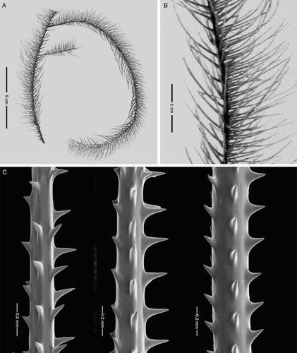
Spines (C) smooth, acute, narrow; distal margin at right angles to axis or inclined distally. On axis 0.16 mm in diameter, polypar spines up to 0.17 mm tall and 0.03–0.04 mm in diameter along mid-section; abpolypar spines 0.03 mm. On axis 0.18–0.20 mm in diameter, polypar spines 0.12–0.15 mm; abpolypar spines 0.04–0.06 mm. On axis 0.25 mm in diameter, polypar spines up to 0.11 mm tall; abpolypar spines c. 0.04 mm. Spines arranged in axial rows, three or four of which visible in lateral view; spaced 0.19–0.28 mm apart in each row; with four to five spines per millimetre. Maximum size of polypar spines in paratypes ranges from 0.16 to 0.18 mm.
Polyps poorly preserved on holotype, but estimated to be about 2 mm in diameter in transverse diameter. On NIWA 11752, polyps are 2–2.5 mm in transverse diameter; those on NIWA 4319 are up to 2.8 mm in transverse diameter.
Etymology
The species name is derived from the Greek, dodeka (twelve) and stichos (row), in reference to the most common number of rows of pinnules along the axis.
Discussion
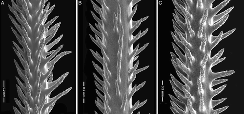
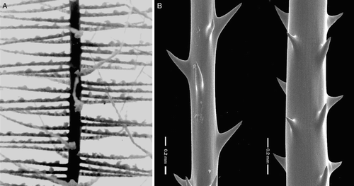
Parantipathes dodecasticha is closely related to P. helicosticha Opresko (Citation1999). Both species form very sparsely branched colonies and have short pinnules (mostly 2 cm in length). The major difference between the two species is in the number of rows of pinnules. In P. helicosticha there are usually six to eight rows total (A). In contrast, in P. dodecasticha, the typical condition is 10 to 12. This difference is also reflected in the number of pinnules per semi-spiral group on each side of the axis; three to four (rarely five) in P. helicosticha and five to six (rarely seven) in P. dodecasticha, and in the total number of pinnules per centimetre (25 per centimetre in P. helicosticha and 35–40 per centimetre in P. dodecasticha). Other differences between the two species are seen in the maximum size and angle of inclination of the spines. In P. helicosticha the spines (B) are up to 0.22 mm tall and are usually inclined distally, whereas in P. dodecasticha, the polypar spines are up to 0.18 mm tall and usually extend out at right angles to the axis (although in some colonies they are directed distally). Although not consistent across all specimens, in P. dodecasticha the abpolypar spines tend to be shorter compared to those in P. helicosticha, and the spines are spaced much closer together (0.19–0.28 mm vs. 0.35 mm or greater).
Figure 12 Parantipathes helicosticha Opresko, holotype, SAM H 903 (schizoholotype USNM 99401/SEM stub 100). A, Close-up view of pinnules; B, spines on pinnules.
Parantipathes robusta n.sp.
(, )
Material examined
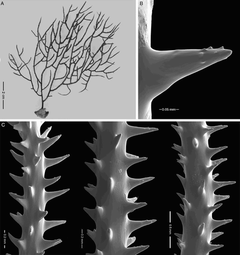
Holotype: NIWA 4318 (schizoholotype USNM 1174706/SEM stub 353), Stn T25, 12 Mar 1981, 48.15°S, 179.34°W, 693 m (see for type locality).
Description
Corallum monopodial or sparsely branched, and pinnulate in a bottlebrush pattern. Holotype (A) consists of an 11 cm long pinnulated section attached at its base at right angles to a short 1.5 cm pinnulated section, suggesting that the stem had been broken at one time and the upper part regenerated as a branch. Pinnules usually arranged biserially in six (rarely seven) rows (only four rows at the top of the corallum), and in semi-spiral groups of three (rarely four) pinnules each (two per group at top of the corallum). Pinnules (C) simple, stiff, inclined distally; up to 2.5 cm long on posterolateral side of axis and up to 1.5 cm on anterior side. Basal diameter of pinnules up to 0.4 mm (excluding spines); midpoint diameter typically about 0.27 mm (excluding spines). Pinnules about 3–5 mm apart in each row; with 15 pinnules total per centimetre (five semi-spiral groups of three each).
Figure 13 Parantipathes robusta n. sp., holotype, NIWA 4318 (schizoholotype, USNM 1174706/SEM stub 353). A, Colony; B, polyps; C, close-up view of pinnules; D, spines on pinnules.
Spines (D) on middle of pinnules smooth, narrow, acute; some forked; clusters of double and triple spines present. On pinnules 0.2 mm in diameter, polypar spines up to 0.24 mm; abpolypar spines 0.06 mm; mutual distance of spines 1.0 to 1.4 mm. Spines shorter and more crowded on thicker basal section of pinnule; on axis 0.38 mm in diameter, polypar spines 0.14 mm, abpolypar spines 0.06 mm; mutual distance 0.60 to 0.77 mm. Spines arranged in axial rows, three or four of which visible in lateral view.
Polyps at base of pinnules about 3 mm in transverse diameter; those near the tip of pinnule up to 4 mm in transverse diameter (B). Polyps arranged uniserially on upper side of pinnules; 2 to 2.5 polyps per centimetre.
Etymology
The species name is derived from the Latin robusta (thick) in reference to the thick, stiff pinnules.
Discussion
This species is distinguished from other species of Parantipathes primarily by the occurrence of forked spines which are not found in other species of the genus. Molodtsova & Pasternak (Citation2005) provide a comprehensive comparative analysis of the seven previously recognised nominal species of the genus. Parantipathes robusta differs from all of these in having forked spines, and in having the largest spines (up to 0.24 mm) and largest polyps (up to 4 mm). In addition, in P. robusta the spines in each axial row are spaced much further apart than in the other species. In other characteristics (i.e. pinnular length, pinnular density, number of pinnules per semi-spiral group), P. robusta overlaps with one or more of the other species.
Associate Editor: Dr Jonathan Banks.
Acknowledgements and funding
The author wishes to thank all the funding agencies and contributors to the voyages during which specimens included in this publication were collected. Special thanks go to Dennis Gordon, Di Tracey, Kareen Schnabel, and Sadie Mills of the National Institute of Water and Atmospheric Research (NIWA) for their hospitality and assistance during numerous visits to Wellington.
Samples from cruises KAH0011, TAN0205 and TAN0413 were supplied by the NIWA Invertebrate Collection and were collected as part of a research programme ‘Seamounts: their importance to fisheries and marine ecosystems’, undertaken by the NIWA and funded by the former New Zealand Foundation for Research, Science and Technology and with additional funding from the Ministry of Fisheries and NOAA Satellite Operations Facility.
The author also wishes to thank S Cairns, W Keel, C Bright, W Moser and P Greenhall for their assistance during visits to the USNMNH. The photomicrographs were prepared in the SEM Laboratory of the USNMNH; SD Whittaker of the USNMNH kindly assisted in the SEM operation. DM Opresko is a Research Associate of the USNMNH, and gratefully acknowledges that affiliation. Funding for this project was provided, in part, by a United States Department of Justice (DoJ) grant to the Smithsonian Institution. The author is very appreciative of the DoJ's support of black coral research at the Smithsonian. Sincere thanks also go to T Molodtsova and M Bo for reviewing the manuscript and providing many useful comments and suggestions, and to Jonathan Banks and Marie Hodgkinson for their editorial assistance. Permission to use the photo of Antipathes densa (see A) was kindly provided by Nikoletta Helidonis of Bayerischen Akademie der Wissenschaften.
Disclosure statement
No potential conflict of interest was reported by the author.
References
- Brook G 1889. Report on the Antipatharia. Reports of the Scientific Results of the Voyage of the Challenger. Zoology 32: 1–222.
- Cairns SD, Gershwin L-A, Brook FJ, Pughet P, Dawsonet EW, Vicenteal OO et al. 2009. The phylum Cnidaria: corals, Medusae and Hydroids. In: Gordon D ed. New Zealand inventory of biodiversity, volume 1, Kingdom Animalia. Christchurch, Canterbury University Press.
- Cooper CF 1909. Antipatharia. Reports of the Percy Sladen Trust Expedition to the Indian Ocean. Transactions of the Linnean Society of London (Zool. Ser. 2) 12: 301–323. doi: 10.1111/j.1096-3642.1909.tb00144.x
- Ehrenberg CG 1834. Die Corallenthiere des rothen Meeres, physiologisch untersucht und systematisch verzeichnet. Berlin, Abhandlung Königlichen Akademie der Wissenschaften. Pp. 153–154.
- Goldberg WM 1991. Chemistry and structure of skeletal growth rings in the black coral Antipathes fiordensis (Cnidaria: Antipatharia). Hydrobiologia 216: 403–409. doi: 10.1007/BF00026493
- Goldberg WM, Grange KR, Taylor GT, Zuniga AL 1990. The structure of sweeper tentacles in the black coral Antipathes fiordensis. Biological Bulletin 179: 96–104. doi: 10.2307/1541743
- Grange K 1985. Distribution, standing crop, population structure, and growth rates of black corals in the southern fiords of New Zealand. New Zealand Journal of Marine and Freshwater Research 19: 467–475. doi: 10.1080/00288330.1985.9516111
- Grange K 1990. Antipathes fiordensis, a new species of black coral (Coelenterata:Antipatharia), an ecological dominant in the southern fiords of New Zealand. New Zealand Journal of Zoology 17: 279–282. doi: 10.1080/03014223.1990.10422603
- Grange K 1991. Mutualism between the antipatharian Antipathes fiordensis and the ophiuroid Astrobrachion constrictum in New Zealand fiords. Hydrobiologia 216/217: 297–303. doi: 10.1007/BF00026478
- Grange KR, Goldberg WM 1993. Chronology of black coral growth bands: 300 years of environmental history? In: Battershill CN et al. eds. Proceedings of the Second International Temperate Reef Symposium, Auckland, New Zealand 7–10 January 1992. Wellington, NIWA—Marine. Pp. 169–174.
- Grange KR, Singleton RJ 1988. Population structure of black coral, Antipathes aperta, in the southern fiords of New Zealand. New Zealand Journal of Zoology 15: 481–489. doi: 10.1080/03014223.1988.10422628
- Grange K, Singleton RJ, Richardson JR, Hill PJ, del Main W 1981. Shallow rock-wall biological associations of some southern fiords of New Zealand. New Zealand Journal of Zoology 8: 209–227. doi: 10.1080/03014223.1981.10427963
- Gray JE 1857. Synopsis of the families and genera of axiferous zoophytes or barked corals. Proceedings of the Zoological Society of London 25: 278–294.
- Miller KJ 1996. Piecing together the reproductive habits of New Zealand's endemic black corals. Water and Atmosphere 4: 18–19.
- Miller KJ 1997. Genetic structure of black coral populations in New Zealand's fiords. Marine Ecology Progress Series 161: 123–132. doi: 10.3354/meps161123
- Miller KJ 1998. Short-distance dispersal of black coral larvae: inference from spatial analysis of colony genotypes. Marine Ecology Progress Series 163: 225–233. doi: 10.3354/meps163225
- Molodtsova TN, Pasternak FA 2005. Redescription of Parantipathes euantha (Pasternak, 1958) (Anthozoa: Antipatharia) from the Kurile-Kamchatka Trench. Invertebrate Zoology 2: 169–179.
- Opresko DM 1999. New species of Antipathes and Parantipathes (Cnidaria: Anthozoa: Antipatharia) from coastal waters of South Australia and Tasmania. Records of the South Australian Museum 32: 143–154.
- Opresko DM 2001. Revision of the Antipatharia (Cnidaria: Anthozoa). Part I. Establishment of a new family, Myriopathidae. Zoologische Mededelingen, Leiden 75: 343–370.
- Opresko DM 2004. Revision of the Antipatharia (Cnidaria: Anthozoa). Part IV. Establishment of a new family, Aphanipathidae. Zoologische Mededelingen, Leiden 78: 209–240.
- Opresko DM 2006. Revision of the Antipatharia (Cnidaria: Anthozoa). Part V. Establishment of the new family Stylopathidae. Zoologische Mededelingen, Leiden 80: 109–138.
- Opresko DM, Cairns SD 1992. New species of black coral (Cnidaria: Antipatharia) from the northern Gulf of Mexico. Northeast Gulf Science 12: 93–97.
- Opresko DM, Tracey D, Mackay E 2014. Antipatharia (black corals) for the New Zealand region. A field guide of commonly sampled New Zealand black corals including illustrations highlighting technical terms and black coral morphology. Wellington, National Institute of Water and Atmosphere.
- Pallas PS 1766. Elenchus Zoophytorum Sistens Generum Adumbrationes Generaliores et Specierum Cognitarum Succinctas Descriptiones cum Selectis Auctorum Synonymis. Hagae-Comitum [The Hague], Petrum van Cleef.
- Parker NR, Mladenov PV, Grange KR 1997. Reproductive biology of the antipatharian black coral Antipathes fiordensis in Doubtful Sound, Fiordland, New Zealand. Marine Biology 130: 11–22. doi: 10.1007/s002270050220
- Pourtalès LFde 1880. Zoological results of the Blake expedition to the Caribbean Sea. Bulletin of the Museum of Comparative Zoology, Harvard 6: 113–118.
- Schultze LS 1902. Die Antipatharien der deutschen Tiefsee-Expedition, 1898–1899. Wissenschaftliche Ergebnisse der deutschen Tiefsee-Expedition auf dem Dampfer. Valdivia 3: 89–100.
- Silberfeld E 1909a. Diagnosen neuer Japanischer Antipatharien sus der Sammlung von Herrn Prof. Doflein. Zoologischer Anzeiger 34: 760–763.
- Silberfeld E 1909b. Japanische Antipatharien. Beitrage zur Naturgeschichte Ostasiens. Abhandlungen der Mathematisch-Physikalischen Klasse der Königlich Bayerischen Akademie der Wissenschaften 7: 1–30.
- Totton AK 1923. Coelenterata. Part III. Antipatharia (and their Cirripede commensals). British Antarctic (Terra Nova) Expedition, 1910–1913. Natural History Report, Zoology 5: 97–120.
- van Pesch AJ 1914. The Antipatharia of the Siboga expedition. Siboga Expeditie Monographie 17: 1–258.