
ABSTRACT
Beaked whales are poorly known cetaceans with a peculiar dentition consisting of one or two pairs of mandibular tusks, sometimes accompanied by dozens of peg-like rudimentary teeth normally concealed in the gum tissue. This study investigated the ultrastructure, chemical and biomechanical properties of vestigial teeth in a specimen of Cuvier's beaked whale deposited in the Otago Museum collections. The juvenile individual had 49 needle-like vestigial teeth in situ on the lower jaws. Scanning electron microscopy images revealed a thin layer of semi-structured prismless enamel covering the teeth. Mechanical properties values were within the range of values reported for other cetaceans, suggesting relaxed biomechanical functional demands. Chemical analyses revealed preservation of the hydroxyapatite composition despite morphological simplification and vestigial nature. The presumed high incidence of vestigial teeth in Ziphius cavirostris, the small size and the degenerate nature of these structures are consistent with their interpretation as vestiges rather than atavisms.
Introduction
Beaked whales (Ziphiidae) have the most peculiar dentitions among odontocetes, representing the extremes of reduced tooth number seen in this clade. Most extant ziphiids lack a functional dentition, having only one or two pairs of mandibular tusks most commonly erupted in adult males (Heyning & Mead Citation1996; Werth Citation2000). These functionally edentulous cetaceans are primarily squid-eaters, capturing their prey using suction-feeding (Heyning & Mead Citation1996; Werth Citation2000).
The fossil record of beaked whales dates from the late early–middle Miocene, and many fossil ziphiids have been reported to have complete sets of upper and lower teeth (e.g. Ninoziphius platyrostris (de Muizon Citation1983)). Among living ziphiids, complete sets of upper and lower teeth are only common in Shepherd's beaked whale, Tasmacetus shepherdi (Mead Citation1989). In addition to the common pairs of tusks, the occasional occurrence of small vestigial teeth has also been reported in several ziphiids (Fraser Citation1936; Boschma Citation1951; Kirino Citation1956; Fordyce et al. Citation1979; Gomerči et al. Citation2006).
Cuvier's beaked whale, Ziphius cavirostris, possesses a single pair of large and bulbous teeth with rounded or conical crowns at the tip of the lower jaw. These tusks are commonly larger and more prominent in males, with females' tusks often being slender and smaller (Boschma Citation1951). The tusks are sometimes accompanied by dozens of peg-like rudimentary teeth, normally concealed in the gum tissue (Fraser Citation1936; Boschma Citation1951; Fordyce et al. Citation1979). Although the presence of these teeth in deceased–stranded animals is not novel, there have been no studies reporting on their ultrastructure and properties. This paper aims to investigate and describe the ultrastructure, chemical and biomechanical properties of vestigial teeth in a specimen of Cuvier's beaked whale, and to compare the findings with what is known for the functional dentition of other odontocete whales.
Methods
Material
The skull and jaws of this specimen are deposited in the vertebrate collection of the Otago Museum, Dunedin, New Zealand (specimen number OMNZ VT2765). The specimen historically belonged to the Department of Zoology, University of Otago, and was donated to the Otago Museum in 2005. Information regarding the sex and provenance of the specimen is unknown.
Skull measurements
Standard skull measurements followed Bianucci et al. (Citation2007). Long measurements of the skulls and mandibles were measured with a flat tape. Smaller skull measurements were taken with a digital calliper (Precision stainless digital calliper; 150 mm, ±2 mm). All measurements were rounded to the nearest mm.
Sample preparation
Previous to this study, the skull and teeth of the specimen were stored dry in a climate-controlled museum environment (18 ± 2 °C, 55 ± 5% relative humidity). One vestigial tooth was removed from the mid region of the lower left jaw. The tooth was cleaned with ethanol and embedded in epoxy resin. After setting, the specimen was longitudinally ground with 300 grit silicon carbide paper until the tooth surface was exposed. Further polishing using 1200, 2400 and 4000 grit silicon carbide papers was performed until reaching the tooth midline. Polishing was performed on a TegraPol-21 polisher (Struers). The embedded tooth was then ultrasonically cleaned before nanoindentation analyses.
Mechanical properties
The polished block was mounted with wax on a metal base using a paralleling machine. Tests were performed using a nanobased indentation system (Ultra Micro-Indentation System, UMIS-2000, CSIRO) with a calibrated Berkovich indenter (three-sided pyramidal) (Synton) under standard laboratory conditions. A series of two lines of 30 indents were performed along the enamel, with the indents spaced 30 µm from each other. Two arrays of 4 × 4 indents were performed on dentine. Indents at the enamel–dentine junction or at the outer enamel edge were excluded. Nanoindentation analyses were performed using a peak load of 50 mN, and elastic modulus (E) and hardness (H) values were calculated using the UMIS software based on the Oliver and Pharr (Citation1992) method.
After mechanical testing, the specimen was repolished with 4000 grit silicon carbide paper and put through a second round of ultrasonic cleaning before being etched with 2 M HCl for 5 s. The specimen was then ultrasonically cleaned with acetone before being surface-coated with 25 nm of carbon before scanning electron microscopy analysis.
Dental ultrastructure and chemical analysis
Scanning electron microscope images were obtained in a Zeiss-Sigma VP FEG scanning electron microscope (Carl Zeiss Inc.) operating at 20 kV in backscattered mode. Chemical analyses using energy-dispersive X-rays were carried out in the same instrument. Analytical settings used were 20 kV accelerating voltage, 60 µm aperture size and a working distance of 8.5 mm. Elemental maps were produced to show the concentration and distribution of major (Ca and P) and minor elements (F, Cl, Sr, Mg, and Na) in selected regions. Scanning time was set to 15 min. Data obtained from energy-dispersive X-rays analysis was processed using AZtec (Oxford Instruments NanoAnalysis).
Results
Skull morphometry and physical maturity
OMNZ VT2765 is a juvenile specimen, as indicated by its open cranial sutures (e.g. maxilla–premaxilla suture, frontomaxillary suture, zygomaticomaxillary suture, parietosupraoccipital suture, supraoccipitalinterparietal suture and sphenoccipital suture), wide right premaxillary sac fossa and relatively short rostrum (A). The mesorostral ossification is also not developed. Skull and mandible morphometric measurements of the specimen are listed in .
Figure 1. A, Lateral view of the skull and jaws of OMNZ VT2765. B, Detail of the small vestigial teeth in the lower jaw. C, Close-up view of vestigial teeth. Scale bar = 1 cm.
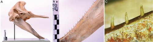
Table 1. Measurements (in mm) of skull of Ziphius cavirostris OMNZ VT2765.
Dental morphology
This specimen presented the alveoli for the expected pair of mandibular tusks, but the tusks have been lost. In all, 49 vestigial teeth were in situ on the lower jaws of the specimen (B). Twenty-two teeth were observed on the left lower jaw and 29 on the right. No vestigial teeth were mounted on the upper jaw; however, it is not known if upper teeth were originally present and lost. The vestigial teeth were not enclosed within alveoli, but rather were loosely attached to a shallow sulcus in the mandible. It is presumed that in the live animals teeth were embedded within the gum tissue. A shallow sulcus was also present in both maxillae.
Vestigial teeth were slender, high crowned and needle-like (C). Teeth were curved lingually and presented a circular cross section. The transition from the crown to the cingulum was poorly defined. On average, vestigial teeth measured 8–10 mm in length and 0.8–1.2 mm in diameter.
Ultrastructure
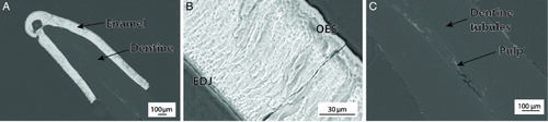
Scanning electron microscope images revealed that the vestigial tooth sampled was covered with a thin layer of enamel (). This layer was 120–140 µm thick on average, except towards the base of the crown where it was less than 10 µm thick. Enamel was prismless from the enamel–dentine junction to the outer surface; however, it appeared pseudoprismatic or slightly structured. Electron backscattered images suggested a higher mineral content in enamel than in dentine. The dentine was mainly featureless, but dentine tubules could be observed radiating from the pulp to the outer surface.
Figure 2. A, Scanning electron microscope image overview of a Ziphius cavirostris vestigial tooth. B, Detailed view of the enamel layer showing a semi-structured prismless layer (EDJ, enamel–dentine junction; OES, outer enamel surface). C, Dentine tubules irradiating from the pulp towards the outer surface.
Mechanical properties
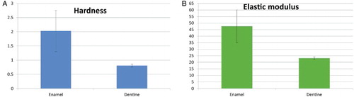
Nanoindentation-derived mechanical properties were calculated for the one vestigial tooth sampled. Hardness values averaged 2.03 ± 0.73 GPa for enamel and 0.81 ± 0.05 GPa for dentine. Elastic modulus values averaged 47.60 ± 12.48 GPa for enamel and 23.21 ± 0.99 GPa for dentine ().
Figure 3. A, Average hardness values in GPa (± SD) for enamel and dentine. B, Average elastic modulus values in GPa (± SD) for enamel and dentine.
Chemical analysis
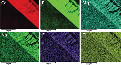
Chemical mapping done via energy-dispersive X-rays provided a qualitative assessment of the distribution of major (Ca and P) and minor (Na, Cl, F, Sr, Mg) elements within enamel and dentine (). Chemical maps showed higher concentrations of Ca and P in enamel compared with dentine. For the minor elements, this also seems to be the trend; however, their lower concentrations did not allow precise resolution.
Figure 4. Qualitative chemical map showing the distribution of major and minor elements in enamel and adjacent dentine.
Discussion
Skull measurements and unfused sutures suggest that OMNZ VT2765 is a juvenile specimen. Kernan (Citation1918) reported condylobasal length values of 854 mm for a young adult female and 435 mm for an unknown sex foetus at term, and Hubbs (Citation1946) reported condylobasal lengths of 716 mm for an immature female. Omura (Citation1972) studied a juvenile specimen of unknown sex with condylobasal length measuring 590 mm. Our specimen, with a condylobasal length of 520 mm, is probably a very young juvenile. The occurrence of at least 49 vestigial teeth in this specimen is in accordance with the findings of Fraser (Citation1936), who observed a reduction in number of vestigial teeth with increase in total body length of the animal. Fraser (Citation1936) reported small and immature animals having about 30 vestigial teeth in each mandible, whereas the larger animals had 10 teeth, or even as few as one tooth. He suggested that absorption of teeth must occur as the animals get older, which might explain the disparity in vestigial tooth numbers between young and older specimens.
Morphology of the vestigial teeth in our specimen is consistent with previous historical reports that describe them as slender, conical and tapering gradually to a sharp point (True Citation1910). Boschma (Citation1951) described the smaller vestigial teeth as conical, slightly curved, circular or oval in cross section, meaning that their shape is often entirely different from that of the larger tusks characteristic of ziphiids. This author reviewed historical records on the occurrence of vestigial teeth in several species of Ziphiidae since the 1700s, and recorded at least 12 previous studies that describe vestigial teeth in Ziphius cavirostris.
Boschma (Citation1951) interpreted the vestigial teeth in Ziphius as rudimentary and functionless. In most of the cases described by him, rows of small teeth were concealed in the gum tissue, with very few teeth protruding above, but with such weak attachment that they could easily be removed. In addition, all teeth studied had closed pulp cavities and sometimes the roots were filled with a bulbous mass of cement beyond the original root apex.
Other beaked whales also commonly possess vestigial teeth in addition to the tusks. Kirino (Citation1956) found a strong tendency to impaction in the genus Berardius, with most of the concealed teeth being located posteriorly rather than anteriorly, and females often had more impacted teeth than males. Kirino (Citation1956) also interpreted the occasional presence of the additional teeth, their rudimentary formation, high variability in size and strong inclination to impaction, as evidence for the atavistic reappearance of an ancestral polydont condition.
Although atavisms have a very low frequency of occurrence and only appear in unusual circumstances within the species (Hall Citation2002), vestigial structures, rudiments or remnants are reduced body parts or organs, often without visible function, that may be fully developed and functioning in primitive members of that phylogenetic lineage (Bejder & Hall Citation2002). These structures are often small in comparison with their size in ancestors or in closely related species, so they are sometimes described as atrophied or degenerate (Bejder & Hall Citation2002). The presumed high incidence of vestigial teeth in Ziphius cavirostris, the small size and degenerate nature of these structures are consistent with their interpretation as vestiges rather than atavisms.
At the ultrastructural level, analysis with scanning electron microscopy revealed a thin layer of enamel covering the crowns of vestigial teeth. The thickness of this layer was similar to values reported for other odontocete cetaceans with thin enamel (Loch et al. Citation2013a); however, the overall tooth size of these vestigial teeth was much smaller compared with normal teeth in, for example, Delphinidae. Most odontocete cetaceans have either prismatic decussating or radial enamel, with few possessing prismless enamel (Ishiyama Citation1987; Loch et al. Citation2013a). The tusks of ziphiids commonly have no enamel or only a thin layer, < 10 µm thin, which easily wears away, exposing the dentine and cementum underneath (Ishiyama Citation1987). The presence of a semi-structured prismless layer reinforces the vestigial nature of the teeth, and could shed light on the structure of the dentition in early ziphiids.
Chemical analysis via energy-dispersive X-rays revealed the presence of the expected major and minor elements in tooth hydroxyapatite as reported in other odontocete cetaceans (see Loch et al. Citation2014), suggesting the maintenance of the chemical composition despite morphological simplification and vestigial nature of the teeth. In terms of values for mechanical properties, Loch et al. (Citation2013b) reported hardness values ranging from 2.36 to 3.86 GPa for the enamel of 10 species of dolphins, whereas dentine values ranged from 0.56 to 0.80 GPa. Elastic modulus values ranged from 13.51 to 69.32 GPa in enamel, and 7.84 to 21.55 GPa for dentine. The values we reported here are within the range of mechanical properties values for dentine in cetaceans; however, hardness values for the enamel were slightly lower. The lower values reported for cetaceans in comparison to other terrestrial and aquatic vertebrates were interpreted by Loch et al. (Citation2013b) as being the result of relaxed biomechanical functional demands in dolphins, which would also explain the lower values in the vestigial teeth analysed here.
This study reported the occurrence of vestigial teeth in a specimen of Cuvier's beaked whale, Ziphius cavirostris, and provided the first characterisation of the ultrastructure and mechanical and chemical properties of vestigial teeth in ziphiid cetaceans. Future structural and developmental studies will shed light on the dental evolution of these elusive and poorly known marine mammals.
Acknowledgements
We would like to acknowledge Emma Burns, Cody Fraser and Kane Fleury (Otago Museum) for facilitating access to the specimen, and Robert Morris (Otago Museum) for the support towards this research. R Ewan Fordyce provided valuable suggestions to early drafts of this manuscript. We are indebted to two anonymous reviewers for their suggestions, which much improved this revised version. Thanks are also extended to Marco Brenna (OCEM, University of Otago) for his help with scanning electron microscopy and energy-dispersive X-rays analyses, and Jules Kieser (in memoriam) for the everlasting inspiration to study unusual and exciting things.
Associate Editor: Dr Jonathan Banks.
Disclosure statement
No potential conflict of interest was reported by the authors.
Additional information
Funding
References
- Bejder L, Hall BK. 2002. Limbs in whales and limblessness in other vertebrates: mechanisms of evolutionary and developmental transformation and loss. In: Raff RA, Wallace A, Carroll SB, Coates MI, Wray G, editors. Evolution and development. Volume 4. Hoboken, NJ: Blackwell Publishing; p. 445–458.
- Bianucci G, Lambert O, Post K. 2007. A high diversity in fossil beaked whales (Mammalia, Odontoceti, Ziphiidae) recovered by trawling from the sea floor off South Africa. Geodiversitas. 29: 561–618.
- Boschma H. 1951. Rows of small teeth in ziphioid whales. Zool Med leiden. 31: 139–148.
- Fordyce RE, Mattlin RH, Wilson GJ. 1979. Stranding of a Cuvier's beaked whale, Ziphius cavirostris. Mauri Ora. 7:73–82.
- Fraser FC. 1936. Vestigial teeth in specimens of Cuvier's whale (Ziphius cavirostris) stranded on the Scottish coast. Scott Nat. 222:153–157.
- Gomerči H, Gomerči MD, Gomerči T, Luci H, Dalebout M, Galov A, Škrti D, Vukovi S, Huber D. 2006. Biological aspects of Cuvier's beaked whale (Ziphius cavirostris) recorded in the Croatian part of the Adriatic Sea. Euro J Wild Res. 52:182–187. doi: 10.1007/s10344-006-0032-8
- Hall BK. 2002. Atavisms. In: Pagel M, editor. Encyclopedia of evolution. Volume 1. New York: Oxford University Press; p. 86–87.
- Heyning JE, Mead JG. 1996. Suction feeding in beaked whales: morphological and observational evidence. Natural History Museum of Los Angeles County. 464:1–12.
- Hubbs CL. 1946. First records of two beaked whales, Mesoplodon bowdoini and Ziphius cavirostris, from the Pacific Coast of the United States. J Mammal. 27:242–255. doi: 10.2307/1375435
- Ishiyama M. 1987. Enamel structure in odontocete whales. Scanning Micros. 1: 1071–1079.
- Kernan JD. 1918. The skull of Ziphius cavirostris. B Am Mus Nat Hist. 38:349–394.
- Kirino T. 1956. On the number of teeth and its variability in Berardius bairdi, a genus of the beaked whale. Okajimas folia anatomica Japonica. 28:429–434. doi: 10.2535/ofaj1936.28.1-6_429
- Loch C, Duncan W, Simões-Lopes PC, Kieser JA, Fordyce RE. 2013a. Ultrastructure of enamel and dentine in extant dolphins (Cetacea: Delphinoidea and Inioidea). Zoomorphology. 132: 215–225. doi: 10.1007/s00435-012-0180-1
- Loch C, Swain MV, Fraser SJ, Gordon KC, Kieser JA, Fordyce RE. 2014. Elemental and chemical characterization of dolphin enamel and dentine using X-ray and Raman microanalyzes (Cetacea: Delphinoidea and Inioidea). J Struct Biol. 185: 58–68. doi: 10.1016/j.jsb.2013.11.006
- Loch C, Swain MV, van Vuuren LJ, Kieser JA, Fordyce RE. 2013b. Mechanical properties of dental tissues in dolphins (Cetacea: Delphinoidea and Inioidea). Arch Oral Biol. 58: 773–779. doi: 10.1016/j.archoralbio.2012.12.003
- Mead JG. 1989. Shepherd's beaked whale Tasmacetus shepherdi Oliver, 1937. In: Ridgway SH, Harrison R, editors. Handbook of marine mammals. Volume 4. London: Academic Press. p. 309–320.
- de Muizon C. 1983. Un Ziphiidae (Cetacea) nouveau du Pliocene inferieur du Perou. Comptes rendus de l'Academie des Sciences de Paris 297: 85–88.
- Oliver WC, Pharr GM. 1992. An improved technique for determining hardness and elastic modulus using load and displacement sensing indentation experiments. J Mat Res. 7: 1564–1583. doi: 10.1557/JMR.1992.1564
- Omura H. 1972. An osteological study of the Cuvier's beaked whale, Ziphius cavirostris, in the northwest Pacific. Scientific Reports of the Whales Research Institute. 24:1–34.
- True FW. 1910. An account of the beaked whales of the family Ziphiidae in the collection of the United States National Museum: with remarks on some specimens in other American museums. Bull US Nat Mus. 73: 1–89.
- Werth AJ. 2000. Feeding in marine mammals. In: Schwenk K, editor. Feeding: form, function and evolution in tetrapod vertebrates. San Diego: Academic Press; p. 487–526.