Abstract
A Hodgson's hawk-eagle (Spizaetus nipalensis) reared by a falconer showed severe weakness with multiple fractures of bone. It had a history of being fed an all-meat diet. Serological examination revealed a hypocalcaemia (72.0 μg/ml), and hypophosphataemia (29.0 μg/ml). Gross and microscopic examinations demonstrated severe osteodystrophia fibrosa (fibrous osteodystrophy) characterized by osteoclastic bone resorption and intertrabecular fibrosis with unmineralized trabecular bone containing a large amount of unmineralized osteoid. There was also hypertrophy and hyperplasia of the parathyroid glands, which is consistent with nutritional secondary hyperparathyroidism.
1 Introduction
Nutritional secondary hyperparathyroidism caused by long term deficiency of vitamin D3, calcium or phosphorus, or by imbalances in the last two nutrients causes excessive production of parathyroid hormone (PTH), resulting in osteoclastic bone absorption (Woodard, Citation1997). Rickets, which has the same pathogenesis, occurs in various kinds of young birds and mammals and is well described (Whitehead & Wilson, Citation1992; Thorp, Citation1994). However, description of osteodystrophia fibrosa (fibrous osteodystrophy) in avian species is limited (Long et al., Citation1983). Here, we describe the disease secondary to nutritional hyperparathyroidism in a Hodgson's hawk-eagle (Spizaetus nipalensis) bred for falconry.
2 Materials and Methods
Necropsy was performed and the liver, kidneys, heart, lungs, intestine, pancreas, adrenal glands, thyroid glands, parathyroid glands, sternum, femur, tibiotarsus and skeletal muscles were routinely fixed in 10% neutral buffered formalin, embedded in paraffin wax and stained with haematoxylin and eosin. Selected samples of the sternum, femur and tibiotarsus were decalcified by formic acid before embedding.
3 Results
A 12-year-old male Hodgson's hawk-eagle (Spizaetus nipalensis) reared by a falconer in Japan was presented to the Akita Omoriyama Zoo with a history of severe weakness, leg paralysis and anaemia (haematocrit 21%). The bird had been fed only commercial eviscerated meat since the falconer had become elderly and could no longer continue traditional hunting because of his own physical limits. Radiography revealed multiple bone fractures and severe thinning of cortical bones, particularly in the skull and long bones. Complete fractures were found radiographically at the distal metaphysis of the left humerus, proximal metaphysis of the left ulna and at the diaphysis of the left tibiotarsus. Incomplete fractures were also observed at the distal metaphysis of the left tibiotarsus, distal bilateral tarsometatarsus, proximal metaphysis of the right radius, diaphysis of the right metacarpal and diaphysis of the right femur. Metaphyses of the long bones adjacent to the elbow, hock and carpal joints and the skull were apparently decalcified and cortical bones in these regions had become thinner. The bird was surgically treated for these fractures by intramedullary pinning and given a vitamin B and calcium compound by oral administration. Biochemical examination eight days later revealed lower serum calcium (72.0 μg/ml) and phosphorus (29.0 μg/ml) and higher alkaline phosphatase (ALP) (3100 IU/l) than normal. Normal ranges of these inorganic substances or enzyme in raptors are 89.3–101.9 μg/ml (calcium), 30.3–43.4 μg/ml (phosphorus) and 31–257 IU/l (ALP), respectively (Samour, Citation2000). The bird died ten days after the initial examination.
Gross examination confirmed that bone was very fragile with multiple complete and/or incomplete fractures. The parathyroid glands were bilaterally enlarged, up to 6 mm in diameter, and they were even larger than thyroid glands.
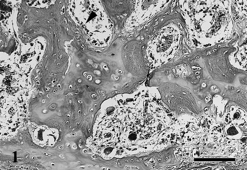
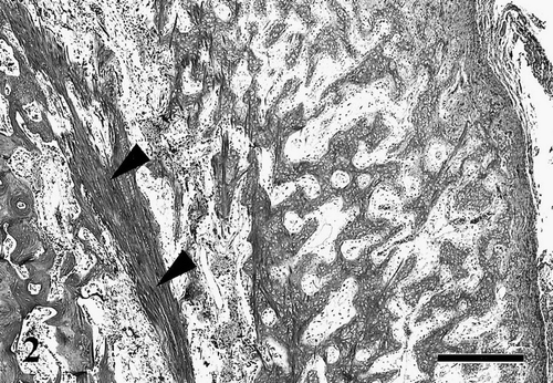
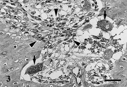
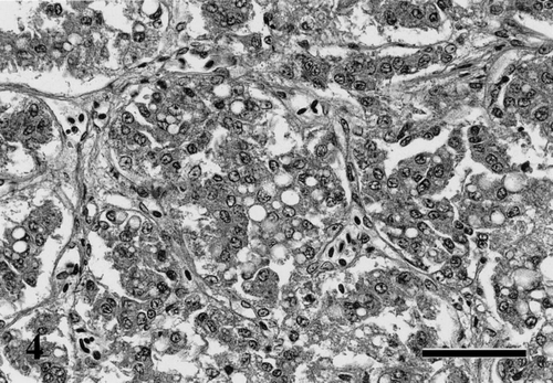
Histological changes of the affected bone mainly consisted of osteomalacic lesions and fractures. The former was characterized by a significant decrease in the amounts of mineralized trabecular bone associated with an increase in the width of unmineralized osteoid (). The osteoid borders were occasionally lined by osteoblasts and distinct osteoid seams were recognized. These changes occurred in the cortical and cancellous bone of the sternum, femur and tibiotarsus. The cortical bones of the diaphyses were also markedly thinned with dilation of the Haversian canals. The femur and tibiotarsus partly showed subperiosteal proliferation of trabeculae and loose fibrous tissue (). In addition, numerous osteoclasts were frequently observed on the surface of the trabeculae, Haversian canals and endosteum and fibrous tissue proliferated between these trabeculae (), suggesting a progression to osteodystrophia fibrosa. Although there were endochondral and membranous ossifications in and around the necrotic bone of each fracture, mineralization was incomplete in these foci. Enlarged parathyroid glands consisted of proliferation of hypertrophic chief cells, which had lightly eosinophilic and vacuolated cytoplasm (). Follicles of thyroid glands were atrophic and were lined by cuboidal epithelium.
Fig. 1 Decalcified section of the sternum. Trabecular bone mainly composed of thick unmineralized osteoid seams occasionally lined by osteoblasts (arrowhead). Haematoxylin and eosin (HE). Bar=100 μm.

Fig. 2 Decalcified section of the tibiotarsus. Marked periosteal bone reaction with proliferation of osteoid trabeculae and loose fibrous tissue at the exterior of thinning cortex bone (arrowheads). HE. Bar=500 μm.

Fig. 3 Decalcified section of the tibiotarsus. Osteoclastic bone resorption on the surface of bone trabeculae (arrows) and proliferation of fibroblasts in the medullary cavity (arrowheads). HE. Bar=30 μm.

Fig. 4 Parathyroid gland showing proliferation of chief cells with cytoplasmic swelling and vacuolation. HE. Bar=50 μm.

Other histopathological findings included moderate hyperplasia of the adrenal cortex, moderate atrophy and degeneration of the pectoral muscles and mild myocardial fibrosis.
4 Discussion
Osteodystrophia fibrosa is the result of continuous and excessive secretion of PTH and is characterized by marked osteoclastic resorption and fibrous replacement (Palmer, Citation1991). Locomotory disorders with fractures in birds of prey including those for falconry, have been reported as "cramps" or "fits"; protracted dietary deficiency of calcium or imbalance of calcium/phosphorus ratio has been considered important causative factors (Wallach & Flieg, Citation1970; Cooper, Citation1975). However, pathological descriptions of osteodystrophia fibrosa due to nutritional secondary hyperparathyroidism have rarely been reported in avian species (Long et al., Citation1983).
The present case was considered nutritional secondary hyperparathyroidism that resulted in severe osteodystrophia fibrosa. The active osteoclastic bone resorption and fibrous tissue proliferation between trabeculae in the examined bones are characteristic findings of osteodystrophia fibrosa. On the other hand, accumulation of osteoid and formation of osteoid seams in the medullary cavity are specific features of osteomalacia, which frequently progresses to osteodystrophia fibrosa when severe hyperparathyroidism develops in the course of the disease (Woodard, Citation1997). Compensating subperiosteal bone proliferation occurs in some cases of osteopoenia (Riddell, Citation1996). However, subperiosteal proliferation of osteoid and fibrous tissues in the present case was considered as unmineralized reactive callus resulting from microscopic fractures, because it was observed only in part of the femur and tibiotarsus.
The calcium: phosphorus ratio (Ca: P) of meat and fish commonly fed to birds of prey range from 1: 17 to 1: 44 whereas the correct Ca: P ratio for avian diets is listed as 1.5: 1 (Wallach & Flieg, Citation1970). As exceptionally high dietary levels of phosphorus in these diets maybe from insoluble salts with calcium and prevent their absorption in addition to an absolute calcium deficiency, these diets result in an increased activity of the parathyroid gland for a long term.
Hyperparathyroidism has been reported in a hawk affected with severe fibrous osteodystrophy (Long et al., Citation1983). The parathyroid glands of the hawk had characteristic oxyphil cells, a variant of chief cells, due to continued hypocalcemia. Although oxyphil cells have been described in hyperparathyroidism of many animal species (Roth & Capen, Citation1974), the cells have not been observed in any other birds, including the present case. This suggests that other factors such as the age of birds, the type of deficient nutrients and the duration of deficiency are more important for the proliferation of oxyphil cells in hyperparathyroidism of avian species.
For a long time, falconry has been maintained throughout the world as a traditional form of hunting and the avian species used include hawks, eagles, buzzards and peregrines. Dietary requirements of raptors have not been sufficiently studied, but artificial diets such as muscle meat and eviscerated prey are known to lack vitamins, calcium and phosphorus and may cause various metabolic bone diseases in raptors (Keymer, Citation1972; De Water, Citation1996). Investigation of the dietary requirements of raptors is essential to preserve these rare birds.
Acknowledgments
The authors would like to thank Dr C. Itakura of the Institute of Physical and Chemical Research, Wako, Japan, for his suggestions.
References
- Cooper , J.E. 1975 . Osteodystrophy in birds of prey . Veterinary Record , 97 : 307
- De Water D.V. Raptor rehabilitation, Diseases of Cage and Aviary Birds 3rd edn, Rosskopf W., Woerpel R. (eds) Williams & Wilkins: Baltimore 1996 1007 1028
- Keymer , I.F. 1972 . Diseases of birds of prey . Veterinary Record , 90 : 579 – 594 .
- Long , P. , Choi , G. and Rehmel , R. 1983 . Oxyphil cells in a red-tailed hawk (Buteo jamaicensis) with nutritional secondary hyperparathyroidism . Avian Diseases , 27 : 839 – 843 .
- Palmer, N. (1991). Bones and joints. In K.V.F. Jubb, P.C. Kennedy & N. Palmer (Eds.), Pathology of Domestic Animals 4th edn Volume 1 (pp. 1–181). San Diego: Academic Press.
- Riddell C. Skeletal system, Avian Histopathology 2nd edn, Riddell C. (ed.) American Association of Avian Pathologists: Kennet Square, PA 1996 45 60
- Roth , S.I. and Capen , C.C. 1974 . Ultrastructural and functional correlations of the parathyroid gland . International Review of Experimental Pathology , 13 : 161 – 221 .
- Samour, J. (2000). Avian Medicine. London: Harcourt Publishers.
- Thorp , B.H. 1994 . Skeletal disorders in the fowl: a review . Avian Pathology , 23 : 203 – 236 .
- Wallach, J.D. & Flieg, G.M. (1970).Cramps and fits in carnivorous birds. In J. Lucas (Ed.), International Zoo Yearbook Volume 10 (pp. 3–4). London: Zoological Society of London.
- Whitehead C.C. Wilson S. Characteristics of osteopenia in hens, Bone Biology and Skeletal Disorders in Poultry Whitehead C.C. (ed.) Carfax Publishing: England 1992 265 280
- Woodard J.C. Skeletal system, Veterinary Pathology 6th edn, Jones T.C., Hunt R.D., King N.W. (eds) Williams & Wilkins: Baltimore 1997 899 946