Abstract
The present study reports the clinical, virological and pathological findings observed in a natural outbreak of highly pathogenic avian influenza in farmed commercial ducks. The ducks developed clinical signs, including mild respiratory distress, depression, mild diarrhoea, loss of appetite and increasing mortality (up to 12%). At necropsy, multifocal mottled necrosis was commonly found in the pancreas with splenomegaly, hepatomegaly, and swollen kidneys. Microscopically, there was necrotized pancreatitis and hepatitis, and lymphocytic meningoencephalitis and myocarditis. Influenza viral antigen was demonstrated in areas closely associated with histopathological lesion. Avian influenza virus was isolated from the caecal tonsil, faeces, and kidney of the domestic ducks. The isolated virus was identified as a highly pathogenic H5N1, with a haemagglutinin proteolytic cleavage site deduced amino acid sequence of … QREKRKKR/GLFGAIAG … In order to determine the pathogenicity of the isolate, eight 6-week-old specific pathogen free chickens were inoculated intravenously with the virus, and all birds died within 24 h after inoculation. This is the first report of an outbreak of highly pathogenic avian influenza with clinical signs in commercial domestic ducks in South Korea.
Influenza Aviaire Hautement Pathogène (H5N1) chez des canards domestiques en Corée du Sud
La présente étude rapporte les résultats des observations clinique, virologique et pathologique réalisées à partir d'un élevage de canards qui a présenté un cas spontané d'influenza aviaire hautement pathogène. Les symptômes développés par les canards incluaient une affection respiratoire bénigne, de la dépression, une légère diarrhée, une perte d'appétit, et une augmentation de la mortalité (jusqu'à 12%). A l'autopsie, une nécrose marbrée multifocale à généralement été observée au niveau du pancréas avec une splénomégalie, une hépatomégalie et des reins gonflés. Sur le plan microscopique, il a été noté une hépatite et une pancréatite nécrosées, ainsi qu'une méningo-encéphalite et une myocardite lymphocytaires. L'antigène viral de l'influenza a été mis en évidence dans des zones étroitement associées aux lésions histopathologiques. L'AIV a été isolé à partir des amygdales cæcales, des fèces et des reins des canards domestiques. Le virus isolé, a été identifié comme étant H5N1 hautement pathogène, avec la séquence déduite en acides aminés du site de clivage de l'hémagglutinine protéolytique : “… QREKRKKR/GLFGAIAG …’. Afin de déterminer la pathogénicité de l'isolat, des poulets, exempts de microorganismes pathogènes spécifiés, âgés de 6 semaines ont été inoculés par voie intraveineuse avec le virus et tous les animaux sont morts dans les 24 heures après l'inoculation. Ceci est le premier rapport d'un cas de HPAI avec des signes cliniques chez des canards d'élevage en Corée du Sud.
Hochpathogene aviäre Influenza (H5N1) in kommerziellen Hausenten in Südkorea
Die vorliegende Studie berichtet über die klinischen, virologischen und pathologisch-anatomischen Befunde im Rahmen eines natürlichen Ausbruchs einer hochpathogenen Influenza (HPAI) bei kommerziell gehaltenen Enten. Die klinischen Symptome der Enten umfassten geringradige Atemnot, Störung des Allgemeinbefindens, milde Diarrhoe, Appetitlosigkeit und erhöhte Mortalität (bis zu 12%). Bei der pathologisch-anatomischen Untersuchung wurden häufig multifokale, marmorierte Pankreasnekrosen, Spleno- und Hepatomegalie sowie geschwollene Nieren gefunden. Histologisch ließen sich nekrotisierende Pancreatitis und Hepatitis sowie lymphozytäre Meningoenzephalitis und Myocarditis feststellen. Virales Influenza-Antigen wurde in Arealen in Nähe der histopathologischen Läsionen nachgewiesen. AIV wurde aus Zäkaltonsillen, Fäzes und Nieren der Hausenten isoliert. Das isolierte Virus wurde als hochpathogenes H5N1 mit der an der Hämagglutinin-proteolytischen Spaltstelle abgeleiteten Aminosäurensequenz “… QREKRKKR/GLFGAIAG …” identifiziert. Zur Pathogenitätsbestimmung des Isolats wurden acht sechswöchige spezifiziert pathogenfreie Hühnerküken intravenös mit dem Virus inokuliert und alle Tiere starben innerhalb von 24 Stunden. Dies ist die Erstbeschreibung eines Ausbruchs von HPAI mit klinischen Symptomen bei kommerziellen Hausenten in Südkorea.
Influenza aviar de alta patogenicidad (H5N1) en patos domésticos comerciales en Corea del Sur
En el presente estudio se describen los hallazgos clínicos, virológicos y patológicos asociados a un brote natural de influenza aviar altamente patógena en patos comerciales domésticos. Los patos presentaron distrés respiratorio leve, depresión, leve diarrea, pérdida de apetito y mortalidad incrementada (hasta el 12%). A la necropsia, se observaron áreas multifocales de necrosis en el páncreas, junto con esplenomegalia, hepatomegalia y riñones tumefactos. Al microscopio se observó pancreatitis necrotizante y hepatitis, meningoencefalitis linfocítica y miocarditis. El antígeno vírico de influenza se detectó en áreas muy relacionadas con las lesiones histopatológicas. Se aisló AIV de tonsilas cecales, heces y riñón de los patos domésticos. El virus aislado se identificó como altamente patogéno H5N1, con una secuencia de aminoácidos del punto de ruptura proteolítica de la hemaglutinina de “…QREKRKKR/GLFGAIAG… .’ Para determinar la patogenicidad del aislado, se inocularon vía intravenosa con el aislado, ocho pollos de seis semanas de edad libres de patógenos específicos y todas las aves murieron a las 24 horas tras la inoculación. Esta es la primera descripción de un brote de HPAI con signos clínicos en patos comerciales domésticos en Corea del Sur.
Introduction
Avian influenza (AI) viruses have been isolated from numerous wild and domestic avian species (Alexander, Citation2000), and the order Anseriformes including ducks, geese and swans are regarded as the primordial reservoir hosts of these viruses (Hinshaw et al., Citation1980). Highly pathogenic avian influenza (HPAI) caused by some viruses of H5 and H7 subtypes produce a fulminating and rapidly fatal systemic disease in poultry such as chickens and turkeys (Swayne & Halvorson, Citation2003). Although HPAI viruses have been reported to infect domestic ducks flocks, no signs of disease were seen and infection was confirmed only following virus isolation (Alexander, Citation2000).
South Korea had experienced an epidemic of HPAI caused by a type A influenza virus of the H5N1 subtype from mid December 2003 to late March 2004. Among the 19 affected establishments of domestic poultry, the virus infected nine domestic duck farms, consisting of eight breeder ducks farms and one commercial meat duck farm. All breeder ducks infected with the AI virus showed clinical signs of decreased egg production and feed consumption rates but without increases in mortality. However, the infected commercial domestic ducks exhibited moderate increased mortality rates and respiratory signs. In this report, we describe clinical and pathological findings observed in commercial domestic meat ducks infected with the H5N1 HPAI virus.
Materials and Methods
Animals
On 20 December 2003, Chonnam Regional Livestock and Veterinary Service submitted 10 dead commercial ducks (14 days old) from a domestic duck farm exhibiting moderate mortality with severe depression to the Avian Disease Division, National Veterinary Research and Quarantine Service, Anyang, South Korea.
Pathology and immunohistochemistry
The birds were surface disinfected and a necropsy was performed. Tissue samples were taken (trachea, lung, heart, liver, spleen, kidney, proventriculus, gizzard, pancreas, intestine, cloacal bursa, brain, skeletal muscle, eyelids and skin) and fixed in 10% neutral buffered formalin, processed and embedded in paraffin blocks. Sections were made at 5 µm and were stained with haematoxylin and eosin. Duplicate sections were stained with a mouse-derived monoclonal antibody specific for a type A influenza virus nucleoprotein (H16-L10-4; kindly donated by Dr Selleck, CSIRO, Australia) as the primary antibody[0]. All reactions were carried out in an automated immunohistochemistry instrument (Ventana ES; Ventana Medical Systems, Tuscon, Arizona, USA). Antigen–antibody reactions were revealed with standardized development times by the instrument, using the avidin–biotin method with copper enhancement and diaminobenzidine as the substrate. The tissue sections were counter stained with Gill's No. 3 haematoxylin (Sigma, St Louis, Missouri, USA).
Virology
For virological examination, 10% homogenates of the caecal tonsils, faeces and kidneys of the birds submitted were inoculated into the allantoic cavity of 9-day-old specific pathogen free embryonated eggs. Allantoic fluids from dead embryonated eggs were harvested and tested by the agar gel precipitin test for type A influenza virus. All agar gel precipitin test positives were subtyped by haemagglutination and neuraminidase inhibition tests with a panel of reference antisera (provided by OIE Reference Laboratory, Veterinary Agency, Surrey, UK). The virulence of the isolate was determined as described by the OIE procedure. In addition, to analyse haemagglutinin cleavage site sequences, RNA was extracted from virus-containing allantoic fluid, followed by cDNA synthesis and polymerase chain reaction, as described earlier (Munch et al., Citation2001).
Bacteriology
In bacterial examination, the liver and spleen were aseptically collected using sterilized cotton-tipped swabs. Each swab collected was streaked on both 5% sheep blood agar (Komed Co., Ltd, South Korea) and MacConkey's agar, and also inoculated into 10 ml trypticase soy broth as growth media. The solid media plates and broths were incubated at 37°C for 24 to 48 h in aerobic and anaerobic conditions.
Results
Case history
On 15 December 2003, a disease characterized by depression, ruffled feathers, diarrhoea, respiratory distress, and loss of appetite occurred in the commercial duck farm located at the southern part of Korea, Chonnam province. The farm consisted of two flocks—flock A (4000 9-day-old birds) and flock B (10 900 10-day-old birds)—but the clinical signs were found in only flock A. Ducklings for flock A were obtained from a hatchery, located in the middle part of Korea, and place on the production farm at 1 day of age. The parent duck flock was later determined, through active surveillance, to be infected with H5N1 HPAI virus.
Increased mortality was first observed in flock A at 9 days of age, and cumulative mortality in the flock was 12% by 14 days of age when the ducks were depopulated. However, no clinical signs and mortality was observed in flock B, which had been obtained from a different hatchery. Ten of the dead birds (14 days old) were submitted for examination, and a final diagnosis of HPAI was made based on laboratory tests, including pathological, immunohistochemical and virological test results.
Gross findings
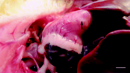
On postmortem examination, the birds were dehydrated and in poor physical condition. In many birds, the pancreas had a mottled appearance indicative of necrosis. The livers were enlarged, friable and congestion. Spleens were increased in size with multifocal white spots. In addition, there were whitish foci of myocardial necrosis (), swollen kidneys, and congested and/or haemorrhagic lungs.
Figure 1. Large white foci of necrosis (arrow) in the heart from a dead duck. Bar = 0.6 cm.
Histology
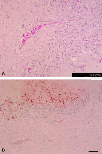
There were multiple confluent foci of coagulative necrosis with mild mixed inflammatory cellular responses in multiple organs, including the heart, brain, pancreas and liver. Extensively multifocal myocardial necrosis with haemorrhages and mononuclear cellular infiltration was observed. The brains contained randomly scattered foci of malacia with gliosis, and lymphocytic perivascular cuffing with oedema (A). In the pancreas, multifocal cellular swelling to necrosis of the pancreatic acinar epithelium was present with mild inflammatory response. There was an increase in sinusoidal cellularity attributed to the combined presence of Kuppfer cell hyperplasia, and increased numbers of mononuclear cells in the portal triads of liver. Discrete foci of tubular epithelial necrosis were found in the kidney. Mildly lymphocytic depletion and necrosis of peri-ellipsoidal and peri-arteriolar sheaths was present in the spleen. In the lung, mildly interstitial pneumonia with congestion was observed.
Figure 2. Photomicrograph of the cerebellum from a dead duck. 2A: focal mononuclear perivascular cuffs with gliosis in the neurophil. Haematoxylin & eosin stain. Bar = 60 µm. 2B: viral antigen detected in the Purkinje neurons, dendrites and granular cell layer. Immunohistochemical stain with haematoxylin counterstain. Bar = 75 µm.
Immunohistochemistry
Systemic localization of AI virus was common to the submitted domestic ducks. There was a strong association between the location of viral antigen and histological lesions. Influenza viral antigen was demonstrated within cardiac myocytes, pancreatic acinar epithelium, peripheral nerves and ganglia, pulmonary endothelial cells, renal tubular epithelial cells, and neurons, Purkinje cells and granular layers of the brain (b), including the cerebral hemispheres, medulla, cerebellum and pons. Viral antigen also was observed in the skeletal myofibres, smooth muscular layer of the intestine, and elastic fibres of tunica media in the artery.
Virology
Type A influenza viruses were isolated from the caecal tonsils, faeces and kidneys in embryonating specific pathogen free chicken eggs. The viruses were subtyped as H5N1 by haemagglutination and neuraminidase inhibition tests, and caused 100% mortality within 24 h in groups of eight intravenously inoculated chickens. The isolate exhibited a deduced amino acid sequence in the region of the genome coding for the cleavage site of the haemagglutinin molecule of … GREKRKKR/GLFGAIAG … , which contained multiple basic amino acids. The high lethality and presence of multiple basic amino acids in the proteolytic cleavage site indicates the virus was highly pathogenic.
Bacteriology
No pathogenic bacteria were isolated on aerobic or anaerobic cultures from liver and spleen samples.
Discussion
The prominent pathologic changes observed at necropsy in this study consisted of haemorrhage in the heart, necrotic foci in the pancreas, and enlargement of the liver and spleen. Histopathology indicated parenchymal necrosis of multiple organs such as the brain, heart and pancreas, similar to findings in chicken, turkey, quail, and ostriches infected with other HPAI viruses (Allwright et al., Citation1993; Manvell et al., Citation1998; Capua et al., Citation2000; Clavijo et al., 2001; Perkins & Swayne, Citation2002). Examination of the pathogenicity test data, sequence analysis of the haemagglutinin cleavage site (RKKR/GLFG) and serological subtyping of the isolated virus from several organs of the dead ducks resulted in the conclusion that the virus was the HPAI virus of the H5N1 subtype. On the basis of the pathological findings, and the characterization of the virus isolated, the disease that occurred in the ducks was determined to be caused by the HPAI virus, although the virus did not produce high mortality in ducks as it did in experimental chickens.
The affected duck farm consisted of two flocks (A and B) that were reared in different houses. The disease signs, including moderate mortality, were present only in ducks of flock A, which were derived from eggs obtained from a duck breeder flock affected with HPAI. Ducks in flock B were not affected by HPAI and were obtained from a non-infected duck breeder farm. Therefore, as a result of an epidemiological investigation, we concluded that ducks in flock A might have been exposed to HPAI virus at the contaminated hatchery or during transport from the hatchery to the duck farm.
The H5N1 Korean HPAI virus isolates from the chicken (CK/Korea/ES/03) and domestic duck (DK/Korea/EDS1/03) were genetically almost identical to each other (99.8% nucleotide identity) (Lee et al., Citation2004). Intranasal inoculation of the Korean virus AI isolated from the chicken into 2-week-old Pekin ducks resulted in a mortality rate of 25%, with a mean death time of 4.0 days in affected birds. The field mortality in the present case (12%) was similar, suggesting the Korean H5N1 HPAI viruses have moderate pathogenicity for ducks. By contrast, some of the H5N1 1997 to 1999 chicken and goose HPAI viruses, and the A/duck/Anyang/AVL-1/01 H5N1 HPAI virus isolated from imported duck meat from China, did not produce clinical signs or death in experimentally inoculated ducks, suggesting a lower pathogenicity potential for ducks than the viruses from the 2003 to 2004 South Korea outbreak (Perkins & Swayne, Citation2002; Tumpey et al., Citation2002).
Previous studies reported that HPAI viruses that were fatal in chickens could replicate in the cells lining intestinal tracts without showing signs of disease in ducks (Webster et al., Citation1978; Hinshaw et al., Citation1980). Similarly, when experimentally infected with H5N1 HPAI virus, viral antigen was detected in the spleen and bursa or skeletal muscles from the inoculated ducks (Perkins & Swayne, Citation2002; Tumpey et al., Citation2002). However, in the present study, the viral antigens were immunohistochemically found in the various organs, especially the brains and peripheral nerves, from commercial ducks that died from HPAI. These findings were very similar to those of mallard ducks experimentally infected with H5N1 HPAI viruses, which were isolated from dead wild aquatic birds in Hong Kong in 2003, and considered to be neurotropic and pathogenic in the ducks—but the Hong Kong virus caused high mortality rates in intranasally inoculated ducks (Sturm-Ramirez et al., Citation2004). By contrast, the eight cases of adult breeder ducks infected with HPAI in South Korea had decreased egg production and feed consumption rates, but without increased mortality (unpublished data). Consequently, domestic ducks may be susceptible to HPAI, but production of disease and death are dependent upon the virus strain, host age and other environmental factors.
Translations of the abstract in French, German and Spanish are available on the Avian Patholgy website.
Related Research Data
References
- Alexander , D.J. 2000 . A review of avian influenza in different bird species . Veterinary Microbiology , 74 : 3 – 13 .
- Allwright , D.M. , Burger , W.P. , Geyer , A. and Terblanche , A.W. 1993 . Isolation of an influenza A virus from ostriches (Struthio camelus) . Avian Pathology , 22 : 59 – 65 .
- Capua , I. , Mutinelli , F. , Marangon , S. and Alexander , D.J. 2000 . H7N1 avian influenza in Italy (1999–2000) in intensively reared chickens and turkeys . Avian Pathology , 29 : 537 – 543 .
- Hinshaw , V.S. , Webster , R.G. and Turner , B. 1980 . The perpetuation of orthomyxoviruses and paramyxoviruses in Canadian waterfowl . Canadian Journal of Microbiology , 26 : 622 – 629 .
- Lee , C.W. , Suarez , D.L. , Tumpey , T.M. , Sung , H.W. , Kwon , Y.K. , Lee , Y.J. , Choi , J.G. , Joh , S.J. , Kim , M.C. , Lee , E.K. , Park , J.M. , Lu , X. , Katz , J.M. , Spackman , E. , Swayne , D.E. and Kim , J.H. 2004 . Characterization of highly pathogenic H5N1 avain influenza A viruses isolated from South Korea . Journal of Virology , 79 : 3692 – 3702 .
- Manvell , R.J. , Jorgensen , P.H. , Nielsen , O.L. and Alexander , D.J. 1998 . Experimental assessment of the pathogenicity of two avian influenza A H5 viruses in ostrich chicks (Struthio camelus) and chickens . Avian Pathology , 27 : 400 – 404 .
- Munch , M. , Nielsen , L.P. , Handberg , K.J. and Jorgensen , P.H. 2001 . Detection and subtyping (H5 and H7) of avian type A influenza virus by reverse transcription-PCR and PCR-ELISA . Archives of Virology , 146 : 87 – 97 .
- Perkins , L.E. and Swayne , D.E. 2002 . Pathogenicity of a Hong Kong-origin H5N1 highly pathogenic avian influenza virus for emus, geese, ducks, and pigeons . Avian Diseases , 46 : 53 – 63 .
- Slavijo , A. , Riva , J. and Pasick , J. 2003 . Pathogenicity of a ratite-origin influenza A H5 virus in ostriches (Struthio camelus) . Avian Diseases , 47 ( 3 Suppl ) : 1203 – 1207 .
- Sturm-Ramirez , K.M. , Ellis , T. , Bousfield , B. , Hissett , L. , Dyrting , K. , Rehg , J.E. , Poon , L. , Guan , Y. , Peiris , M. and Webster , R.G. 2004 . Reemerginf H5N1 influenza viruses in Hong Kong in 2002 are highly pathogenic to ducks . Journal of Virology , 78 : 4892 – 4901 .
- Swayne , D.E. and Halvorson , D.A. 2003 . “ Influenza ” . In Diseases of Poultry , 11th edn , Edited by: Saif , Y.M. , Barnes , H.J. , Glison , J.R. , Fadly , A.M. , McDougald , L.R. and Swayne , D.E. 135 – 160 . Ames : Iowa State University Press .
- Tumpey , T.M. , Suarez , D.L. , Perkins , L.E.L , Senne , D.A. , Lee , A.J , Lee , Y.J. , Mo , I.P. , Sung , H.W. and Swayne , D.E. 2002 . Characterization of a highly pathogenic H5N1 avian influenza A virus isolated from duck meat . Journal of Virology , 76 : 6344 – 6355 .
- Webster , R.G. , Yakhno , M. , Hinshaw , V.S. , Bean , W.J. and Murti , K.G. 1978 . Intestinal influenza: replication and characterization of influenza viruses in ducks . Virology , 84 : 268 – 278 .