Abstract
Plasma biochemical and haematological parameters were examined in 4-week-old to 12-week-old game birds. Healthy, uninfected pheasants and partridges had similar levels of total protein, albumin, osmolality, Na+, Cl−, K+, Mg2+ and glucose. Triglyceride, globulin and Ca2+ were significantly higher and PO4 3− was lower in the partridges. Pheasants carrying a light to moderate infection with Spironucleus had significantly lower total protein, albumin, osmolality, Na+, Cl–, Ca2+ and PO4 3−. In severely affected pheasants, the osmolality, Na+ and Cl− fell further. Triglyceride and glucose were significantly lower than in healthy birds, and Mg2+ was higher. Similar data were obtained from infected partridges. Red cell parameters rose significantly in pheasants severely affected by spironucleosis, and the percent of heterophils was significantly higher and lymphocytes and basophils lower in their blood smears. The breast and leg muscle wet weight from severely affected pheasants was 22.2 and 37.7% that of uninfected birds, although the water content of the breast muscle was significantly higher.
Hématologie et biochimie chez des jeunes faisans et perdrix rouges en bonne santé et effets de la spironucléose sur ces paramètres
Les paramètres hématologique et biochimique du plasma ont été étudiés chez le gibier à plumes âgé de 4 à 12 semaines. Des perdrix et des faisans non infectés et en bonne santé ont présenté des niveaux similaires en qui concerne les protéines totales, l'albumine, l'osmolalité, le Na+, le Cl−, le K+, le Mg++ et le glucose. Chez les perdrix, les triglycérides, les globulines et le Ca++ ont été significativement plus élevés et le PO 4 −−− moins élevé. Les faisans ayant une infection modérée à Spironucleus présentaient des valeurs significativement inférieures en ce qui concerne les protéines totales, l'albumine, l'osmolalité, le Na+, le Cl−, le Ca++ et le PO4 −−−. Chez les faisans sévèrement atteints l'osmolalité, le Na+ et le Cl− ont chuté davantage. Les triglycérides et le glucose ont été significativement inférieurs à ceux des animaux en bonne santé, et le Mg++ plus élevé. Des données similaires ont été obtenues à partir des perdrix infectées. Les valeurs des paramètres des globules rouges ont augmenté significativement chez les faisans sévèrement atteints par la spironucléose et le pourcentage des hétérophiles a été significativement supérieur, celui des lymphocytes et basophiles inférieur au vu des frottis. Le poids des muscles du bréchet et des pattes des faisans sévèrement atteints ont été respectivement 22,2% et 37,7% de ceux des animaux non infectés bien que la teneur en eau du muscle du bréchet ait été significativement supérieure.
Hämatologie und Biochemie von gesunden jungen Fasanen und Rothühnern (Alectoris rufa) und Einfluss einer Spironukleose auf diese Parameter
Bei 4-12 Wochen alten Wildgeflügel wurden verschiedene biochemische und hämatologische Plasmaparameter untersucht. Gesunde, nicht infizierte Fasanen und Rothühner wiesen hinsichtlich Gesamteiweiß, Albumin, Osmolarität, Na+, Cl−, K+, Mg++ und Glukose ähnliche Werte auf. Bei den Rothühnern waren die Triglycerid-, Globulin- und Ca++- Werte signifikant höher und die PO4 −−−Werte signifikant niedriger. Fasanen mit einer gering- bis mittelgradigen Infektion mit Spironucleus hatten signifikant niedrigeres Gesamteiweiß und Albumin sowie signifikant niedrigere Osmolaritäts-, Na+-, Cl−-, Ca++- und PO4 −−−-Werte. In hochgradig betroffenen Fasanen fielen die Osmolaritäts-, Na+- und Cl−-Werte noch weiter. Ähnliche Werte wurden bei den infizierten Feldhühnern festgestellt. Die Parameter des roten Blutbildes stiegen bei den hochgradig von Sprionukleose betroffenen Fasanen signifikant an und in ihren Blutausstrichen war der Prozentsatz der Heterophilen signifikant höher, der der Lymphozyten und Basophilen signifikant niedriger. Das Frischgewicht der Brust- und Beinmuskulatur der hochgradig erkrankten Fasanen betrug 22,2 % bzw. 37,7 % dessen der nicht infizierten Tiere, obwohl der Wassergehalt der Brustmuskulatur signifikant höher war.
Hematología y bioquímica en faisanes jóvenes y perdices rojas sanos y efecto de la espironucleosis en estos parámetros
Se evaluaron parámetros bioquímicos y hematológicos plasmáticos en aves de caza. Faisanes y perdices no infectados y sanos tenían niveles parecidos de proteína total, albúmina, osmolaridad, Na+, Cl−, K+, Mg++ y glucosa. Los triglicéridos, globulina y Ca++ eran significativamente mayores y el de PO4 −−− menor en las perdices. Los faisanes que padecían una infección de leve a moderada contenían un nivel significativamente menor de proteína total, albúmina, osmolaridad, Na+, Cl−, Ca++ y PO4 −−−. En los faisanes afectados gravemente la osmolaridad, Na+ y Cl− caían aún más. Los triglicéridos y la glucosa eran significativamente menores que en las aves sanas, el nivel de Mg++ mayor. Se obtuvieron resultados similares en las perdices infectadas. Los parámetros de células rojas incrementaron significativamente en los faisanes afectados de manera grave por la espironucleosis y en frotis sanguíneos el porcentaje de heterófilos era significativamente mayor, mientras que el de linfocitos y basófilos era menor. El peso húmedo de la musculatura pectoral y de la pata de los faisanes afectados gravemente fue el 22.2% y 37.7% del peso en aves no infectadas, aunque el contenido de agua en la musculatura pectoral fue significativamente mayor.
Introduction
A syndrome where farmed game birds up to some 3 to 4 months of age in the rearing or release pens show diarrhoea, a stilted gait, severe loss of muscle tissue (presenting with a very thin to “knife-bone keeled” breast) and deaths of 1 to 50% or more has been associated with the presence of Spironucleus in their small intestine. This protozoan in the Diplomonadida: Hexamitidae was first described as Hexamita meleagridis, a cause of serious disease in turkeys in North America and elsewhere (McDougald, Citation1998a Citationb). Electron microscopic studies in fish and mice moved several Hexamita spp. to the genus Spironucleus (Burgerolle et al., Citation1973 Citation1980; Poynton & Sterud, Citation2002), and studies suggest the same for the species in pheasants and partridges (Cooper et al., Citation2004; Lloyd, unpublished observations). Similar parasites now have been described in association with disease in other hosts, including mice and a variety of birds (pheasants, partridges, pigeons, wild parrots) and fish (Flatt et al., Citation1978; Eisenbrandt & Russell, Citation1979; Swarbrick, Citation1990; Harper, Citation1991; Philbey et al., Citation2002). The specific relationship between the parasites in the various groups of hosts remains undetermined, although Spironucleus in fish recently has been shown to comprise a number of different species (Poynton et al., Citation2004). The structure of the intestine was described as histologically normal at the time of spironucleosis by Hussain (Citation2001). Conversely, Cooper et al. (Citation2004) described villous pathology and invasion of Spironucleus trophozoites into the epithelium of adult chukar partridges, but this flock also was infected with a number of other pathogens. In addition, although present in diseased birds or animals, Spironucleus can be found in apparently normal birds (Lloyd, unpublished observations) so the mechanisms whereby Spironucleus causes disease remain uncertain.
Recent studies demonstrated that fluid absorption by the small intestine was deficient in severely affected birds (Lloyd et al., Citation2005). This suggests that electrolyte therapy may be beneficial. Little is known, however, of the haematology and biochemistry of game birds and the effects of disease on these. Limited information is available for zoo/pen-reared adult pheasants (Phasianus colchicus), adult chukar or rock partridges (Alectoris chukar, Alectoris graeca) and young adult red-legged partridges (Alectoris rufa) (Balasch et al., Citation1973; Rico et al., Citation1977; Woodard et al., Citation1983). This study was designed to examine a number of haematological and plasma biochemical parameters and breast and leg muscle parameters to establish values for young farmed pheasants (P. colchicus) and red-legged partridges (A. rufa) in rearing and release pens and to investigate any effect of Spironucleus infection.
Materials and Methods
Birds
Birds aged 4 to 12 weeks were presented by gamekeepers for diagnosis of disease or to confirm the absence of parasitic infection from their flocks. Many of the birds had been used in a study examining fluid absorption from their small intestine (Lloyd et al., Citation2005). The birds were killed by cervical dislocation and immediately bled either from the jugular veins or heart. Blood for biochemistry was collected into lithium heparin from both pheasants and partridges; that for haematology into ethylenediamine tetraacetic acid from pheasants only. Blood from an individual bird was used either for haematology or for biochemistry. Muscle samples were collected from these and additional pheasants within 5 min of euthanasia.
Parasitology examination
The small intestine was examined for parasites grossly through its length and in smears at the curvature of the duodenum, and at three approximately equidistant sites along the jejunum and ileum plus the caecum midway along its length. The birds were divided into three groups based on the level of infection with Spironucleus and clinical presentation; namely, activity, diarrhoea and emaciation (none or thin or very thin to “knife-bone keeled” breast) as described by Lloyd et al. (Citation2005). Briefly, group 1 pheasants were considered normal in that no Spironucleus or other parasites bar very light coccidial or light caecal Trichomonas were found in some. Birds with heavier Trichomonas or coccidial infections were excluded. Groups 2 and 3 birds were divided clinically and on the basis of numbers of Spironucleus in their small intestine. Group 2 birds contained light to moderate Spironucleus infections and were active and sometimes thin but never “knife-bone”. Group 3 birds were heavily infected with Spironucleus, more than about 500 Spironucleus per 20×field (850 µm2 diameter) with many parasites in crypts, and were clinically affected, being very thin to knife-bone and sometimes moribund. Again birds with more than very light coccidial or Trichomonas infections were excluded. Group 2 included some birds that had been in a flock affected for several days and sometimes treated with therapeutic levels of dimetridazole in water over and above the supposedly preventative levels in food. Sometimes group 2 birds came from the same farm as group 3 birds.
Haematology
Duplicate blood samples were centrifuged at 10000×g in a microhaematocrit centrifuge and read with a Hawksley reader. Total red blood cell (RBC) and white blood cell (WBC) counts were determined in a Neubauer chamber using Natt–Herrick's solution as diluent, 5 and 30 min after preparation, respectively. Differential counts were made on Giemsa-stained blood smears and the mean of 4×100 WBC was taken. Haemoglobin was measured on some samples in a Cell-Dyn 3500R (Abbot Labs). Mean corpuscular volume (MCV) and Mean corpuscular haemoglobin concentration (MCHC) were calculated by standard formulae.
Blood biochemistry
Plasma was recovered from heparinized blood, usually immediately and always within 1 h. Twelve parameters were determined on most samples. K+ results were discarded from any plasma sample that was more than slightly haemolysed. Biochemical analyses were carried out using a Beckman Coulter Synchron CX5 (Beckman Coulter, High Wycombe, UK), and osmolality in an Advanced Micro-osmometer 3MO (Vital Scientific, Sussex, UK).
Muscle
A piece of breast muscle was preserved in buffered formalin, and histological sections were stained with haematoxylin and eosin and phosphotungstic acid haematoxylin. Another sample was weighed immediately and after drying at 100°C for up to 48 h until constant. Moisture is expressed as the percentage weight lost on drying. From a small number of birds known to be between 7.5 and 8.5 weeks of age, all the muscle from the breasts and both legs was collected to determine the total wet weight.
Results
Haematology
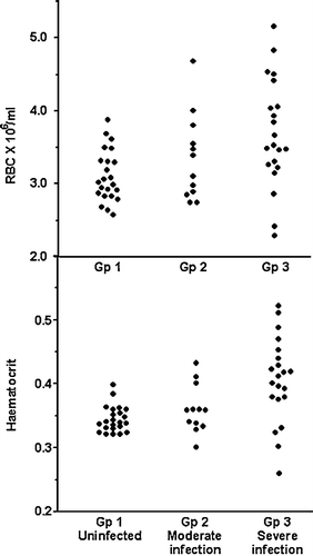
Haematological values were examined in pheasants only and are presented in . The mean RBC, haematocrit and haemoglobin values all increased but the data were significant only in the group 3 severely infected birds. Values in this group did vary greatly. Of three birds from one outbreak, two had very low blood values and the third also was below the mean values of uninfected birds (). Values in some other birds were very high. There was good correlation between the haematocrit and RBC counts (r=0.83) and between the haematocrit and haemoglobin values (r=0.91). Total leukocyte counts were similar between the groups. Differential counts were examined for only a limited number of uninfected (group 1, n=7); moderately infected (group 2, n=6) and severely infected (group 3, n=9) pheasants. The latter two groups showed an increase in the percentage of heterophils and a decrease in lymphocytes. This did not translate into changes in the absolute numbers of heterophils and lymphocytes, possibly due to the high variability in total WBC numbers and small number of differentials counted. Some but not all the uninfected birds showed high basophil counts, but only two basophils were seen in all the group 2 and group 3 blood smears (). Monocytes did not differ and ranged from 2 to 9%. Only three eosinophils were seen.
Table 1. Comparison of haematological values between young, farmed pheasants that are uninfected and those that are moderately or severely affected by spironucleosis
Biochemistry
The plasma biochemistry parameters of uninfected, healthy group 1 pheasants and partridges are compared in . Total protein and albumin levels were similar in both species although globulin levels were significantly higher in partridges, so altering the Alb:Glob ratio. Osmolality, Na+, Cl−, K+, Mg2+ were similar, but Ca2+ was significantly higher and PO4 3− significantly lower in partridges—giving a higher Ca2+: PO4 3− ratio in partridges. Glucose levels were similar but plasma triglyceride was considerably higher in partridges compared with pheasants.
Table 2. Comparison of plasma biochemistry values between healthy, young, farmed pheasants and partridges
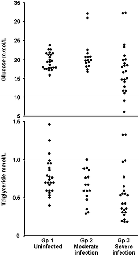
There were many significant differences in biochemical values between uninfected pheasants and the groups of Spironucleus-infected pheasants (). Levels of K+ and globulin were similar and there was little difference in the Alb:Glob ratio. Total protein and albumin were significantly lower in group 2 pheasants compared with uninfected pheasants, but mean values did not fall further in the severely affected group 3 birds as values in these were very variable with very high protein values in some birds. Osmolality, Na+ and Cl− levels declined significantly in relation to the severity of infection, while Ca+ and particularly PO4 3− levels, and so the Ca+: PO4 3− ratio, all were low in infected birds. Glucose () and triglyceride were very variable and significantly lower only in the group 3 birds, while Mg2+ was high in these birds. Overall, although the number of infected partridges examined was smaller, the biochemistry results () were very similar to those from pheasants.
Figure 2. Effect of Spironucleus infection on plasma glucose and triglycerides levels in pheasants. Gp, group.

Table 3. Comparison of plasma biochemistry values between young, farmed pheasants that are uninfected and those that are moderately or severely affected by spironucleosis
Table 4. Comparison of plasma biochemistry values between young, farmed partridges that are uninfected and those that were moderately or severely affected by spironucleosis.
Muscle
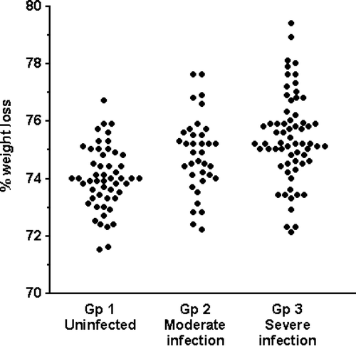
The wet weights (mean±standard error) of the breast and both leg muscles dissected from group 3 pheasants (n=8) were 19.2±1.2 and 26.6±1.1 g, respectively. This was 22.2 and 37.7% of the weight of the respective muscles recovered from uninfected group 1 pheasants (n=7), from which 86.6±6.5 and 70.6±3.6 g muscle, respectively, were recovered (P<0.001). Conversely, the water content of muscle from infected pheasants was significantly greater (). The water content of the group 1 birds’ muscle (n=52) was 74.0±0.15%, compared with 74.7±0.22% in the moderately infected group 2 birds (n=38) (versus group 1, P<0.01) and 75.4±0.19% in the severely affected group 3 birds’ muscle (n=65) (versus group 1, P<0.0001; versus group 2, P<0.05). Histologically, haematoxylin and eosin staining revealed no obvious necrosis, inflammation or other change in the breast muscle of group 3 compared with uninfected birds. Phosphotungstic acid haematoxylin staining of myofibrils did reveal some internal fragmentation, swelling and loss of striations.
Discussion
The red cell parameters of uninfected pheasants, where they can be compared, are similar to those reported previously (Ritchie et al., Citation1994). Haemoconcentration, indicating dehydration, was evident in many of the group 3 severely affected birds. Spironucleus infection does induce diarrhoea (Swarbrick, Citation1990) and reduced absorption of water by the intestines of comparably affected birds (Lloyd et al., Citation2005). Two of three birds from one farm that was severely affected with spironucleosis were anaemic but no evidence of haemorrhage was apparent due to coccidiosis or other cause. Possibly the anaemia was the result of dietary deficiency or previous disease. The red cell values of group 2 birds did not differ significantly from uninfected birds, although one or two birds did show elevated values. This is consistent with the fact that some may have been incubating the disease while others possibly were recovering. Also, absorption of water from comparable birds had been variable with some low, others normal and some showing elevated absorption (Lloyd et al., Citation2005). WBC counts were high but haematological values do vary widely in different birds species (Balasch et al., Citation1973) and the counts are not markedly higher than the 31.0 to 39.0 and 10.0 to 46.0×10⁹ reported for domestic and wild turkeys (Malewitz & Calhoun, Citation1957; Bounous et al., Citation2000) and the 36.9×10⁹ reported for adult red-legged partridges (Rico et al., Citation1977). The percentage of heterophils increased and lymphocytes decreased in the severely affected birds. This seems to be disease related as, while the stress of transport was shown to alter differential counts in pigeons (Scope et al., Citation2002), all the pheasants used here were caught and transported before examination. A high percentage of basophils has been described in galliformes, including pheasants (Ritchie et al., Citation1994), but, as the function of these in birds is not understood, reasons for their disappearance in diseased birds cannot be given.
The levels of eight of the biochemical parameters were similar in both uninfected pheasants and partridges. Partridges had higher levels of triglyceride, globulin and calcium, but lower phosphate, and so a higher Ca2+:PO4 3− ratio than did pheasants. These differences could be species related, as Rodriguez et al. (Citation2004) also found relatively high mean triglyceride values in red-legged partridges. Alternatively, as the partridges used for biochemical values originated from only one farm over a 2-year period while the uninfected pheasants originated from six farms, the differences could reflect different management and nutrition. Total protein, albumin and globulin values in these young, farmed pheasants are lower than previous values given for smaller groups of adults of the same species (Balasch et al., Citation1973; Rico et al., Citation1977). This may be age related as similar differences are described between 6-week-old broiler chickens and adult chickens (Ross et al., Citation1978; Mitruka & Rawnsley, Citation1977). Overall the plasma electrolyte and mineral values are relatively similar to those reported in adult game birds and in young and adult chickens—other than the potassium levels in the young pheasants examined here being considerably higher than described in adults by Balasch et al. (Citation1973), and glucose and triglyceride values are higher in this study than in chickens, no information being available for game birds.
Changes in plasma biochemical values in Spironucleus-infected birds are similar to those described in other animals (i.e. calves) affected by diarrhoea (Groutides & Michell, Citation1990). Infected birds generally had lower levels of plasma proteins, particularly albumin, but there were no significant differences between the moderately and severely infected birds as some of the latter showed signs of haemoconcentration with high values for total protein and globulins. Infected birds had low levels of albumin, osmolality and a number of electrolytes, and some of these declined in relation to severity of infection. There is probably an electrolyte loss due to the diarrhoea. Also, severely infected birds have malabsorption of water, and therefore presumably electrolytes. Triglyceride levels in birds have been shown to fall during a short period of starvation (Kurima et al., Citation1994) and, although some Spironucleus-infected birds did have food in their intestine, many had not been feeding in the hours before postmortem examination. The low glucose could be due to starvation, lack of absorption or use by the protozoa. Trophozoites of Spironucleus are numerous in heavily infected birds and are unceasingly moving, at least in vitro, presumably utilizing nutrients. Low plasma glucose is known to induce coma, and so on, and many, but not all, of the most severely affected birds were among those with the lowest blood glucose. The variability in these field cases could account for the individual variability in values of glucose, Mg2+, and so on, from low to elevated among this group. Comparable variability has been described in diarrhoeic calves compared with terminally ill calves (Groutides & Michell, Citation1990).
There was a marked loss of muscle mass. This is known to occur in as little as 10 days in severely affected birds (Lloyd, unpublished observations). However, this loss in muscle was not due to any dehydration related to the diarrhoea and the reduced intestinal absorption of water (Lloyd et al., Citation2005). In fact, there was overhydration of muscle tissue possibly related to the low plasma osmolality, although whether the increase in water content was intracellular or intercellular was not determined. There was no marked pathology in the muscles and the changes of swelling of myofibrils and loss of striations that were present possibly are due to the increased intracellular water content. Additional studies are needed to determine the mechanism(s) of weight loss.
Acknowledgments
This work was supported by the Game Conservancy Trust. The authors would like to thank the gamekeepers and Peter Dalton's Game Consultancy.
References
- Balasch , J. , Palacios , L. , Musquera , S. , Palomeque , J. , Jiminez , M. and Alemany , M. 1973 . Comparative haematological values of several galliformes . Poultry Science , 52 : 1531 – 1534 .
- Burgerolle , G. , Joyon , L. and Oktem , N. 1973 . Contribution a l’étude cytiloguique et phylétique des diplozaires (Zoomastigophora, Diplozoa, Dangeard 1910). II. Étude ultrastructurale du genre Spironucleus (Lavier 1936) . Parasitologica , IX : 405 – 502 .
- Burgerolle , G. , Kunsstyr , L. , Senaud , J. and Friedhoff , K.T. 1980 . Fine structure of trophozoites and cysts of the pathogenic diplomonad Spironucleus muris . Zeitschrift fur Parasitenkunde , 62 : 47 – 61 .
- Bounous , D.I. , Wyatt , R.D. , Gibbs , P.S. and Kilburn , J.V. 2000 . Normal haematological and serum biochemical reference intervals for juvenile wild turkeys . Journal of Wildlife Diseases , 36 : 393 – 396 .
- Cooper , G.L. , Charlton , B.R. , Bickford , A.A. and Nordhausen , R. 2004 . Hexamita meleagridis (Spironucleus meleagridis) infection in chukar partridges associated with high mortality and intracellular trophozoites . Avian Diseases , 48 : 706 – 710 .
- Eisenbrandt , D.L. and Russell , R.J. 1979 . Scanning electron microscopy of Spironucleus (Hexamita) muris infection in mice . Scanning Electron Microscopy , 3 : 23 – 27 .
- Flatt , R.E. , Halvorsen , J.A. and Kemp , R.L. 1978 . Hexamitiasis in a laboratory mouse colony . Laboratory Animal Science , 28 : 62 – 65 .
- Groutides , C.P. and Michell , A.R. 1990 . Changes in plasma components in calves surviving or dying from diarrhoea . British Veterinary Journal , 146 : 205 – 210 .
- Harper , F.D.W. 1991 . Hexamita species present in some avian species in South Wales . Veterinary Record , 128 : 130
- Hussain , A.Z. 2001 . Morphometric examination of the small intestines and caeca of pheasants infected with Hexamita and Trichomonas . Veterinary Record , 148 : 484 – 485 .
- Kurima , K. , Bacon , W.L. and Vasilatos-Younken , R. 1994 . Effect of somatostatin in plasma growth hormone and metabolite concentrations in fed and feed-deprived young female turkeys . Poultry Science , 73 : 714 – 723 .
- Lloyd , S. , Irvine , K.L. , Eves , S.M. and Gibson , J.S. 2005 . Fluid absorption in the small intestine of healthy game birds and those infected with Spironucleus spp . Avian Pathology , 34 : 252 – 257 .
- Malewitz , T.D. and Calhoun , M.L. 1957 . The normal haematological picture of turkey poults and blood alterations caused by enterohepatitis . American Journal of Veterinary Research , 18 : 396 – 399 .
- McDougald , L.R. 1998a . Intestinal protozoa important to poultry . Poultry Science , 27 : 1156 – 1158 .
- McDougald , L.R. 1998b . “ Hexamitiasis ” . In The Merck Veterinary Manual , Edited by: Aiello , S.E. 1897 – 1898 . Whitehouse Station : New Jersey Merck & Co .
- Mitruka , B.M. and Rawnsley , H.M. 1977 . Clinical Biochemical and Haematological Reference Values in Normal Experimental Animals , New York : Masson Publishing .
- Philbey , A.W. , Andrew , P.L. , Gestier , A.W. , Reece , R.L. and Arzey , K.E. 2002 . Spironucleosis in Australian king parrots (Alisterus scapularis) . Australian Veterinary Journal , 80 : 154 – 160 .
- Poynton , S.L. and Sterud , E. 2002 . Guidelines for species descriptions of diplomonad flagellates from fish . Journal of Fish Diseases , 25 : 15 – 31 .
- Poynton , S.L. , Fard , M.R. , Jenkins , J. and Ferguson , H.W. 2004 . Ultrastructure of Spironucleus salmonis n. comb. (formerly Octomitis salmonis Moore 1922, Davis 1926, and Hexamita salmonis sensu Ferguson 1979) with a guide to Spironucleus species . Diseases of Aquatic Organisms , 60 : 49 – 64 .
- Rico , A.G. , Braun , J.P. , Bernard , P. and Burgat-Sacaze , V. 1977 . Biometry, haematology, plasma biochemistry and plasma and tissues enzymology of the red partridge (Alectoris rufa) . Annales de Recherches Veterinaries , 8 : 251 – 256 .
- Ritchie , B.R. , Harrison , G.J. and Harrison , L.R. 1994 . Avian Medicine. Principles and Application , Lake Worth: Florida : Wingers Publishing Inc .
- Rodriguez , P. , Tortosa , F.S. , Millan , J. and Gortazar , C. 2004 . Plasma chemistry reference values from captive red-legged partridges (Alectoris rufa) . British Poultry Science , 45 : 565 – 567 .
- Ross , J.G. , Christie , G. , Halliday , W.G. and Morley Jones , R. 1978 . Haematological and blood chemistry “comparison values” for clinical pathology in broilers . Veterinary Record , 102 : 29 – 31 .
- Scope , A. , Filip , T. , Gabler , C. and Resch , F. 2002 . The influence of stress from transport and handling on haematologic and clinical chemistry blood parameters of racing pigeons (Columba livia domestica) . Avian Diseases , 46 : 224 – 229 .
- Swarbrick , O. 1990 . Hexamitiasis and an emaciation syndrome in pheasant poults: clinical aspects and differential diagnosis . Veterinary Record , 126 : 265 – 267 .
- Woodard , A.E. , Vohra , P. and Mayeda , B. 1983 . Blood parameters of one-year-old and seven-year-old partridges (Alectoris chukar) . Poultry Science , 62 : 2492 – 2496 .

