Abstract
An adult free-living European robin (Erithacus rubecula) with a large, firm, subcutaneous mass on the pectoral muscle was examined. The bird was unable to fly and died spontaneously. Necropsy revealed a yellowish, bilobate mass almost completely replacing the pectoral muscles with extensive osteolysis of the keel bone. Histopathology revealed a poorly demarcated, highly cellular sarcomatous tumour with metastases to the lungs, pulmonary blood vessels and heart. Immunohistochemistry was negative for neuron-specific enolase, S-100 protein and the p-27 major capsid protein of avian leukosis viruses. The homogeneously positive immunolabelling for vimentin and scattered positivity for myoglobin and desmin suggested a diagnosis of rhabdomyosarcoma. A retrospective examination of the records for 194 birds of the thrush family, including 64 robins submitted over a 20-year period, showed no diagnoses of neoplasia.
Rhabdomyosarcome des muscles pectoraux d'un rouge-gorge familier (Erithacus rubecula) vivant en liberté
Un rouge-gorge familier (Erithacus rubecula) vivant en liberté avec une masse sous-cutanée importante et ferme sur le muscle pectoral a été examiné. L'oiseau n’était plus capable de voler et est mort spontanément. L'autopsie a révélé une masse jaunâtre bilobée remplaçant presque complètement les muscles pectoraux avec une ostéolyse extensive de l'os du bréchet. L'histologie a révélé une tumeur cellulaire fortement sarcomateuse, peu démarquée, avec des métastases aux poumons, dans les vaisseaux sanguins des poumons et au cœur. L'Immunohistochimie a été négative pour l’énolase spécifique des neurones, la protéine S-100 et la protéine majeure de capside p-27 des virus de la leucose aviaire. L'immunomarquage a été positif de façon homogène pour la vimentine et positif de façon dispersée pour la myoglobuline et la desmine suggérant un diagnostic de rhabdomyosarcome. Un examen rétrospectif des enregistrements de 194 oiseaux de la famille des grives incluant 64 rouges-gorges soumis à examen au cours d'une période de 20 ans a montré qu'il n'y avait pas eu de diagnostic de néoplasie.
Rhabdomyosarkom in der Pektoralismuskulatur eines freilebenden Rotkehlchens (Erithacus rubecula)
Ein adultes freilebendes Rotkehlchen (Erithacus rubecula) mit einer großen, festen subkutanen Umfangsvermehrung im Pektoralismuskel wurde untersucht. Der Vogel war flugunfähig und verstarb spontan. Bei der Sektion stellte sich die Umfangsvermehrung als gelbliche, zweilappige Masse dar, die fast vollständig den Pektoralismuskel ersetzt und eine erhebliche Osteolyse des Sternums verursacht hatte. Histopathologisch war ein gering abgegrenzter, hoch zellulärer sarkomatöser Tumor mit Metastasen in den Lungen, den pulmonalen Blutgefäßen und dem Herzen erkennbar. Die immunhistochemische Untersuchung war hinsichtlich des Nachweises neurospezifischer Enolase, des Protein S-100 und des p-17 Hauptkapsidproteins der aviären Leukoseviren negativ. Die positive homogene Immunfärbung auf Vimentin und die verstreut liegenden Positivreaktionen auf Myoglobin und Desmin führten zur Diagnose Rhabdomyosarkom. Die retrospektive Durchsicht der Befundaufzeichnungen über 194 Vögel aus der Drosselfamilie einschließlich 64 Rotkehlchen aus den letzten 20 Jahren erbrachte keinen einzigen Fall einer Neoplasie bei diesen Vögeln.
Rabdomiosarcoma de los músculos pectorals de un European robin (Erithacus rubecula) salvaje
Se examinó un Petirrojo Europeo (Erithacus rubecula) adulto salvaje con una gran masa dura subcutánea en la región del músculo pectoral. El ave era incapaz de volar y murió espontáneamente. La necropsia mostró una masa bilobular amarillenta que prácticamente reemplazaba los músculos pectorales con ostelisis del hueso de la quilla. La histopatología mostró un tumor de tipo sarcomatoso altamente celular y poco definido con metástasis en los pulmones, vasos sanguíneos pulmonares y corazón. La inmunohistoquímica fue negativa para la enolasa específica de neuronas, la proteína S-100 y la proteína principal de la cápside p-27 del virus de la leucosis aviar. La tinción homogéneamente positiva para la vicentina y la positividad aislada para la mioglobina y desmina sugirieron un diagnóstico de rabdomiosarcoma. En un examen retrospectivo de 194 casos clínicos de aves de la familia Muscicapidae remitidos durante un periodo de 20 años y que incluían 64 Petirrojos Europeos no se encontró ningún diagnóstico de neoplasia.
Introduction
Primary tumours of the skeletal muscle are occasionally observed in avian species. Properly constructed surveys are needed to determine their incidence. Most cases are fibromas, fibrosarcomas, rhabdomyomas and rhabdomyosarcomas (RMS), which are reported in chickens usually as a consequence of infection with avian retroviruses (alpharetroviruses), the avian leukosis virus group (Riddel, Citation1996; Fadly & Payne, Citation2003). As for pet and free-living birds, rare reports of skeletal muscle tumours exist (Latimer, Citation1994; Friend & Thomas, Citation1999; Schmidt et al., Citation2003). Generally, malignant muscle neoplasms are reported twice as frequently as their benign counterparts (Latimer, Citation1994). A site predilection for these tumours is not observed (Schmidt et al., Citation2003). Grossly, RMS are often elevated, lobulated, relatively firm subcutaneous swellings. Microscopically, these neoplasms are composed of a pleomorphic population of fusiform to elongated cells (Latimer, Citation1994). The histopathologic diagnosis of RMS may be difficult; and confirmation of the diagnosis requires the use of electron microscopy or immunohistochemistry. Immunohistochemical characterization of these tumours requires a panel of antibodies. For this reason, antibodies against vimentin, desmin, actin, myoglobin, myosin and titin have proven to be useful in mammals (Cooper & Valentine, Citation2002) and in avian species as well (Frazier et al., Citation1993; Duncan & Fitzgerald, Citation1997; Hafner et al., Citation1998; Ijzer et al., Citation2002).
We describe a primary RMS in the pectoral muscles of a free-living European robin (Erithacus rubecula) with metastases to the lungs and myocardium. We also examined the findings for birds of the thrush family submitted to our institute over the past 20 years to determine the incidence of neoplasia.
Materials and Methods
Case history
An adult free-living European robin was presented to our laboratory in July 2007. The bird was found in a garden unable to fly and it was easily caught by hand. The clinical examination revealed a firm, non-ulcerated, bilobate, subcutaneous mass that blended into and almost completely replaced the pectoral muscles. The bird died spontaneously a few hours after submission and was immediately necropsied.
Samples of the mass and of the lung, testes, heart, liver, intestine, spleen, kidneys, and brain were fixed in 4% neutral buffered formalin, processed routinely, sectioned at 4 µm and sections were stained with haematoxylin and eosin. In addition, immunohistochemical stains were carried out on sections of the tumour. The primary antibodies, antigen retrieval methods, and incubation conditions used are described in . Biotinylated horse anti-mouse or goat anti-rabbit antibodies (as appropriate) were used as secondary antibodies, followed by the avidin–biotin complex immunoperoxidase procedure (Hsu et al., Citation1981). Diaminobenzidine (Dako, Milan, Italy) with 0.5% H2O2 in imidazol buffer (pH 7.2, 0.1 M) was used as the chromogen. Replicate sections of neoplastic tissue were similarly immunostained using normal rabbit serum (p-27, S-100 protein, myoglobin) and normal mouse serum (neuron-specific enolase [NSE], vimentin, desmin, α-smooth muscle actin) instead of the primary antiserum, as negative controls. Unaffected pectoral muscle included in the sections was used as positive control for myoglobin, vimentin, desmin, and α-smooth muscle actin. Sections of mouse nervous system were used as positive controls for NSE and S-100. Section of multiple organs from a subgroup J avian leukosis virus J-infected chicken was used as a positive control for p-27. Replicate sections of tumour were immunostained using normal rabbit and mouse serum for use as negative controls for the respective polyclonal and monoclonal antibodies. To determine the incidence of neoplasia, the caseload of our laboratory was checked since 1987. The findings for 194 birds of the thrush family, including 64 robins, were examined.
Table 1. Primary antibodies, antigen retrieval methods and dilutions used for immunohistochemistry
Results and Discussion
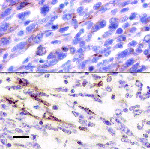
The bird was in poor body condition. The left pectoral muscle was completely effaced by a bulging, yellowish mass (2.5 x 2 cm) that invaded the keel bone and more than one-half of the right pectoral muscle. The wing joints were unaffected. The firm cut surfaces of the neoplastic lobules revealed necrotic centres surrounded by small, multifocal haemorrhages (). The testes were inactive. Microscopically, the tumour was densely cellular, unencapsulated and infiltrated the skeletal muscle. Large areas of necrosis and scattered haemorrhages were also detectable. The tumour was composed of streams and bundles of spindle cells (20 to 30 µm in length) with a small amount of fibrous stroma. Neoplastic cells exhibited indistinct cell borders, marked anisocytosis–anisokaryosis, a high nuclear to cytoplasmic ratio, a moderate amount of eosinophilic cytoplasm, and plump oval to elongated, centrally located nuclei coarsely clumped to vacuolated chromatin and one or two prominent magenta nucleoli. A few multinucleated giant cells with up to seven nuclei were also observed. Mitoses were six to seven per high-power field (x40) and were frequently bizarrely configured. Small metastases of the aforementioned neoplastic cells were observed in the lungs, in pulmonary blood vessels () and in the myocardium. It is conceivable that the bird died from the metastases possibly causing cardiac arrhythmia and circulatory changes. Additionally, several small granulomatous lesions centred on remnants of helminth parasites were detected in the liver and intestine. Immunolabelling for vimentin was homogeneous and intense (). About 30% of neoplastic cells were immunopositive for myoglobin (), 15% of neoplastic cells were immunopositive for desmin, while immunostaining for α-smooth muscle actin was intense for perivascular and interstitial cells but less intense and rare in neoplastic cells. Staining for p-27, NSE, and S-100 was absent in the tumour cells but was strongly positive in all control tissues.
Figure 1. Bulging, yellowish mass almost completely effacing the pectoral muscles of a robin. Cut surfaces reveal necrotic centres surrounded by multifocal haemorrhages.
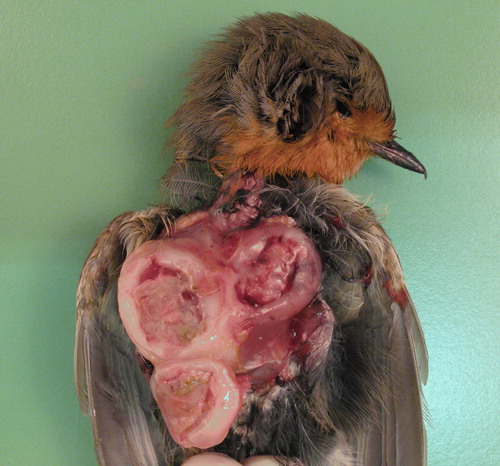
Figure 2. Micrograph showing metastases of the tumour in the lung parenchyma. Haematoxylin and eosin. Bar = 20 µm. Inset: neoplastic embolus in a pulmonary blood vessel. Haematoxylin and eosin. Bar = 35 µm.
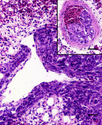
Figure 3. Upper: vimentin immunolabelling in the majority of the neoplastic cells. Lower: myoglobin immunoreactivity in scattered neoplastic cells. Avidin–biotin complex method, Mayer's haematoxylin counterstain. Bar = 10 µm.
Histologically, this tumour showed undifferentiated sarcomatous characteristics that can be shared by tumours including leyomyosarcoma, RMS, fibrosarcomas or even schwannomas. In similar cases, immunohistochemistry is routinely used to discriminate among the possible diagnoses (Cooper & Valentine, Citation2002). It is important to use a panel of antibodies for immunohistochemical characterization of these tumours because the antigens are variably expressed depending upon the degree of differentiation of the neoplastic cells. Moreover, care is required in interpreting the results of immunohistochemistry as staining, even though specific, may be focal (Cooper & Valentine, Citation2002). Lack of immunostaining for S-100 and NSE suggested a non-neural differentiation of this tumour, but homogeneous and intense vimentin positivity suggested the mesenchymal origin. Although scattered, the positive immunostaining for the myogenic components myoglobin and desmin led to the diagnosis of RMS. These results may be interpreted as a sign of poor differentiation. Vimentin is expressed early in rhabdomyocytes differentiation but is later lost. Desmin is also expressed early but persists; myoglobin is expressed later than desmin. Therefore, poorly differentiated RMS may express desmin but lack myoglobin (Cooper & Valentine, Citation2002). Our results partially agrees with previous reports of avian RMS. Fernandez-Bellon et al. (Citation2003) found positive and homogeneous immunostaining for vimentin and myoglobin in a RMS of a racing pigeon but neoplastic cells were negative for desmin. In contrast, Gulbahar et al. (Citation2005) reported intense immunolabelling for both vimentin and desmin but immunostaining for myoglobin was not performed. As for α-smooth muscle actin, rare positive immunostaining is not surprising in RMS as indicated previously (Cooper & Valentine, Citation2002; Gulbahar et al., Citation2005). Finally, negative immunostaining for p-27 excluded the involvement of avian leukosis viruses infection, which in chickens, and rarely in other birds, may be associated with the onset of similar tumours (Fadly & Payne, Citation2003).
Free-ranging wild birds are commonly afflicted with numerous parasites that occasionally cause illness and death (Cole & Friend, Citation1999). The parasitic granulomas observed in the sections of liver and intestine of the robin, although numerous, were considered incidental findings. The granulomatous reaction and fixation artefacts prevented identification of the parasites remnants by histopathological examination.
In conclusion, a case of RMS in a robin is described. Primary tumours of skeletal muscle are infrequent in avian species (Reece, Citation2003; Schmidt et al., Citation2003) particularly in free-living birds. Over a 20-year period, we have examined 194 birds belonging to the thrush family including 64 European robins, and this was the only tumour found. Curiously, the bird was an adult, male robin found on the outskirts of Milan in summertime. In Northern Italy robins are used to spending the breeding season in mountain forests up to an altitude of 2000 m, whereas they are common in the plains and in urban areas during fall and winter (Bottoni & Massa, Citation1992). It is therefore possible to speculate that the tumour prevented the bird from reaching the breeding areas at the proper time as it led to a progressive inability to fly.
References
- Bottoni , L. and Massa , R. 1992 . “ Pettirosso (Erithacus rubecula) ” . In Atlante degli uccelli svernanti in Lombardia , Edited by: Fornasari , L. , Bottoni , L. , Massa , R. , Fasola , M. , Brichetti , P. and Vigorita , V. 232 – 233 . Milano : Regione Lombardia—Università degli Studi di Milano .
- Cole , R.A. and Friend , M. 1999 . “ Miscellaneous parasitic diseases ” . In Field Manual of Wildlife Diseases—General Field Procedures and Diseases of Birds Resource Publication 167 , Edited by: Friend , M. and Franson , J.C. 249 Washington , DC : US Department of The Interior Fish and Wildlife Service .
- Cooper , B.J. and Valentine , B.A. 2002 . “ Tumors of muscle ” . In Tumors in Domestic Animals , 4th edn , Edited by: Meuten , D.J. 319 – 363 . Ames : Iowa State Press .
- Duncan , A.E. and Fitzgerald , S.D. 1997 . Clinical challenge: multiple primary rhabdomyosarcomas within the myocardium of a vulture . Journal of Zoo and Wildlife Medicine , 28 : 501 – 503 .
- Fadly , A.M. and Payne , L.N. 2003 . “ Neoplastic diseases ” . In Diseases of Poultry , 11th edn , Edited by: Saif , Y.M. , Barnes , H.J. , Glisson , J.R. , Fadly , A.M. , McDougald , L.R. and Swayne , D.E. 405 – 407 . Ames : Iowa State Press .
- Fernandez-Bellon , H. , Martorell , J. , Rabanal , R. and Ramis , A. 2003 . Rhabdomyosarcoma in a racing pigeon (Columba livia) . Avian Pathology , 32 : 613 – 616 .
- Frazier , K.S. , Herron , A.J. , Hines , M.E. , Miller , C.L. , Hensley , G.T. and Altman , N.H. 1993 . Metastasis of a myxoid leiomyosarcoma via the renal and hepatic portal circulation in a sarus crane (Grus antigone) . Journal of Comparative Pathology , 108 : 57 – 63 .
- Friend , M. and Thomas , N.J. 1999 . “ Miscellaneous Diseases ” . In Field Manual of Wildlife Diseases—General Field Procedures and Diseases of Birds Resource Publication 167 , online edn , Edited by: Friend , M. and Franson , J.C. 261 – 267 . Washington , DC : US Department of The Interior Fish and Wildlife Service .
- Gulbahar , M.Y. , Ozak , A. , Guvenç , T. and Yarim , M. 2005 . Retrobulbar rhabdomyosarcoma in a budgerigar (Melopsittacus undulatus) . Avian Pathology , 34 : 486 – 488 .
- Hafner , S. , Goodwin , M.A. , Smith , E.J. , Fadly , A. and Kelley , L.C. 1998 . Pulmonary sarcomas in a young chicken . Avian Diseases , 42 : 824 – 828 .
- Hsu , S.M. , Raine , L. and Fanger , H. 1981 . Use of avidin–biotin–peroxidase complex (ABC) in immunoperoxidase techniques: a comparison between ABC and unlabeled antibody (PAP) procedures . The journal of histochemistry and cytochemistry , 29 : 577 – 580 .
- Ijzer , J. , Dorrestein , G.M. and van der Hage , M.H. 2002 . Metastatic subcutaneous sarcoma and abdominal carcinoma in a peach-faced lovebird (Agapornis roseicolis) . Avian Pathology , 31 : 101 – 104 .
- Latimer , K.S. 1994 . “ Oncology ” . In Avian Medicine: Principles and Application , Edited by: Ritchie , B.W. , Harrison , G.J. and Harrison , L.R. 647 – 648 . Lake Worth , FL : Wingers Publishing, Inc .
- Reece , R.L. 2003 . “ Neoplastic diseases ” . In Diseases of Poultry , 11th edn , Edited by: Saif , Y.M. , Barnes , H.J. , Glisson , J.R. , Fadly , A.M. , McDougald , L.R. and Swayne , D.E. 541 – 564 . Ames : Iowa State Press .
- Riddel , C. 1996 . “ Muscular System ” . In Avian Histopathology , 2nd edn , Edited by: Riddell , C. 66 Saskatchewan , , Canada : American Association of Avian Pathologists .
- Schmidt , R.E. , Reavill , D.R. and Phalen , D.N. 2003 . “ Muscoskeletal system ” . In Pathology of Pet and Aviary Birds , Edited by: Schmidt , R.E. , Reavill , D.R. and Phalen , D.N. 153 Ames : Iowa State Press .