Abstract
Exposure of a virulent isolate of Newcastle disease virus (NDV) and two highly pathogenic avian influenza (HPAI) viruses, one of H7N1 subtype and the other H5N1 subtype, to a continuous ultraviolet B flux of approximately 90µW/cm2, which models solar ultraviolet radiation, resulted in an exponential decline in infectivity with time. The time taken for a reduction in titre of 1 log10 median tissue culture infectious dose for each virus was: NDV, 69 min; H7N1 HPAI virus, 158 min; and H5N1 HPAI, virus 167 min.
Introduction
Virulent Newcastle disease viruses (NDVs) and highly pathogenic avian influenza (HPAI) viruses cause devastating disease outbreaks with major welfare and economic impacts on the poultry industry. HPAI viruses, especially the H5N1 viruses responsible for the outbreaks occurring across Asia and into Europe and Africa since 1996, also present a zoonotic threat (WHO, Citation2012a). During outbreaks of Newcastle disease and HPAI diseases in poultry flocks, large amounts of virus may be released into the environment, contaminating poultry houses, vehicles and other fomites. Most control measures enforced for these diseases require cleansing and disinfection of buildings, vehicles and equipment; despite this, however, the possibility remains that some virus may escape disinfection or contaminate environmental water, or land in the case of free-range farms.
Following an outbreak of disease with one of these viruses, cleansing and disinfection methods are applied, followed by a period of time before restocking or use of equipment and vehicles that may have been contaminated can take place. In the European Union this period is 21 days (CEC, Citation1992, Citation2006) but it is not clear that this would be sufficient time for the natural inactivation of any remaining virus. Certainly HPAI viruses have been reported to persist for much longer periods in media such as water (Stallknecht et al., Citation1990). Ambient temperature and sunlight (as a source of ultraviolet [UV] light) are likely to be the two most important factors affecting virus survival in a clean environment.
It is well known that UV radiation acts as a natural environmental virucide because it causes photodimer formation between the pyrimidine bases in DNA and RNA, resulting in conformational changes that interrupt the viral replication process. The UV spectrum is divided into three categories: UVA, UVB and UVC (WHO, Citation2012b). The most active antimicrobial UV wavelengths are in the range of 200 to 280 nm as these are most highly absorbed by nucleic acids. This range corresponds to the lower ranges of UVB (280 to 320 nm) and UVC (185 to 280 nm), while UVA (320 to 400 nm), which is the most abundant in sunlight, has a much lower effect on viruses and other microbial agents. Additionally, the shorter UVC wavelengths are strongly absorbed by ozone in the Earth's atmosphere, leaving UVB as the principal environmentally effective virucidal radiation.
There have been a number of publications on the effects of UVC radiation on the infectivity of NDV or avian influenza viruses (Qayyum et al., Citation1999; Chumpolbanchorn et al., Citation2006; Sagripanti & Lytle, Citation2007; Shahid et al., Citation2009; Fasina et al., Citation2010; Lenes et al., Citation2010; Cutler et al., Citation2011) but none of these have attempted to model environmental UV radiation, specifically by assessing the virucidal effect of exposure to UVB radiation. In the present study we monitored the effect of continuous exposure to UVB radiation on the infectivity of virulent NDV, H7N1 HPAI and H5N1 HPAI subtypes.
Materials and Methods
Virus and cell lines
All viruses were supplied by the International Reference Laboratory for Avian Influenza and Newcastle Disease Virus at the Animal Health and Veterinary Laboratories Agency, Weybridge, UK. Amplified virus stocks in allantoic fluid for use in the study were quantified in 9-day-old to 11-day-old embryonated specific pathogen free fowls' eggs, and the titre is expressed as median egg infectious doses (EID50 values). The virus strains used were APMV-1/chicken/UK/1453/96 (NDV, 109.3 EID50/ml), A/ostrich/Italy/984/00 (HPAI H7N1, 108.3 EID50/ml) and A/turkey/Turkey/1/05 (HPAI H5N1, 109.2 EID50/ml). All work using these viruses was completed in high-containment laboratory facilities (DEFRA SAPO 4). Materials generated in the study were quantified using tissue culture; titres are expressed as the median tissue culture infectious dose (TCID50). NDV samples were quantified in Madin–Darby bovine kidney cells and influenza samples in Madin-Darby canine kidney cells.
Ultraviolet source and experimental design
The UV source was provided by a Reptisun 10.0 (Zoo Med Laboratories, Inc. San Luis Obispo, CA, USA) compact fluorescent lamp housed within a dome reflector and clamped to a lamp stand inside a class I/III microbiological safety cabinet. The UV source was set 14 cm above a polystyrene 96-well flat-bottomed microtitre plate (Cellstar; Greiner, Stonehouse, Gl UK) containing virus, to deliver a continuous UVB flux of approximately 90 µW/cm2 with the lid removed during the course of the experiment. A 90 µW/cm2 reading using a UVB specific meter (Solarmeter 6.2 UVB and 5.7 UVA + B; Solartech Inc.Harrison Township, MI USA) equated to a 240 µW/cm2 reading using a combined UVA and UVB meter, and thus an incidental 150 µW/cm2 of UVA flux is also administered to the sample during the experiment. This level of UVA flux was considered too low to contribute to photodimer formation. The stock virus was diluted 1:10 in 0.1 M phosphate-buffered saline (pH 7.2) and 200 µl added to each well in the microtitre plate. A control plate was placed within the same cabinet and shielded by a metal screen to represent an unexposed time course. Measurements taken through the plate both with and without the virus matrix present showed that the matrix column absorbed an average 12 µW/cm2 UVB. An ambient temperature of 19 to 21°C was maintained throughout the study. Triplicate samples were taken at specific time points over a 24-h period; samples were removed from both exposed and unexposed plates and stored at −70°C until quantification.
Prior to quantification, samples were diluted 1:10 in Eagle's minimal essential medium without serum and standard antibiotics, and the TCID50 titre determined in Madin–Darby bovine kidney (NDV) and Madin–Darby canine kidney (HPAI) cell lines. After incubation at 37°C for up to 7 days, infected tissue culture plates were stored at −70°C prior to testing for haemagglutination activity using the standard procedures (Swayne, Citation2012). Positive wells were recorded and the virus titre calculated using the Spearman–Karber formula. The log10 TCID50 values for triplicate samples were used for statistical analysis using GraphPad Prism v 6.00 (GraphPad Software, San Diego, California, USA).
Results
For each of the three viruses tested, exposure to a continuous UVB flux of approximately 90 µW/cm2 resulted in a decline in infectivity, demonstrated using regression analysis to plot lines of best fit ().
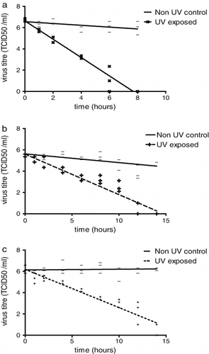
Some loss of infectivity was detected in the controls that had not undergone UVB exposure, but this was marginal in comparison, as follows. After 10 h of UVB exposure, the NDV titre had declined from 6.8 TCID50/ml to undetectable while the control virus titre had fallen to 5.7 TCID50/ml. For the influenza viruses at 14 h the titres for the H7N1 UVB exposed virus had fallen from 5.5 TCID50/ml to undetectable, but only to 4.9 TCID50/ml for the control, while for the H5N1 virus the initial titre of 6.2 TCID50/ml had declined to 1 TCID50/ml but the control virus titre was estimated at 6.3 TCID50/ml. T-tailed analysis shows for each of the viruses that the control and UV exposed slopes were significantly different (P < 0.0001). Comparisons between viruses of the UV exposed slopes show that the influenza viruses are not significantly different to each other (P = 0.5182) whereas the NDV slope significantly differs from either of the influenza slopes (P < 0.0001).
The time needed to reduce the titre of each virus by 1 log10 (90%) is also referred to as the D value; this was calculated for each virus and the values obtained were: NDV, 69 min; H7N1 HPAI, 158 min; and H5N1 HPAI, 167 min.
Discussion
From the data presented, one can conclude that exposure to UVB light increases the loss of virus infectivity for each virus tested when compared with unexposed virus samples. From the slope of the linear decline in infectivity () one could conclude that the two HPAI viruses were similarly sensitive to UVB, but the decline in infectivity of the NDV isolate was more than 2.5 times faster. An explanation for the difference in sensitivity of NDV to UVB radiation could be related to the non-segmented RNA genome of NDV compared with the eight RNA gene segments of the influenza viruses. On exposure to UVB, a photodimer reaction in part of the continuous ∼15 kb NDV genome may be sufficient to prevent replication—whereas UVB exposure of influenza viruses might result in photodimer formation affecting only one or more of the gene segments, but not the entire genome with viable genes remaining. Partially disabled influenza virus particles may be able to infect cells and reassort with other partially, but differently, disabled influenza virus particles to give a full complement of viable genes and allow replication to take place, increasing survival of the avian influenza viruses compared with NDV.
In studies of the virucidal effects of UV radiation, especially if they are to be a help in understanding the field situation, it is important to take into account not just the variations that may occur with the pathogen, but also the effect of the matrix or medium in which the pathogen is suspended. In the present study the experimental conditions were designed so that UVB radiation could readily penetrate the medium to reach virus. Other experimenters used peptone water (Shahid et al., Citation2009) or turbid, diluted faecal material (Chumpolbanchorn et al., Citation2006). They concluded that under the specific conditions described in their studies UVB radiation did not reduce viral infectivity. However, it seems possible that their study design may have been a contributing factor to this observation. For example, the presence of a physical glass barrier (Fasina et al., Citation2010) is likely to have prevented UV radiation reaching the virus. Radiant energy emitted by a UV source is readily blocked by dust and other contaminating particulates, and the present study was designed to minimize the presence of particulates and shadows for the duration of the UVB exposed time course.
The use of sunlight, or an artificial UVB source, as an addition component of a post-outbreak decontamination routine could be implemented at minimal cost. Any residual virus present on an exposed surface of equipment or fomites may have its infectivity greatly reduced by this treatment; however, the inactivating properties of UVB require direct, unimpeded exposure to the radiation source, which is difficult to ensure in the field.
From the data obtained in the present study we conclude that UVB light does have a marked inactivating effect on the infectivity of a NDV and two HPAI viruses suspended in a clear liquid solution and a clean environment. The NDV is more than twice as sensitive to the inactivating effects of UVB light than the HPAI viruses used in this experiment.
Acknowledgement
Funding from Defra project SE4009.
References
- CEC. (1992). Council directive 92/66/EEC introducing community measures for the control of Newcastle disease. Official Journal of European Communities, L260, 1–20.
- CEC. (2006). Council directive 2005/94/EC community measures for the control of avian influenza. Official Journal of European communities, L10, 16–65.
- Chumpolbanchorn, K., Suemanotham, N., Siripara, N., Puyati, B. & Chaichoune, K. (2006). The effect of temperature and UV light on infectivity of avian influenza virus (H5N1, Thai field strain) in chicken fecal manure. The Southeast Asian Journal of Tropical Medicine and Public Health, 37, 102–105.
- Cutler, T., Wang, C., Qin, Q., Zhou, F., Warren, K., Yoon, K.J., Hoff, S.J., Ridpath, J. & Zimmerman, J. (2011). Kinetics of UV254 inactivation of selected viral pathogens in a static system. Journal of Applied Microbiology, 111, 389–395. 10.1111/j.1365-2672.2011.05046.x
- Fasina, F.O., Egbuji, A.N., Gado, D.A., Nyam, D.C, Olawuyi, A.K., Meseko, C.A., Oladokun, A.T., Ularamu, H.G., Ponman, S. & Nwagbo, I. (2010). Non-attenuation of highly pathogenic avian influenza H5N1 by laboratory exposure to ultraviolet rays. African Journal of Clinical and Experimental Microbiology, 11, 129–136.
- Lenes, D., Deboosere, N., Menard-Szczebara, F., Jossent, J., Alexandre, V., Machinal, C. & Vialette, M. (2010). Assessment of the removal and inactivation of influenza viruses H5N1 and H1N1 by drinking water treatment. Water Research, 44, 2473–2486. 10.1016/j.watres.2010.01.013
- Qayyum, R., Naeem, M. & Muhammad, K. (1999). Effect of physico-chemical factors on survival of Newcastle disease virus. International Journal of Agriculture & Biology, 1, 42–44.
- Sagripanti, J.L. & Lytle, C.D. (2007). Inactivation of influenza virus by solar radiation. Photochemistry & Photobiology, 83, 1278–1282. 10.1111/j.1751-1097.2007.00177.x
- Shahid, M.A., Abubakar, M., Hameed, S. & Hassan, S. (2009). Avian influenza virus (H5N1); effects of physico-chemical factors on its survival. Journal of Virology 6, 38. 10.1186/1743-422X-6-38
- Stallknecht, D.E., Kearney, M.T., Shane, S.M. & Zwank, P.J. (1990). Effects of pH, temperature, and salinity on persistence of avian influenza viruses in water. Avian Diseases, 34, 412–418. 10.2307/1591429
- Swayne, D. (2012). Avian influenza. Chapter 2.3.4. In World Organisation for Animal Health (OIE). (Ed.). Manual of diagnostic tests and vaccines for terrestrial animals (pp.1–17). Paris: OIE.
- WHO. (2012a). Cumulative number of confirmed human cases for avian influenza A(H5N1) reported to WHO, 2003–2012. Retrieved from http://www.who.int/influenza/human_animal_interface/EN_GIP_20120810CumulativeNumberH5N1cases.pdf
- WHO. (2012b). Ultraviolet light and the Intersun Programme. Retrieved from http://www.who.int/uv/intersunprogramme/en/