
ABSTRACT
Currently, turkey coronaviruses (TCoV) are isolated from homogenized intestines of experimentally infected embryos to ensure a maximum recovery of viral particles from all components of the intestines. However, the process of homogenization also ensures release of a significant amount of cellular RNAs into the sample that hinders downstream viral genome sequencing. This is especially the case for next generation sequencing (NGS) which sequences molecules at random. This characteristic means that the heavily abundant cellular RNA in the sample drowns out the minority viral RNA during the sequencing process and, consequently, very little to no viral genome data are obtained. To address this problem, a method was developed, in which 10 descendent isolates of the European strain of TCoV were recovered uniquely from the intestinal lumen without homogenization of the tissue. For nine out of 10 samples, NGS produced viral RNA reads with good coverage depth over the entire TCoV genomes. This is a much-needed new, simple and cost effective method of isolating TCoV that facilitates downstream NGS of viral RNA and should be considered as an alternative method for isolating other avian enteric coronaviruses in the interest of obtaining full-length genome sequences.
Introduction
Turkey coronaviruses (TCoV) belong to the Coronaviridae family, subfamily Orthocoronavirinae, genus Gammacoronavirus, subgenus Igacovirus, and are important viral agents involved in multifactorial enteritis in turkeys (Guy, Citation2020). Isolating these viruses has been achieved by inoculation of field samples into the intra-amniotic cavity of turkey embryonated eggs (Guionie et al., Citation2013; Guy, Citation2015). In this procedure, intestines of embryos are collected four days post-inoculation, homogenized and clarified by centrifugation to produce a virus stock. Whilst this technique means that there is a maximum recovery of infectious virus from the tissue, it also means that there is a massive release of cellular nucleic acid into the virus stock, which can vastly outnumber viral nucleic acids. In terms of “classical virology” (purification, titration, antigen preparation, etc. ) this does not pose a problem. However, a sample of this type is not optimal for next generation sequencing (NGS) of viral genome molecules (Flageul et al., Citation2021). This is because NGS is based on random nucleotide sequencing, and thus nucleic acids at the higher ratio in the sample (in this case cellular RNAs) are preferentially sequenced and viral RNA sequencing output is drastically reduced.
To circumvent this problem, enrichment methods that increase the proportion of a targeted genome can be used. One way is to amplify the targeted genome using polymerase chain reaction (PCR) (Zhou et al., Citation2009). However, this technique is sensitive to (i) PCR efficiency in amplifying the target molecule and to (ii) the incorporation of erroneous nucleotides that lead to erroneous downstream sequences. A second way is to remove undesired genetic material (in this case cellular), either by purifying viral particles, using ultracentrifugation (Kuo et al., Citation2016), gel exclusion size (Loa et al., Citation2002), or micro-fluidic isolation (Han et al., Citation2015), or by purifying viral genomes using specific nucleotide primers coupled to magnetic beads (Xing et al., Citation2016). The major problem with these purification methods is that they all require sufficient amounts of starting sample to overcome an important loss of biological material. This makes them unsuitable when dealing with limited amounts of sample, or samples in which the target is at low abundance. Also, all of these methods are costly.
In the present study, a novel method of isolating avian enteric coronaviruses in ovo, in the interest of improving downstream NGS, was developed. The method focused on removing cellular nucleic acids by eliminating the process of tissue homogenization described in “classical” methods (Guionie et al., Citation2013) and on creating an efficient protocol for recovery of viral particles uniquely from the intestinal lumen. Samples prepared using this method were subjected to iron torrent NGS to analyse the efficacy in recovering sequence data along the full-length genome with good coverage depth.
Materials and Methods
Samples
The liquid contents from the jejunum, ileum and caeca of nine intestinal samples that had been collected at day 2 or 3 post-infection from nine different turkeys infected with TCoV 080385d during a previous experimental trial (Environmental and Occupational Health & Safety’s (ANSES) ethical committee authorization code APAFIS#10839-2017073115311452v4) were harvested by compressing the tissue with sterile tweezers. Contents were then mixed with phosphate buffered saline (1 g/ml), vortexed and centrifuged (3000 × g, 4°C, 15 min). The supernatants were then collected.
Novel virus isolation from the intestinal lumen of turkey embryos
Each supernatant described above was diluted 1/10 in phosphate buffer saline supplemented with penicillin (10 U/µl), streptomycin (10 µg/µl) and amphotericin B (250 µg/µl). One hundred microlitres of each dilution were then inoculated into the amniotic cavity of 19-day-old embryonated SPF turkey eggs (provided by the Anses department for breeding and experimentation in poultry and rabbits of the laboratory of Ploufragan-Plouzané-Niort) and incubated at 37°C for 4 days. Five eggs were used per sample, resulting in the inoculation of 135 eggs. Embryos that died during the first 24 h of incubation were discarded. After incubation, the intestines from 23-day-old embryos were harvested in 400 µl of PBS before being vortexed for one minute at maximum speed (2500/min) and centrifuged (3300 × g, 4°C, 15 min). Supernatants were collected and constituted the luminal virus stocks (LVS).
RNA extraction
RNAs were extracted from LVS using the King Fisher automatic extractor (Thermo Fisher Scientific, Illkirch, France). Briefly, 200 µl of viral stock were lysed with 300 µl of RLT buffer (QIAgen) supplemented with 3 µl of β-mercaptoethanol (Sigma, Saint-Quentin-Fallavier, France) and 16 µg of glycogen (Invitrogen Villebon Sur Yvette). After lysis, 20 µl of magnetic beads suspension and 400 µl of isopropanol were added for RNA precipitation. Three washing steps were performed using washing buffers and ethanol 80% (Thermo Fisher Scientific, Illkirch, France). Finally, RNAs were eluted using 100 µl of elution buffer. Beads, washing and elution buffers were provided in the NucleoMag Vet kit (Macherey-Nagel, Hoerdt, France).
Detection of viral RNA
Extracted RNAs were tested using a pan avian coronavirus (AvCoV) RT-qPCR as previously described (Callison et al., Citation2006)
Next generation sequencing
Ten AvCoV RNA- positive LVS were chosen. From each of these samples, a maximum concentration of RNA was used respecting an upper ceiling of 500 ng in a maximum volume of 20 µl and was treated with 4 units of DNase (Turbo DNA-free™ kit from Life Technologies Villebon Sur Yvette, France). Following this treatment, ribosomal RNAs were removed (ribodepletion) using Low Input RiboMinus™ Eukaryote System v2 (Life Technologies,Villebon Sur Yvette, France). RNA libraries were then prepared with Ion Total RNA-seq kit 2 (Life Technologies,Villebon Sur Yvette, France). Briefly, this process involved enzymatic fragmentation of RNA and ligation of adaptors to the fragmented RNA to generate cDNA from RNA molecules by reverse transcription. Resulting cDNAs were then amplified and barcoded for identification. Finally, library quality was checked on a Bioanalyzer from Agilent (High Sensitivity DNA Kit and RNA 6000 Pico Kit, Agilent, Courtaboeuf, France) and submitted to Ion-Torrent sequencing. For the bioinformatic process, read trimming was achieved with Trimmomatic (v0.38) using default parameters (Bolger et al., Citation2014) and then the total number of reads obtained for each LVS were randomly normalized to one million using the tool Seqtk. The genomic composition of the samples was determined by aligning clean NGS data on the NCBI nt database (last update, November 2018) using Megablast (2.2.26) with default parameters (Morgulis et al., Citation2008) and visualised with KronaTools (v2.7) (Ondov et al., Citation2011). Additionally, reads were aligned on the previously published genome of TCoV-080385d (GenBank accession number KR822424.1, Brown et al., Citation2016) using BWA (v0.7.17-r1188) and Samtools (v1.9.9).
Results and discussion
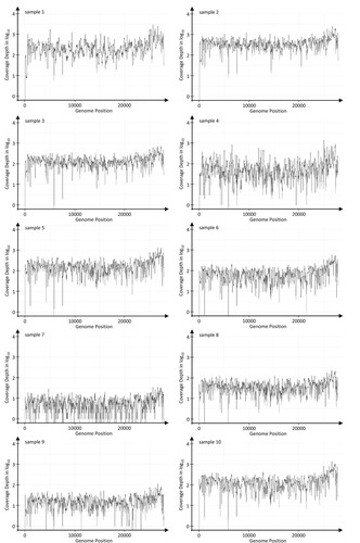
In total, 87 RNA samples were positive for AvCoV RNA (data not shown), and 10 of these samples were selected for NGS. After aligning reads over the reference genome, coverage depth was evaluated. The median coverage depths for the 10 LVS were 242, 391, 148, 57, 180, 79, 6, 42, 18 and 150, and sequence reads along the full-length TCoV genome were obtained in 9/10 LVS (LVS 1, 2, 3, 4, 5, 6, 8, 9 and 10 ).
Figure 1. TCoV RNA coverage depths obtained for 10 LVS after total NGS reads obtained for each had been randomly normalized to one million using the tool Seqtk and then aligned to the TCoV-080385d reference genome (GenBank accession number KR822424.1). Results were formatted and visualised using the R package ggplot2 (v3.1.0).
The random sequencing approach of NGS allows sequencing of all nucleic acid molecules present within a sample. However, in practice, this random technique of sequencing can make the characterization of minority nucleic acid chains difficult since most of the sequenced data would be determined from the most represented nucleic acid chains. Thus, a complete picture of the minority viral genomic nucleic acid composition of a sample of interest is seldom obtained. In this study, a method has been developed that demonstrates for the first time that (i) TCoVs can be isolated from the lumen of the intestines of inoculated turkey embryos without the need for homogenization of the intestinal tissue itself and (ii) avoiding tissue homogenization provides a sample much more suited to NGS.
This new method is a much-needed in ovo alternative for avian enteric coronavirus propagation that allows successful downstream NGS. Furthermore, compared to other purifying or amplification techniques, this simple and cost-effective method, incorporating well practiced in ovo methods, does not require significant amounts of starting sample, expensive and specific equipment or reagents.
Finally, these results suggest that if a virus replicating in vivo can be collected from cellular secretions, and tissue homogenization can be avoided, then nonspecific NGS sequencing of their genomes should yield better results.
Disclosure statement
No potential conflict of interest was reported by the authors.
Additional information
Funding
References
- Bolger, A.M., Lohse, M. & Usadel, B. (2014). Trimmomatic: a flexible trimmer for Illumina sequence data. Bioinformatics, 30, 2114–2120.
- Brown, P.A., Touzain, F., Briand, F.X., Gouilh, A.M., Courtillon, C., Allée, C., Lemaitre, E., De Boisséson, C., Blanchard, Y. & Eterradossi, N. (2016). First complete genome sequence of European turkey coronavirus suggests complex recombination history related with US turkey and guinea fowl coronaviruses. Journal of General Virology, 97, 110–120.
- Callison, S.A., Hilt, D.A., Boynton, T.O., Sample, B.F., Robison, R., Swayne, D.E. & Jackwood, M.W. (2006). Development and evaluation of a real-time Taqman RT-PCR assay for the detection of infectious bronchitis virus from infected chickens. Journal of Virological Methods, 138, 60–65.
- Flageul, A., Lucas, P., Hirchaud, E., Touzain, F., Blanchard, Y., Eterradossi, N., et al. (2021). Viral variant visualizer (VVV): a novel bioinformatic tool for rapid and simple visualization of viral genetic diversity. Virus Research, 291, 198201.
- Guionie, O., Courtillon, C., Allee, C., Maurel, S., Queguiner, M. & Eterradossi, N. (2013). An experimental study of the survival of turkey coronavirus at room temperature and +4°C. Avian Pathology, 42, 248–252.
- Guy, J.S., 2020. Turkey coronavirus enteritis. In D.E. Swayne (Ed.). Diseases of Poultry, 14th ed. pp. 402–408. Wiley Blackwel.
- Guy, J.S. (2015). Isolation and propagation of coronaviruses in embryonated eggs. In H.J. Maier, E. Bickerton & P. Britton (Eds.), Coronaviruses: Methods and Protocols, Methods in Molecular Biology (pp. 63–71). New York, NY: Springer.
- Han, H.-S., Cantalupo, P.G., Rotem, A., Cockrell, S.K., Carbonnaux, M., Pipas, J.M. & Weitz, D.A. (2015). Whole-genome sequencing of a single viral species from a highly heterogeneous sample. Angewandte Chemie International Edition, 54, 13985–13988.
- Kuo, L., Hurst-Hess, K.R., Koetzner, C.A. & Masters, P.S. (2016). Analyses of coronavirus assembly interactions with interspecies membrane and nucleocapsid protein chimeras. Journal of Virology, 90, 4357–4368.
- Loa, C.C., Lin, T.L., Wu, C.C., Bryan, T.A., Thacker, H.L., Hooper, T. & Schrader, D. (2002). Purification of turkey coronavirus by Sephacryl size-exclusion chromatography. Journal of Virological Methods, 104, 187–194.
- Morgulis, A., Coulouris, G., Raytselis, Y., Madden, T.L., Agarwala, R. & Schäffer, A.A. (2008). Database indexing for production MegaBLAST searches. Bioinformatics, 24, 1757–1764.
- Ondov, B.D., Bergman, N.H. & Phillippy, A.M. (2011). Interactive metagenomic visualization in a web browser. BMC Bioinformatics, 12, 385.
- Xing, N., Guan, X., An, B., Cui, B., Wang, Z., Wang, X., Zhang, X., Du, Q., Zhao, X., Huang, Y. & Tong, D. (2016). Ultrasensitive detection of porcine epidemic diarrhea virus from fecal samples using functionalized nanoparticles. PLoS One, 11, e0167325.
- Zhou, B., Donnelly, M.E., Scholes, D.T., St. George, K., Hatta, M., Kawaoka, Y. & Wentworth, D.E. (2009). Single-reaction genomic amplification accelerates sequencing and vaccine production for classical and swine origin human influenza A viruses. Journal of Virology, 83, 10309–10313.